Irritative changes in the EEG are a form of general disturbances in biopotentials. They are more often observed in meningovascular neoplasms, which are intimately connected to the vessels of the meninges. To record the electrical activity of the brain, neurophysiologists at the Yusupov Hospital perform EEG using the latest equipment from world manufacturers.
The results of the study are deciphered by candidates of medical sciences. Leading experts in the field of neurology and neurophysiology analyze EEG data using a computer program. If there are EEG changes that can be interpreted ambiguously, at a meeting of the expert council, professors and doctors of the highest category discuss the results of the study and collectively make a decision regarding the diagnosis and treatment tactics of the patient.
Irritative changes on the EEG
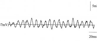
In the presence of irrigative changes against the background of disorganization of the alpha rhythm with a pointed shape and uneven amplitude of alpha oscillations, the voltage of beta oscillations increases 2-3 times. Pathological changes may appear in combination with diffuse epileptoid activity. In some patients, sharp waves are consistently recorded on the EEG, which coincide with the rhythm of the electrocardiogram. The totality of these EEG changes, expressed equally in all areas of the hemispheres, reflects irritative phenomena in the cerebral cortex. They are caused by an excessive influx of afferent impulses from the angioreceptive zones and from the richly innervated meninges, which are constantly exposed to the effects of a slowly growing tumor.
When EEG is recorded in such patients over time, as the tumor grows, the amplitude of frequent rhythms decreases and low-amplitude delta waves appear, equally pronounced in all areas of both hemispheres of the brain. The stage of irritative cerebral disturbances of biopotentials is more often observed when vascular neoplasms are located in the sagittal, perisagital and anteriobasal regions of the brain. In these areas, tumor nodes are directly connected to the venous sinus.
If patients suffering from brain tumors have symptomatic epilepsy, irritative cerebral changes are also recorded in the EEG in the early stages of the disease. They manifest themselves as a combination of sharpened waves of the alpha rhythm, increased beta oscillations and epileptoid diffuse potentials. Against the background of a general disturbance of cortical rhythm, the EEG may record an epileptogenic focus in the area of the cortex that is directly affected by the tumor. A mildly irritative type of EEG indicates minor damage to brain structures.
Reasons for changes
Diffuse changes in the bioelectrical activity of the brain are not inherited and do not arise out of nowhere. These anomalies are a consequence of disruption of certain brain processes, and sometimes damage to neural connections. What else leads to disturbances in the functioning of the central nervous system:
- Anemia (anemia). The brain receives little oxygen, and cells—neurons—starve.
- Atherosclerosis of cerebral vessels.
- Inflammation due to infection (meningitis, encephalitis, arachnoiditis).
- Associated disorders. Often the cause of this condition is a persistent metabolic disorder and lack of sleep.
In case of gross changes in brain activity, examination usually reveals:
- necrotic processes;
- scarring;
- brain swelling.
The causes of such serious conditions are injuries and bruises. A neurologist should strictly monitor all changes. Such a disease cannot be left to chance.
Functions of Brain Waves
The brain is an electrochemical organ. Electrical activity in the brain manifests itself in the form of brain waves. Four types of waves are recorded on the EGG:
- beta waves (the fastest oscillations with large amplitude, the frequency of which is in the range of 15–40 Hz) are generated by the awake brain, actively involved in mental activity;
- alpha waves are the opposite of beta waves, their amplitude is larger and their frequency is 9-14 Hz;
- for theta waves, the amplitude is even greater, and the frequency is 5-8 Hz, they are generated by the brain of a person who is almost asleep;
- delta waves have a maximum amplitude and frequency of 1.5-4 Hz.
If the frequency of theta waves on the ECG drops to zero, this means that brain death has occurred. Deep, dreamless sleep is characterized by a theta wave frequency of 2-3 Hz. When a person lies down in bed and reads for a few minutes before going to sleep, he or she is in a “low beta” state. The moment we put down a book, turn off the light and close our eyes, the brain waves successively pass through the stages of beta, alpha, theta, and ultimately delta.
The four types of brain vibrations are common to all people, regardless of gender, age, nationality, culture and nationality. The results of EEG studies show that although one frequency always dominates in brain oscillations, the remaining three, depending on the level of activity of the person, are also always present.
Diffuse changes: symptoms
How do mild diffuse changes in the bioelectrical activity of the brain manifest themselves? Symptoms of changes will be noticeable even with the slightest changes in the normal activity of the biocomputer. As a rule, they are as follows:
- dizziness;
- slowness, weakness.
- With increasing changes, headaches and cramps appear.
The psyche also changes under the influence of changes in the brain. A person experiences sudden changes in mood, his behavior begins to seem hysterical to others. The circle of interests narrows, the motivation to act disappears. It becomes increasingly difficult for the patient to remember new information.
If these symptoms persist for a long time, you should urgently consult a doctor and get a diagnosis. Diffuse changes in the bioelectrical activity of the brain are a very serious disease. More precisely, its harbinger. If nothing is done, the condition worsens very quickly.
EEG interpretation
Decoding an electroencephalogram is the process of interpreting it, taking into account the clinical symptoms that the patient has. When analyzing the EEG, neurophysiologists at the Yusupov Hospital take into account:
- basal rhythm;
- the level of symmetry in the electrical activity of brain neurons in the right and left hemispheres;
- commissure activity;
- EEG changes during functional tests (hyperventilation, photostimulation, opening and closing eyes).
Neurologists-neurophysiologists make the final diagnosis only taking into account certain clinical signs of the disease that worry the patient.
Changes in the alpha rhythm on the EEG are the following signs:
- constant recording of the alpha rhythm in the frontal lobes of the brain;
- violation of sinusoidal waves;
- interhemispheric asymmetry above 30%;
- unstable frequency;
- paroxysmal or arc-shaped rhythm;
- rhythm index less than 50%;
- amplitude is less than 20 µV or more than 90 µV.
Severe interhemispheric asymmetry may be evidence of a tumor, brain cyst, heart attack, stroke, or scar at the site of an old hemorrhage. High frequency and instability of the alpha rhythm can appear after traumatic brain injury. A disorganized type of EEG (impaired organization of the alpha rhythm or its complete absence) indicates acquired dementia.
In children, delayed psychomotor development is indicated by:
- alpha rhythm disorganization;
- moving the focus of activity from the occipital and parietal regions;
- increased synchrony and amplitude;
- excessive response to hyperventilation;
- weak short activation reaction.
A decrease in the amplitude of the alpha rhythm on the EEG, a weak activation reaction, and a shift in the focus of activity from the back of the head and crown are signs of psychiatric pathology. Excitable psychopathy is manifested by a slowdown in the frequency of the alpha rhythm against the background of normal synchrony. Inhibitory psychopathy is characterized by EEG desynchronization, low frequency and alpha rhythm index. Increased synchronization of the alpha rhythm in all parts of the brain, a short activation reaction are a sign of neuroses.
In patients, neurophysiologists determine the following pathological types of beta rhythm:
- paroxysmal discharges;
- low frequency, distributed along the convexital surface of the brain (adjacent to the frontal, temporal, parietal and occipital bones of the skull);
- amplitude more than 7 µV;
- violation of symmetry between the hemispheres in amplitude;
- sinusoidal type of beta rhythm.
Beta rhythm disturbances on the EEG indicate brain pathology. The presence of diffuse beta waves with an amplitude no higher than 50-60 μV indicates a concussion. Short spindles in the beta rhythm indicate encephalitis. Beta waves with a frequency of 16–18 Hz and high amplitude in the central and anterior parts of the brain are signs of delayed psychomotor development of a child.
Normally, the theta rhythm and delta rhythm can only be recorded on the EEG of a sleeping person. In a state of wakefulness, such slow waves appear in the presence of degenerative processes in the brain tissue, which are combined with compression, high blood pressure and lethargy. Paroxysmal theta and delta waves in a patient while awake are recorded when the deep parts of the brain are damaged.
High amplitude delta waves are evidence of a tumor. The predominance of theta and delta waves on the EEG with maximum activity in the occipital region, flashes of bilaterally synchronous waves, the number of which increases with hyperventilation, are a sign of delayed psychomotor development of the child.
Traumatic subarachnoid hemorrhage
Traumatic subarachnoid hemorrhage is an accumulation of blood under the arachnoid membrane of the brain, the most common form of intracranial hemorrhage in traumatic brain injury (TBI).
Traumatic damage can be caused by both direct damage to vessels located in the subarachnoid space (pial arteries and veins) and severe vasomotor disturbances accompanying the course of TBI. Typically, the development of traumatic subarachnoid hemorrhage accompanies brain contusions. Therefore, the detection of blood in the cerebrospinal fluid (CSF) in patients with TBI is considered as one of the signs of brain damage.
Editorial: Hereditary Niemann-Pick disease
Symptoms of traumatic subarachnoid hemorrhage
- The clinical picture of traumatic subarachnoid hemorrhage is characterized by a combination of cerebral, meningeal and focal neurological symptoms.
- In addition to disturbances of consciousness, all patients experience intense headaches, often accompanied by dizziness, nausea, and vomiting.
- General cerebral symptoms are often accompanied by psychopathological symptoms in the form of psychomotor agitation, disorientation and confusion.
- Meningeal symptoms (photophobia, painful restriction of movements of the eyeballs, stiff neck, Kernig's, Brudzinski's symptoms, etc.) are detected in most patients. Their severity largely depends on the massiveness of the traumatic subarachnoid hemorrhage. Meningeal symptoms usually increase during the first few days after injury.
In case of massive traumatic subarachnoid hemorrhages, focal neurological symptoms can be clear and persistent, while its severity depends on the extent and localization of brain damage. The course of traumatic subarachnoid hemorrhage is often accompanied by autonomic disorders, manifested in changes in peripheral and central hemodynamics, thermoregulation, etc. Most patients experience an increase in temperature within 7-14 days, which is the result of irritation of the hypothalamic thermoregulation center and the meninges by gushing and disintegrating blood.
Treatment of traumatic subarachnoid hemorrhage
Therapeutic measures for traumatic subarachnoid hemorrhage should be pathogenetically determined. Their main goal is to stop bleeding, correct complications of traumatic subarachnoid hemorrhage, intensive sanitation of the CSF, and also prevent purulent complications.
1. Brain changes during injury 2. Clinical picture 3. Coma recovery 4. Prognosis and prospects 5. End of the vegetative state