Features and classification
One of the known variants of cardiomyopathy is ventricular dilatation.
Expansion of cavities occurs in many patients for no apparent reason. As a result, the superficial function of the myocardium is disrupted, which leads to a rapid increase in its size. The appearance of dysfunction is associated with a decrease in the strength of contractions of the muscular wall of the ventricles. At the same time, there is a decrease in the release of blood into the aorta. During the examination of some patients, the thickness of the heart wall does not change when the cavities are dilated. The following options for dilatation of the left ventricle of the heart are distinguished:
- tonogenic;
- myogenic.
With tonogenic dilatation, expansion of the heart cavity is noted due to increased blood flow to them and increased pressure. The myogenic form is characterized by an irreversible change in the volume of the chamber. It appears against the background of elongation of fibers and their stretching with a simultaneous lack of contractility.
The latter variant of dilatation is most often combined with a decrease in wall tone. It is divided into primary and secondary. The first form develops with myocarditis in acute or chronic stages, cardiosclerosis caused by atherosclerosis. During primary expansion, the cavity grows uniformly in size. The function of myocardial contraction is significantly reduced. The pulse and heart rate become weak and difficult to feel.
The secondary form occurs against the background of established myocardial hypertrophy. The size of the heart is significantly increased in comparison with the primary one.
There are many factors that have a negative effect on the myocardium, but there are certain conditions that contribute to dilatation of the left ventricular cavity:
- Pathology associated with damage to the myocardium itself.
- Excessive load.
Some patients are characterized by an asymptomatic course of the disease against the background of complete health. Over time, if it is impossible to compensate for the condition, signs of the disease appear. This is typical for dilated cardiomyopathy. Other causes are inflammation, arterial hypertension, which over time make the muscle wall weak. This condition leads to loss of elasticity and excessive extensibility, which leads to dilatation of the cavity.
Overload of the left chamber of the heart occurs when the functioning of the valve that opens into the aorta is disrupted. The narrowing creates an obstruction to the flow of blood, which over time leads to stretching of the heart tissue and dilatation of the cavity.
This condition is observed in people with defects in which a large volume of blood enters the ventricle.
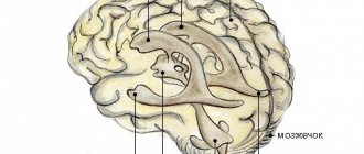
Causes of enlarged ventricles
If the initial examination revealed that the ventricles of the brain in a newborn are slightly enlarged, then do not despair, since in most cases this condition requires only observation during the first years of life, and the prognosis is favorable.
Initially, a slight discrepancy between indicators and norms may be genetically determined and be a feature of the structure of the brain, while pathological changes occur due to a chromosomal malfunction during fetal formation.
There are a number of factors that provoke asymmetry and dilatation (enlargement) of the ventricular cavity:
- infectious diseases during pregnancy (in particular, infection of the fetus with cytomelalovirus);
- blood poisoning, sepsis;
- complications caused by chronic maternal diseases;
- premature birth;
- acute hypoxia during fetal development caused by insufficient blood supply to the placenta;
- varicose veins feeding the fetus;
- long anhydrous period and prolonged labor;
- rapid birth;
- birth injuries, hypoxia caused by umbilical cord entanglement;
- deformation of the cranial bones;
- entry of foreign objects into the brain structures;
- cysts, neoplasms of various nature;
- hemorrhages;
- ischemic and hemorrhagic stroke.
Also, dilation of the ventricles can be caused by cerebral hydrocele of unknown etiology and other congenital diseases.
This is what Evgeniy Komarovsky, a well-known pediatrician in the post-Soviet space and a doctor of the highest category, says about the expansion of the ventricles.
Right ventricular dilatation
One of the reasons for the expansion of the cavity is considered to be insufficient functioning of the valve apparatus. A similar condition is typical for patients who have suffered endocarditis or rheumatism, where damage to the leaflet structures was a complication. Dilatation of the right ventricle occurs in the absence of the pericardium, which occurs in some patients.
The result of this pathology is gradually stretching of the muscle fibers. The altered septum between the atria leads to dilation of the artery in the lungs. An increase in pressure in this vessel indicates an increase in pressure in the cavity of the right ventricle.
Defects also negatively affect the chamber of the heart. They contribute to increased pressure in the pulmonary artery. This process ends with dilatation due to a deficiency of compensatory functions of the body.
Causes of dilatation
Enlargement or dilatation of the lateral ventricles of the brain occurs due to increased production of cerebrospinal fluid. This leads to the fact that it cannot be excreted normally.
This, in turn, leads to disruption of the flow of cerebrospinal fluid. This disease most often occurs in premature babies, but is observed in people of any age.
What causes the disorder in newborns?
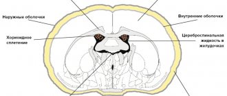
This is how dilatation of the lateral ventricles looks schematically
Dilatation of the lateral ventricles of the brain in infants is often a sign of hydrocephalus, and can also be caused by a number of other reasons.
In newborns, asymmetry is caused by trauma or space-occupying lesions in the brain. Regardless of the possible cause, urgent consultation with a neurosurgeon is required.
Mild asymmetry may be a congenital disorder that does not cause symptoms. In this case, only constant monitoring is required so that the difference between the ventricles does not change.
The main causes of dilatation include:
- viral and other diseases of a woman during pregnancy;
- oxygen starvation of the fetus;
- premature birth;
- birth injuries;
- malformations of the central nervous system.
Ventricular asymmetry can also result from hemorrhage. This pathology occurs due to compression of one of the ventricles by an additional volume of blood. Due to hemorrhage, the ventricles of the brain in an infant may be enlarged for the following reasons:
- various maternal diseases, for example, type I diabetes or heart defects;
- intrauterine infections;
- a long time between the water breaking and the baby being born.
The most common cause of dilatation is hypoxia. Other causes account for less than 1% of cases. It is hypoxia that leads to the accumulation of cerebrospinal fluid, which, in turn, increases intracranial pressure. This leads to expansion of the cavity of the lateral ventricles.
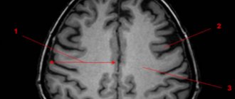
Risk zone for adult patients
A change in the size of the lateral ventricles leads to disruption of the circulation of cerebrospinal fluid. Asymmetry of the lateral ventricles of the brain in adults occurs for the following reasons:
- difficulty in the outflow of cerebrospinal fluid;
- excessive production of cerebrospinal fluid;
- neuroinfections;
- cysts and neoplasms;
- skull injuries;
- hematomas;
- stroke;
- hydrocephalus;
- vascular thrombosis.
Symptoms
Moderate expansion of one or two chambers in the heart may not manifest itself for a long time. Often, pathology is discovered by chance, during a routine examination or treatment of another disease. Severe dilatation of the cavity leads to a decrease in pumping function, which leads to the appearance of signs of heart failure or arrhythmia. These include the following:
- Palpable palpitations.
- Dyspnea.
- Blueness of the nasolabial triangle, lips, earlobes, fingertips.
- As the severity of the course worsens, cyanosis spreads to the skin.
- Swelling in the arms and legs.
- Memory impairment.
- Fatigue and weakness that persists after rest.
- The appearance of discomfort when lying down.
- Dizziness.
- Headache.
- Feeling of interruptions in the heart.
Shortness of breath in the compensation stage appears only with excessive physical exertion. With gradual wear and tear of the myocardium, the condition worsens. Shortness of breath begins to bother you with slight exertion, and then at rest.
With chronic exposure to unfavorable factors, a change occurs in the myocardium, which leads to a gradual expansion of the cavity and thickening of the walls. Dilatation of the left ventricle of the heart in the absence of timely therapy increases the risk of complications. Most often, thrombosis and fibrillation of the ventricles or atria are observed.
In some patients, the valve apparatus is affected, which is manifested by expansion of the ring, deformation of structures and ends in the formation of acquired heart disease.
After the transition from the stage of compensation to decompensation, fluid appears in the abdominal cavity (ascites), and the size of the liver increases (hepatomegaly). The skin of such patients becomes damp and cold to the touch. Systolic blood pressure decreases. Tachycardia is noted.
When auscultated, wheezing is heard in the lungs. Determination of the boundaries of the heart shows cardiomegaly (increase in heart size), the rhythm is disturbed.
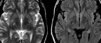
Symptoms and diagnosis of the disorder
In adults, ventricular asymmetry rarely causes symptoms. However, in some cases, this anomaly can cause the following symptoms:
- nausea and vomiting;
- dizziness;
- headache;
- feeling of heaviness and fullness of the head;
- apathy;
- feeling of anxiety.
In addition to these symptoms, the picture of the disease can be supplemented by symptoms of diseases that caused ventricular asymmetry.
Such symptoms include cerebellar disorders, paresis, cognitive impairment or sensory disorders.
In infants, symptoms depend on the severity of the pathology. In addition to general discomfort, symptoms such as throwing back the head, regurgitation, increased head size and others may occur.
Symptoms of the pathology also include strabismus, refusal to breastfeed, frequent crying, anxiety, tremors, and decreased muscle tone.
However, quite often the pathology does not cause characteristic symptoms and can only be detected after an ultrasound scan.
Causes
Dilation of the chamber in the left ventricle often occurs under the influence of several provoking factors. This condition is directly related to the patient’s age, heredity, and the presence of excess body weight. The reasons that negatively affect the myocardium are:
- Congenital heart defects. Exposure to unfavorable environmental factors occurs already during pregnancy. If the lesion becomes extensive, the fetus dies. In the case of a slightly pronounced lesion, a defect is formed.
- Inflammatory diseases, which include myocarditis, pericarditis, endocarditis. The risk group includes children and adolescents, in whom cases of this pathology are often recorded.
- Chronic diseases of the cardiovascular system. These include arterial hypertension, angina pectoris, ischemia.
- Metabolic syndrome, the basis of which lies in the presence of excess body weight and diabetes mellitus in the patient.
- Chronic pathology of lung tissue.
- Diseases of the kidneys, endocrine and hematopoietic systems.
- Genetic predisposition.
- Autoimmune disorders.
One of the common factors of dilatation is chronic intoxication with alcohol and nicotine.
This group also includes side effects from medications. Pheochromocytoma is the most common endocrine pathology. It is a benign or malignant form of tumors. It is characterized by excessive production of adrenaline.
Forecast
Every patient with left ventricular dilatation, already knowing what it is, must follow all medical recommendations. With this diagnosis, early diagnosis and initiation of treatment are important. In advanced forms, there is a high probability of developing heart failure. In these same patients, the valve apparatus is deformed, which leads to mitral insufficiency. This diagnosis significantly affects the quality of life and reduces its duration. The prognosis for patients is unfavorable.
The average survival rate for left ventricular dilatation is 10 years. If the course is asymptomatic, then life expectancy is on average 5 years. Patients with chronic heart failure observed in the hospital survive up to 50% of those admitted.
It is important for each patient to remember that the first symptoms are not considered normal and require a set of diagnostic procedures. Timely treatment will reduce the risk of complications, and treatment will prolong life for many years.