What is a craniotomy?
Sometimes accessing a brain tumor is more difficult than removing it. Therefore, some additional steps must be added to help the surgeon access the cancer in the brain. One such method is craniotomy surgery, which involves the removal of bone in addition to the levels of other brain surgeries.
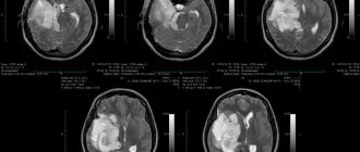
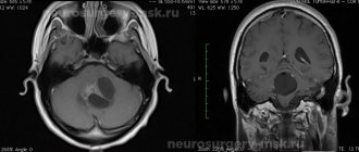
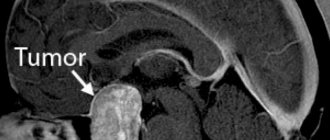
In a craniotomy, a neurosurgeon removes part of the skull bone to gain access to the tumor. Skull resection may require computer assistance and imaging to find and reach the exact location of the cancer. Naturally, magnetic resonance imaging (MRI) or computed tomography (CT) are the methods that come first.
The removed portion of the skull bone is called a “bone flap,” which is placed in place after craniotomy excision of the tumor. Typically, the bone flap is replaced at the end of the procedure with tiny plates and screws. There are cases where the bone flap does not come back, but this is another brain surgery called a "craniectomy", which will be discussed further in the FIT article.
The name of the craniotomy operation comes from the cranium (skull) to be removed. The size and complexity of the craniotomy depends on the patient's problem, which can range from brain swelling to removing foreign objects such as bullets.
Brain tumor removal
Therapy for neoplasms is complex, always complex and quite expensive. For example, in Russia, surgical treatment of a brain tumor is estimated at approximately 350 thousand Russian rubles. You can find out the final cost of treatment in one of the best oncology centers in the Russian Federation after consulting a neurosurgeon. Prices abroad are much higher. In Germany, for such therapy you will need to pay about 28,000-30,000 US dollars, in Israel it is several thousand cheaper, so it is important to take this fact into account when choosing a clinic. You need to pay attention to the nuances. For example, the equipment in foreign clinics is often better, although the treatment methods and results are not fundamentally different, but in the event of an unfavorable outcome, Western specialists will not have to deal with paying local insurance, so they easily take foreign patients who pay for everything themselves.
Note. The main treatment is a radical surgical operation; other methods used in combination act as auxiliary ones (chemotherapy, radiotherapy and other methods).
Brain cancer can only be removed through surgery. It is performed for nodular tumors and is impossible for diffuse neoplasms due to deep infiltration into nearby structures. In such circumstances, brain tumor surgery is performed to remove some of the abnormal tissue, relieve pressure inside the skull, and prevent dropsy. The size of the tumor and the stage of the cancer play a decisive role in deciding whether to remove the tumor.
Radiotherapy involves repeated exposure of cancer cells to radiation, which leads to blockade of division and death of the latter. With significant doses of radiation, pathogenic cells stop developing and most of them die.
At the same time, treatment involves the use of chemotherapy through oral and intravenous administration of cytostatics (toxic substances for pathogenic cells). These therapies have many negative effects. Recently, medicine has been producing more and more unique chemical drugs that selectively target tumor cells, which can significantly reduce the negative impact of poisons on the body.
At the same time, medications are prescribed to eliminate unfavorable signs that can be caused by both the tumor and side effects from the proposed treatment.
Surgery
Surgeries to remove tumors in the brain are the only effective treatment. This type of treatment is very complex. The possibility and effectiveness of the operation depends on many factors, among which important are the stage of cancer, the nature of the spread of pathogenic tissues, the presence of metastases and secondary foci, as well as the localization of pathogenic formations in brain tissue.
Any neurosurgical operation is quite complicated, because the doctor must remove a malignant tumor located between healthy brain tissue. Therefore, treatment of this kind is traumatic, and in some cases impossible. For example, cancer removal is not carried out if the tumor is large, when it affects vital parts of the brain, or if the disease is at a terminal stage. In addition, the person’s health status, the presence of chronic diseases, their stages, the patient’s age and other features are taken into account.
In each specific case, the choice of technique and method of tumor removal is selected individually. At the moment, doctors have a lot of specialized surgical equipment in their arsenal, for example, tomographs, laser and ultrasound equipment. This significantly increases the efficiency of operations to remove tumors.
Most often, surgical removal of brain cancer involves trepanation and resection of the skull. The methodology is as follows:
- At the beginning of the operation, the surgeon dissects the skin layers above the cranial vault (a scalp incision is made).
- After turning away the cut skin, the doctor moves the muscle layers aside.
- At the next stage of the operation, the required part of the bone tissue of the skull is removed so that the brain tumor becomes accessible for manipulation.
- Then the pathological focus is excised and the extracted bone tissue is installed in its original place (this is called trepanation). If part of the skull is not returned to its original place, then such an operation is called resection.
- At the final stage of the operation, the soft tissues covering the skull are restored and sutures or staples are applied.
After resection, the hole in the skull is closed with a special material or mesh. Removing pathogenic tissue reduces intracranial pressure, freeing up space for swollen tissue afterwards. In addition, craniotomy is necessary if the tumor process has spread to bone tissue. After removal, the extracted biological material is sent for histological analysis.
Reasons for performing a craniotomy
Why does a person need to have a craniotomy and cranial resection? As briefly mentioned above, reasons for craniotomy surgery vary and include, but are not limited to:
- Access to the treatment site for diagnosis or removal of tumors
- Reducing intracranial pressure by eliminating damaged or swollen areas of the brain may be caused by traumatic injuries or strokes
- Implantation of a device for purposes such as treating movement disorders such as Parkinson's disease or dystonia (a type of movement disorder), or refractive epilepsy
- Aneurysm clipping or repair
- Removing blood or blood clots from leaky blood vessels
- Draining a brain abscess (an abscess is an infected pocket of pus in the tissue)
- Repair of skull fractures
- Repairing a tear in the membrane lining the brain (dura mater)
- Removing a foreign object, such as a bullet.
Craniotomy and resection are not only used for tumors, as explained above. Whenever the brain is under too much pressure, such as when it is swollen, has blood clots, or has an abscess, craniotomy surgery is the choice. If a craniotomy to remove a tumor, blood clots, a bullet in the head, or for any other reason is a treatment, how should the patient prepare?
Our prices
| Services | Cost, rub.) |
| Removal of a brain tumor in Moscow. The cost depends on where the education is located, what its size is, etc. And also on who performs the operation and where. | from 250,000 rubles |
| Removal of a brain tumor. Price for microsurgical removal of tumors of the cerebral hemispheres. | from 233 280 |
| Removal of a large brain tumor | from 345 600 |
| Brain tumor price of microsurgical removal of a tumor of deep localization with neurophysiological monitoring and intraoperative fluorescence microscopy/endoscopy | from 586 440 |
| Microsurgical removal of tumors of the meninges | from 256 230 |
| Microsurgical removal of neoplasms of the meninges with plastic surgery of the dura mater and venous sinuses using auto- or artificial implants using intraoperative fluorescence microscopy/endoscopy | from 642 780 |
You can find other prices in the section Prices for operations.
Preparation for craniotomy
Before craniectomy surgery, the patient should stop taking any non-steroidal anti-inflammatory drugs and blood thinners for at least a week beforehand. However, if there are medications explicitly prescribed for surgery, they must be carefully taken as scheduled. In addition, the patient should not eat or drink the night before surgery.
Smoking and drinking alcohol are also prohibited from one week to two weeks after surgery. If an MRI or CT scan is required, fiducials may be placed on the patient's forehead and behind the ears to help align the preoperative MRI with the image guidance system. In many cases, the doctor must perform blood tests to make sure the patient is healthy.
Skull resection is performed under general anesthesia, regardless of the type of operation. Signing the consent form is mandatory and the patient must read the pathway carefully and know what they are experiencing. They should also know the risks associated with such brain surgeries.
Healing factors
Low-level laser therapy has been successfully used in the treatment of headaches for more than 30 years.
The therapy is painless, does not cause complications or side effects, and does not require recovery or preliminary preparation. The laser emits red and infrared light. The rays penetrate the superficial and deep subcutaneous layers. Tissues and cells absorb their energy - under the influence of heat, it is converted into biochemical energy.
As a result, the inflammatory process and pain go away. Laser energy accumulates in the laser affected areas gradually, so most patients feel the effect of treatment after 3-6 procedures.
Under the influence of a laser beam:
- relieves tension in the neck muscles;
— the autonomic nervous system is restored;
- sleep is normalized;
- anxiety goes away;
- Chronic fatigue syndrome disappears.
A low-intensity laser beam acts on the brain through the cranial walls, as a result of which the regeneration of nerve cells is accelerated and the process of neuron death is inhibited.
Risks of craniotomy
After tumor removal or craniotomy for other reasons, the patient may experience swelling and bruising of the face or around the eyes, headache, dizziness, and problems with memory, speech, or vision. Thus, the neurosurgeon determines how many days the patient should spend in the intensive care unit (ICU), where heart rate, blood pressure, and nerve signs are constantly monitored. Despite all these precautions, the patient may still be exposed to postoperative risks.
Common post-operative risks are also present here: bleeding, infection, blood clots and reactions to anesthesia. In the case of craniotomy surgery, some other dangers are also added, depending on the type of procedure:
- Strikes
- Convulsions
- Brain swelling
- Nerve damage
- CSF or cerebrospinal fluid leak, which is the fluid that surrounds and protects the brain
- Loss of some mental functions
- Pneumonia, which is a type of lung infection
- Unstable blood pressure
- Muscle weakness
Additionally, some risks are technique-specific and do not occur in all craniotomy patients. These depend on the severity and complexity of the problem and include:
- Memory problem
- Difficulty with speech
- Paralysis
- Impaired balance or coordination
- Coma
Post-operative care can prevent or reduce some risks, but usually the patient must make a compromise and undergo surgery knowing that this may happen. In many cases, it is a matter of life or death, and the patient has no other choice.
Other treatments
In the future, depending on the indications, radiation therapy or chemotherapy may be necessary. Radiation therapy can be used either if surgery is not possible or after surgery to kill any remaining abnormal cells. Depending on the individual situation, external or internal radiotherapy may be prescribed. With internal, a chip with radioactive material is installed directly into the malignant formation and remains there for the required amount of time. As for chemotherapy, it can also be either the main method of treatment or an additional one, in combination with surgery. The drugs can be administered intravenously, taken orally, or injected directly into the cerebrospinal fluid.
Different types of craniotomy
Like any other type of brain surgery, craniotomy also has different types, ranging from right frontal craniotomy for tumor resection to translabyrinthine craniotomy. The model is selected based on the patient's condition, the purpose of the operation and overall benefits. Some of the main types of craniotomy are as follows:
Extended Bifrontal Craniotomy When minimally invasive approaches do not meet surgical needs, the surgeon may choose an extended bifrontal craniotomy. Here an incision is made on the scalp behind the hairline and the bone that forms the outline of the orbits, and the forehead is removed.
The most common goal is to reach complex tumors in the front of the brain. This is a traditional approach to the base of the skull with the core belief that removing excess bone is safer than unnecessary manipulation of the mind. Typically, meningiomas, esthesioneuroblastomas, and malignant tumors of the skull base are treated with extended bifrontal craniotomy. Eventually the bone flap is replaced.
Minimally Invasive Supraorbital» Brow» Craniotomy
A supraorbital craniotomy, commonly known as a brow craniotomy, is another procedure for removing brain tumors. As the name suggests, this is a minimally invasive approach and involves making a small incision in the eyebrow to access tumors at the front of the brain. Rathke's cleft cysts, skull base tumors, and some pituitary tumors can also be treated with this method.
Because it is a minimally invasive procedure, it may be less painful than an open craniotomy. The recovery period may be shorter and there will not be a large scar at the surgical site.
Retrosigmoid" Keyhole" Craniotomy
A retrosigmoid craniotomy, often known as a “keyhole” craniotomy, is another minimally invasive surgical procedure for removing brain tumors. After a small incision behind the ear provides access to the cerebellum and brain stem, the surgeon can remove the tumor from the base of the skull. Meningiomas, acoustic neuromas, and metastatic tumors of the brain or spine are other targets for keyhole craniotomy.
Like other minimally invasive approaches, retrosigmoid craniotomy also requires less recovery time, while it leaves a minimal surgical scar and less pain compared to various types of brain surgeries.
Orbitozygomatic Craniotomy
Orbitozygomatic craniotomy is the second traditional approach to the skull base presented in our article. Like extended bifrontal craniotomy, orbitozygomatic craniotomy is also used to access and operate on complex tumors and aneurysms. This approach is based on the same belief as extended bifrontal craniotomy: removing excess bone is safer than unnecessary manipulation of the brain.
When minimally invasive approaches fail, the bone that forms the outline of the orbit and cheek is removed for replacement at the end for the purpose of craniotomy excision of the tumor. Some cases that can be treated with this type of surgery include craniopharyngiomas, pituitary tumors, and meningiomas. The neurosurgeon can reach deeper and access hard-to-reach areas of the brain, minimizing serious brain damage .
translabyrinthine craniotomy
In a translabyrinthine craniotomy, an incision is made in the scalp behind the ear to remove the mastoid bone and part of the inner ear bone. The significant portion of the inner ear bone that is removed here are the semicircular canals containing receptors for balance. Sometimes the entire tumor cannot be removed due to the severe damage that the removal procedure can create. However, neurosurgeons usually try to remove all or as much of the tumor as possible.
The biggest problem with this method is hearing loss. When the semicircular canals are removed, hearing is sacrificed for access and tumor removal. However, the risk of facial nerve damage may be reduced.
In all of these methods, the removed bone flap is replaced and held in place with screws and plates. This does not always happen with a bone graft, and sometimes the surgeon does not replace the bone.
Treatment
The main methods of treating brain cancer:
- classical neurosurgical operation - involves craniotomy and tumor removal;
- endoscopic surgery - involves inserting instruments and a camera through small holes in the skull (performed for pituitary tumors, which are removed through the nose, as well as superficially located tumors of some parts of the cerebral hemispheres);
- laser ablation of tumors under MRI control;
- radiosurgical methods - destruction of small tumors with precisely directed beams of radiation;
- radiation therapy;
- chemotherapy;
- targeted therapy.
Our expert in this field:
Sergeev Pyotr Sergeevich
Oncologist, surgeon, chemotherapist, Ph.D.
Call the doctor Reviews about the doctor
Craniotomy, Craniectomy and Cranioplasty
Craniotomy, craniectomy and cranioplasty sound similar, but are three different methods of brain surgery. As the names suggest, the cranium (skull) is involved in all three methods. As mentioned earlier, in a craniotomy, part of the head is temporarily removed to allow access to deeper parts of the brain to remove tumors, blood clots, brain tumors, foreign bodies and treat some other problems as mentioned in previous sections of the article. The bone flap is placed in place at the end of the operation and secured with titanium plates and screws.
On the other hand, with a craniectomy, the bone flap is not replaced immediately. Typically, neurosurgeons prefer this method when the problem is traumatic brain injury and other head injuries that involve swelling. The removed portion of the skull is kept away to relieve pressure and compression on certain areas of the brain.
In the third technique, cranioplasty, a bone flap is removed, just like the previous two methods. However, it never reattaches because the removed part of the skull itself has problems: it is damaged or cancerous. Thus, the removed part is replaced with titanium, acrylic or a synthetic bone substitute. Installation of the new part must also be performed by a neurosurgeon.
Relieving pressure in an emergency is one of the main advantages and specific characteristics of cranioplasty. It can save lives with this particular benefit. Another specific benefit of cranioplasty is that the new piece of bone can help restore the natural contour of the skull. Sometimes the removed part distorts the skull, but cranioplasty can help restore the natural shape. Titanium implants are especially preferred for cranioplasty due to their many benefits.
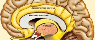
Types of brain cancer
By its nature, brain cancer is quite diverse. The etiology of the disease is based on mutation of DNA cells, when an error occurs in normal cells, division and abnormal growth of pathogenic tissues begins, followed by the formation of a tumor. Cell division can occur in any part of the brain. The classification of pathology depends on the histological structure of the formation, localization and growth pattern.
- Gliomas are a common type of tumor, occurring in 60% of patients. They combine several groups of neoplasms originating in glial cells (brain matter). These tumors include glioblastomas, astrocytomas, and ependymomas. They can have a malignant or benign course.
- Meningiomas develop from the outer layer of the meninges. They occur with almost the same frequency as gliomas. The risk of developing meningioma increases with age, and this tumor occurs 2 times more often in women than in men.
- Medulloblastomas arise from neuroectodermal cells of the cerebellum. They are characterized by rapid growth. More common in children than in adults.
- Ependymoma is a type of glial tumor process. Develops from a thin epithelial membrane, the ventricles of the brain. It is malignant and prone to metastasis. Ependymomas are classified as intracerebral tumors and account for 10% of all cases.
- Astrocytoma is a malignant tumor that originates from astrocytglia (auxiliary cells of the brain). Diagnosed in men.
- Oligodendroglioma - develops from cells of nervous tissue. It is very similar to astrocytoma.
- Hemangioma - arises from cerebral vascular cells.
- Schwannomas (neurilemmomas) – the primary origin is in Schwann cells, which surround and separate the cranial nerves. They are benign, but can affect any cranial nerve.
- Craniopharyngiomas are slow-growing tumors that often cause hormonal disorders and vision problems.
- Secondary tumors (cancer metastases to the brain).
Any formation is dangerous to human health and life, it can compress surrounding tissues, cause the death of neurons, impair blood circulation, and spread to other parts of the brain.
Based on location, brain tumors are divided into:
- intracerebral;
- intraventricular;
- extracerebral.
Like all oncological diseases, tumor-like processes can have a benign or malignant course.
Benign tumors, unlike malignant ones, are not capable of dividing and do not grow into neighboring tissues. Their danger lies in the fact that such formations can compress blood vessels, cause stagnation of venous blood, and degenerate into cancer. Malignant tumors grow rapidly; pathological cells divide and penetrate into other parts of the brain and internal organs. Such tumors often lead to the death of a person and even after they are completely removed, they can reappear.
Brain tumors are divided into two categories:
- Primary – the source of inflammation begins directly in the structures of the brain.
- Secondary - a tumor-like process is present in another organ, but metastasizes to the brain.
Use of titanium implants in Cranioplasty
As mentioned in the previous section, with cranioplasty, the removed part of the skull cannot be put back. It is either damaged, malignant, or has other problems and should not be replaced. Therefore, it is necessary to use an implant made of titanium, PEEK, acrylic or a synthetic bone substitute.
Titanium is one of the most common materials used for surgical purposes and implants. It is also becoming increasingly popular in cranioplasty as it prevents cosmetic deformity, reduces the vulnerability of exposed brain tissue, and minimizes the risks and costs associated with additional surgeries and procedures. .
Brain surgery can be a minimally invasive approach such as a brow craniotomy, a right frontal craniotomy for tumor resection, or cranioplasty, where part of the skull is discarded and a piece of titanium or other materials replaces it. However, the patient should know enough about the operation, its preparation and its risks to minimize post-operative dangers as much as possible. .