Causes
Hypoplasia occurs due to problems with metabolism. But it is incorrect to say that this pathology occurs only due to problems with metabolism and demineralization of the enamel. The condition of a woman during pregnancy is of great importance. Research results suggest that the anomaly develops more often in children whose mothers suffered during pregnancy:
- Rubella.
- ARVI.
- Toxoplasmosis.
- Severe forms of toxicosis.
Also at risk are premature babies and those who suffer from diseases leading to calcium metabolism disorders:
- Encephalopathy.
- Atopic dermatitis.
- Rickets
NOTE: AFTER CARIES, ENAMEL HYPOPLASIA IS THE MOST COMMON DISEASE OF THE HARD TISSUE OF TEETH FORMED BEFORE THEIR ERUPTION. ACCORDING TO STATISTICS, IN THE RUSSIA ABOUT 10% OF CHILDREN HAVE HYPOPLASIA OF MILK TEETH, AND MORE THAN 25% HAVE PATHOLOGY OF MARRIAL INCISERS.
Dental hypoplasia - symptoms and treatment
Hypoplasia is a malformation of the hard tissues of one or an entire group of teeth [13]. It can be congenital or acquired. It develops when the rudiments of milk or permanent teeth are formed. It is not a carious lesion.
Hypoplasia is accompanied by a heterogeneous color of the enamel, its thinning and changes in relief - depressions of various shapes and sizes (from dots to grooves and pits). When teeth erupt, they immediately differ: there are white, yellow or brown spots on their surface.
Otherwise, such dental damage is called hereditary hypoplasia, aplasia, dysplasia or brown enamel dystrophy, and the affected teeth are grooved or plaster [1].
Statistics
Hypoplasia is the most common non-carious lesion of hard tooth tissues - enamel and dentin. It occurs in 50% of children with chronic metabolic disorders that arise before or after birth [10].
As a dental examination of the Russian population in 1999 showed, the prevalence of non-carious lesions among children 12 years old is 43.5%. At the same time, systemic enamel hypoplasia accounts for 36.7% [1].
The most common form of the disease is systemic hypoplasia - damage to all teeth in the permanent or primary dentition. It accounts for 90.6% of all types of hypoplasia [1]. More often, the defect affects the cutting edges or cusps of the teeth. In three out of five cases it develops in children under 9 months of age.
In foreign literature, hypoplasia of molars and incisors (lateral and anterior teeth) is called Molar Incisor Hipomineralisation (MIH) [1]. This form of the disease is common throughout the world [18]. The increase in its incidence of MIH can be explained by stress, deterioration of the general condition of the body, and the negative influence of the environment and food [17].
Causes and risk factors
The development of hypoplasia is associated with metabolic disorders at the time of formation of tooth buds. This is facilitated by external and internal negative factors that affect the body of the expectant mother or her already born child.
Intrauterine factors include:
- Rh conflict (incompatibility of the negative Rh factor of the mother and the positive Rh factor of the child);
- toxicosis and other disorders in the body of a pregnant woman;
- rubella, toxoplasmosis during pregnancy;
- taking tetracycline by a pregnant woman (or a child) [5].
Internal factors associated with child diseases:
- Down syndrome, cerebral palsy and other lesions of the central nervous system (they are both the cause of hypoplasia and associated pathologies) [4];
- rickets or infectious diseases in a child (measles, scarlet fever);
- hypo- and vitamin deficiency;
- changes in mineral and protein metabolism in dyspepsia (digestive disorders) and diseases of the endocrine glands (usually the thyroid gland) [3];
- penetration of infection from the inflammatory focus of a baby tooth into the germ of a permanent tooth.
External factors:
- prematurity, low birth weight of the newborn [16];
- artificial feeding of a child;
- local impact on the tooth germ, for example, birth trauma;
- trauma to the jaws and rudiment at the time of tooth formation.
If pathological factors affect the fetus, hypoplasia of baby teeth develops. If this happens in early childhood, the rudiments of permanent teeth are already affected.
Forms of hypoplasia
Hypoplasia can be local or systemic. In the first case, we are talking about damage to several incisors, and in the second, about larger-scale violations. Both milk and molar teeth can suffer. In the first case, problems with enamel arise due to abnormalities in the mother's body. Hypoplasia of permanent teeth is more common. It occurs when pathology occurs in the child’s body. The first manifestations of the disease, which is systemic in nature, can be detected when the baby is 5–6 months old.
The localization and level of damage to the enamel coating greatly depends on the age at which the disease, which is a pathogenetic link, occurred:
- Diseases at the age of 5–6 months lead to damage to the cutting edges of the central teeth.
- At 8–9 months, the pathology also affects the erupted lateral incisors and the cutting edges of the canines.
- By the age of one year, all teeth suffer, but most of all those that erupted earlier.
If the pathology suffered has led to serious disturbances in metabolic processes, hypoplasia can spread over the entire surface of the teeth. The characteristics of the disease can be determined by the enamel. The more severe the form of hypoplasia, the more pronounced the uneven structure.
Symptoms
Enamel hypoplasia of primary teeth can have two course options - local and systemic. The first form is more favorable, since it affects only a couple of teeth. In case of systemic disease, absolutely all teeth are affected.
The systemic form has symptoms:
- the enamel is underdeveloped and its thickness does not correspond to the norm. Sometimes it may be completely absent;
- hypoplasia of permanent teeth is characterized by a change in the color of the teeth. Spots with a clear outline form on the frontal surface. Their color varies from white to light yellow. Areas of demineralization do not cause pain.
Local form:
- not only the enamel is affected, but also the rudiments of permanent teeth;
- abnormal development of tooth enamel. You can see various streaks and dents on the tooth. But the patient does not experience any painful sensations;
- aplasia is a congenital pathology. The top protective layer may be partially or completely absent on the teeth. In this case, the patient complains of pain and discomfort that occurs when teeth come into contact with irritants.
Treatment of dental enamel hypoplasia is recommended to be carried out when the first signs are identified that indicate the presence of damage to the upper layer.
Patchy hypoplasia
One of the most common forms of hypoplasia is spotted, when white or yellow spots with clear boundaries form on the front and side groups of teeth. As a rule, the lesions are symmetrical. The spots may be shiny or dull. In the first case, we are talking about minor lesions characterized by demineralization of the subsurface layer. As for dull spots, they occur during the final stages of enamel formation, so only the outer layer is affected.
Peculiarities:
- In the area of stain formation, the thickness of the enamel, as a rule, does not change.
- There is no pain or irritation when drinking hot drinks.
- X-ray examination does not reveal the disease.
- The disease has similar features to the spotted form of fluorosis and initial caries, and therefore requires differentiated diagnosis.
Grooved shape
In this case, one or more grooves are formed on the vestibular (front) surface parallel to the cutting edge of the incisors. The depth of damage may vary. As a rule, teeth of the same name are affected symmetrically. As in the case of the erosive form, the anomaly can be seen on the radiograph even before the incisors erupt (horizontal lesions).
Here are some more types of hypoplasia:
- Linear - manifests itself in the form of numerous wavy stripes on the vestibular surface.
- Aplastic is a severe disorder characterized by severe damage to the enamel. Occurs in the case of amelogenesis imperfecta.
- X-ray examination does not reveal the disease.
- Mixed - most often we are talking about a combination of erosive and spotted forms.
Forms of systemic hypoplasia
There are several specific forms of systemic hypoplasia, named after the scientists who described them:
- Fournier's teeth - characterized by damage to the central incisors of the upper jaw, which acquire a barrel-shaped shape.
- Hutchinson's teeth - affects the lower or upper jaw. The incisors are also barrel-shaped, but there are still lunate notches on the cutting edges.
- Pflueger's teeth - in this case, the first molars suffer. Tubercles form on them, and the crowns take on a conical shape.
Another type of systemic hypoplasia is “tetracycline” incisors. Pathology occurs if a woman used tetracycline during pregnancy or the child was given this antibiotic at an early age. It manifests itself in the form of darkening and destruction of tooth enamel (up to aplasia).
Hypoplasia of primary teeth: what is it and its causes
Hypoplasia is characterized by thinning of the enamel and a pronounced change in relief. On the crown of a baby or permanent tooth there are depressions, dots, pits, and grooves. Affected teeth differ from healthy ones already at the moment of eruption - the enamel is covered with white, yellowish or brown spots.
Hypoplasia of primary and permanent teeth is observed in 40% of patients. And the number of children with this pathology is growing steadily. Scientists associate the development of the anomaly with poor ecology, lack of a balanced diet, and hereditary factors. And all because the development of the defect occurs already at the stage of formation of the tooth germ.
A tooth—deciduous or permanent—is made up of pulp, dentin, enamel, and other tissues. Special cells, ameloblasts, are responsible for the proper development of enamel. In the absence of pathologies, one ameloblast forms one enamel prism. And it is these “prisms” that make up the outer tissue of the erupted tooth. As soon as the ameloblasts complete the task assigned to them, the destruction of the building cells occurs. After this, the tooth enamel of the erupted unit loses its ability to recover.
Ameloblasts are only the first stage in creating strong enamel of a child’s tooth. After eruption, it takes about a year for another stage of formation - final mineralization - to be completed. If all stages of development are successful, the child pleases his parents with strong, snow-white, healthy teeth.
Failure at any stage means potential enamel hypoplasia.
Causes of tooth enamel hypoplasia in children
There are three risk factors for the development of hypoplasia:
- features of fetal development during pregnancy;
- illnesses of the child himself;
- external circumstances.
The health of the expectant mother is the basis for the child’s excellent well-being. The risk of hypoplasia can be provoked by:
- Rh conflict - negative Rh factor of the mother and positive Rh factor of the child;
- severe pregnancy, toxicosis;
- acute viral infections or toxoplasmosis during pregnancy;
- taking tetracycline drugs during pregnancy;
- smoking, drinking alcohol.
Among the diseases of the child himself, the risk of defects in the formation of teeth increases:
- CNS lesions, Down syndrome;
- rickets, vitamin deficiency;
- digestive disorders;
- diseases of the endocrine system.
External factors include poor nutrition of the child and injuries to the jaws, which prevent the normal development of tooth germs.
Intrauterine factors provoke hypoplasia of primary teeth. The rest have an equally negative impact on both temporary and indigenous units.
Plaksina Margarita
My patients really like dental remineralization. Pastes rich in minerals have a pleasant taste and smell, and the procedure itself is painless and even pleasant.
Possible complications
In the early stages, the disease manifests itself in the form of small cosmetic defects without causing serious discomfort. If the spots are clearly visible, this indicates more intense destruction of the enamel. In the second case, treatment must be carried out as quickly as possible, since ignoring the problem can cause serious consequences, such as:
- Increased wear of incisors.
- Tissue destruction.
- Deformation and loss of damaged teeth.
- Development of malocclusions.
Treatment of hypoplasia
There are several ways to treat this disease. In the initial stages, as a rule, conservative therapy is sufficient, the purpose of which is to eliminate metabolic problems. The method involves the use of vitamins and medicines containing minerals such as calcium, phosphorus and fluorine.
If we are talking about systemic hypoplasia with severe damage to the enamel coating, mineralization will not be enough. In this case, the doctors at the Nutcracker pediatric dentistry also use the whitening method. The essence of the technology is etching and grinding the enamel with a boron, as well as subsequent polishing. Whitening allows you to highly effectively eliminate dark spots and other defects on the outside of your teeth.
Hypoplasia
- failure in the laying of embryonic petals;
- insufficient volume of amniotic fluid;
- malposition;
- drinking alcohol, drugs and smoking during pregnancy;
- abdominal injuries;
- past infectious diseases;
- exposure to various toxic substances.
Doctors have not yet learned to quickly and timely detect all pathologies of intrauterine development.
That is why it is important for women to undergo all routine examinations. Some of the violations may become indications for termination of pregnancy. There are different types of hypoplasia, which are usually divided according to organ systems.
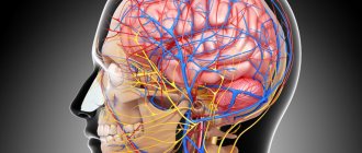
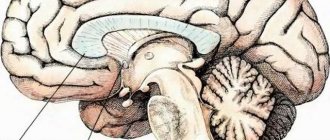
CNS and brain
One of the heaviest varieties. Leads to partial or complete non-viability of the child. It can be noticed even in intrauterine development. Almost always detected in the first hours of life after birth. Symptoms depend on the specific organ that has undergone pathological development.
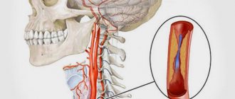
The cardiovascular system
Most often it manifests itself as underdevelopment of blood vessels. If these are small capillaries, their deficiency is compensated by a well-branched mesh. Large vessels can cause problems with organs that they do not sufficiently supply with blood.
Cardiac hypoplasia is quite rare. Most often, this may be underdevelopment of one of the ventricles or atria.
Endocrine system
Hypoplasia of even one of the endocrine glands is systemic in nature. The hormones produced in the body are closely interrelated with each other and completely affect its functioning. Even the absence of one of them can lead to a number of diseases. For example, an underdeveloped pancreas that produces insulin causes congenital diabetes mellitus. And this is a metabolic disorder, due to which other organs and systems will suffer.
Respiratory organs
Complex types of hypoplasia that do not always have a favorable outcome. Human lungs cannot work at full capacity. This means that there will not be enough oxygen in the body, which is why the development of all systems and organs, including the central nervous system, will suffer.
And this is only a small part of the possible pathologies.
Features of treatment
Hypoplasia is a congenital disease. Medicine is still learning to correct pathologies during intrauterine development. Therefore, treatment in this period is more preventive in nature.
After childbirth, therapy is carried out depending on the organ that has not developed sufficiently.
Is it worth carrying out the silvering procedure?
Some dental clinics eliminate the initial stages of hypoplasia using the silvering method. The essence of this therapy is that a layer of active metals is applied to the teeth. Upon contact with enamel, crystals of silver phosphate and potassium fluoride are formed, protecting the enamel from destruction.
The Nutcracker Clinic does not use silver plating. We abandoned this method for several reasons:
- Poor efficiency. To remove plaque and restore the nervous structure of the incisors, you need to carry out about 5 procedures over six months.
- The drugs used often cause allergic reactions.
- In case of advanced caries, the procedure is contraindicated.
- After silvering, the enamel darkens, which causes psychological discomfort in the child.
Prevention
You need to think about preventing non-carious dental diseases and, in particular, hypoplasia even before the birth of the child. This is due to the fact that enamel malformations in most cases occur during fetal development.
To minimize the likelihood of the formation of this pathology, you need to follow these tips:
- Every trimester of pregnancy, a woman should be examined by a dentist.
- Good nutrition is the key to normal metabolism in mother and baby.
- During pregnancy, you should never take medications without first consulting a doctor.
- The baby needs to be breastfed for at least a year and ensure that it does not injure the oral cavity.
- If the first signs of pathology develop, you should immediately consult a doctor.
Etiology
Hypoplasia of tooth enamel in most wedges. situations become a consequence of enamel demineralization. Also, the disease is caused by defective formation of the upper layer during embryonic development. Other reasons due to which hypoplasia of primary teeth progresses include:
- traumatization of the child as it passes through the mother’s birth canal;
- Rhesus conflict;
- birth of a baby earlier than expected;
- dystrophy;
- the presence of ailments affecting the gastrointestinal tract;
- poor nutrition;
- the presence of disruptions in the metabolic process;
- the presence of a somatic disease;
- the course of illnesses of an infectious nature;
- traumatization of the maxillofacial area.
Professional approach to the treatment of hypoplasia
The doctors of the Nutcracker Clinic are candidates and doctors of medical sciences who, over the years of active practice, have encountered many oral problems in children and successfully solved them. By contacting us, you can count on the following benefits:
- We use professional equipment and drugs, which ensures gentle treatment without adverse reactions.
- We individually select therapy for each child or adult, taking into account the characteristics of the pathology.
- We offer affordable prices (you can view them in the price list on the website) and provide discounts on certain services.
Our company's medical centers are located in Moscow and Krasnogorsk. Clinic staff have been successfully treating oral diseases for more than 10 years. We have all the necessary certificates and permits confirming the professionalism of doctors.
Contact the Nutcracker Clinic and have no doubt that your health or the health of your baby will be in good hands! Whatever form of hypoplasia we are talking about, we will be able to choose an effective approach, cure the disease in a short time and give some expert advice that will help keep your teeth in perfect condition.