Reasons you need to know
Factors influencing the occurrence of a neoplasm are mechanical traumatic brain injuries, problems with the cardiovascular system, viral and radiation exposure, and otitis media.
The reasons vary depending on the type of benign tumor: Doctors still do not know why a unilateral neuroma appears. The most likely reason is the high level of radiation around. As a result, the myelin sheath of the nerve fibers is destroyed and a tumor forms.
Bilateral neoplasm in most cases is caused by a hereditary predisposition. The pathology is transmitted according to an autosomal dominant principle. If you do not intervene in time, after 30 years the patient will have hearing problems.
It is believed that if both parents have potential problems that cause acoustic neuroma, then there is a 50% chance that the disease will be passed on to the child. Smoking, excessive alcohol consumption, and unfavorable environmental conditions have a detrimental effect on health.
3.Causes of the disease
There are two types of acoustic neuroma: the sporadic form and the form associated with neurofibromatosis syndrome type II.
Neurofibromatosis type 2 is an inherited disease characterized by the growth of noncancerous tumors in the nervous system. Acoustic neuromas are the most common type of these tumors and often occur in both ears in people over 30 years of age. Neurobromatosis is a rare disease. It accounts for only 5% of all acoustic neuromas. Most acoustic neuromas are sporadic. What causes it is not known. But one known risk factor is exposure to high doses of radiation.
About our clinic Chistye Prudy metro station Medintercom page!
Types of neoplasms and their symptoms
In addition to the fact that the acoustic neuroma is divided into two types based on location (unilateral and bilateral), scientists identify 3 more types of tumors that differ in size:
- up to 25 mm (mild clinical signs, dizziness is possible);
- from 30 to 35 mm (incoordination of movements, whistling in the ears, distortion of facial expressions appear);
- more than 40 mm (accompanied by hydrocephalus, unsteadiness of gait, visual disturbances).
Symptoms of the disease include headache, attacks of nausea, double vision, decreased sensitivity of the cornea of the pupil, impaired swallowing function, loss of taste on the front of the tongue, pain in the face. If the tumor is too large, intracranial pressure increases and affects the brain.
Specialists of the Department of Neurosurgery of the City Clinical Hospital named after. Eramishantsev is advised not to ignore even minor manifestations of neuroma. The longer you wait to go to the hospital, the more likely it is that the problem will get worse.
Diagnosis of the disease
There are 5 common methods for diagnosing acoustic neuroma, with specific examination methods selected individually. Today, magnetic resonance imaging, computed tomography, electronystagmography, audiography, and X-rays of the temporal bones in transverse projection are used.
The doctor decides on the diagnostic method during consultation, but, as practice shows, magnetic resonance imaging has been and remains the most informative of them. The study is carried out in different projections, and to achieve maximum effect, the patient is injected intravenously with a contrast agent. As a result, the specialist will know the exact location of the tumor, its size and stage of development.
When receiving frontal and axial projections, the patient will know with 100% certainty whether the ear canal is widened, whether the tumor has grown deeply into the cranial cavity, which structures the tumor has already affected and which have not yet been affected. A neurological examination is also important, the purpose of which is to find abnormalities in the functioning of the nervous system. A physical examination and examination of the patient's general health are also important in making a diagnosis.
If it is necessary to conduct a histological examination of the tumor, a biopsy is prescribed. This manipulation involves taking tissue for laboratory analysis. The procedure does not take much time and is performed exclusively in a hospital setting.
Main services of Dr. Zavalishin’s clinic:
- consultation with a neurosurgeon
- treatment of spinal hernia
- brain surgery
- spine surgery
Acoustic neuroma treatment
Surgical intervention to remove an acoustic neuroma is associated with significant risks: both in terms of the body’s readiness to endure radical treatment and recovery after it, and with the possible consequences of traditional neurosurgery associated with damage to functional brain tissue during access to the removed acoustic neuroma.
The most progressive method of treating acoustic neuroma (acoustic neuroma), given the small size of this tumor, is non-contact radiosurgery using CyberKnife, which allows you to destroy tumor cells without resorting to surgery.
Treatment of neuroma using the CyberKnife system
Radiosurgical system CyberKnife G4
CyberKnife operates remotely, without contact with the skin, without violating the integrity of body tissues; radiosurgical treatment does not require the use of anesthesia. That is why, throughout the world, radiosurgical treatment of acoustic neuroma is preferred in most cases. The most accurate method of radiosurgery for acoustic neuroma is CyberKnife.
In the process of preparing treatment on CyberKnife, with the help of instrumental diagnostics, a spatial model of the relative location of the tumor and healthy tissue is formed, which will be the volume in which, with the help of numerous individual thin beams of ionizing radiation, a dose of radiation that destroys the acoustic neuroma will be generated. A high-precision software and hardware complex is responsible for delivering the dose, which operates strictly according to a given treatment plan, which makes it possible to protect healthy tissues from radiation as much as possible and uniformly deliver a significant dose to the volume of the acoustic neuroma.
Acoustic neuroma (acoustic neuroma): plan for radiosurgical treatment on CyberKnife. The tumor volume is highlighted in red - this is where the maximum dose of ionizing radiation will be delivered
In some cases, neurosurgical intervention may be indicated. Often, neurosurgery can be supplemented with radiation therapy for tumor volumes that cannot be removed without significant risk. Also, radiation treatment of acoustic neuroma can be used as an independent method.
IMRT radiation therapy in the treatment of neuroma
Elekta linear accelerator
Given the small size of a tumor such as acoustic neuroma, dose accuracy is critical to prevent tumor progression and preserve surrounding healthy tissue. The most effective radiation treatment is IMRT (intensity modulated radiation therapy), based on a 3D imaging system (IGRT).
Unlike linear accelerators of outdated design, such a system makes it possible to deliver the required dose of ionizing radiation precisely into the tumor volume, without affecting the healthy tissue adjacent to the tumor. This results in an increase in the single dose of radiation, which reduces the number of sessions and, therefore, the treatment time.
The radiation treatment plan is calculated on a powerful computer, and the treatment procedure involves multi-level quality control of treatment by a medical physicist, radiation therapist and attending physician. In addition to efficiency, the advantage of radiation treatment for acoustic neuroma is the absence of the need for anesthesia.
Alternative Treatments
Modern methods of radiation treatment make it possible to use CyberKnife, Gamma Knife and linear accelerators for the treatment of brain tumors, which use the principle of delivering a high dose of ionizing radiation to the tumor, which leads to the destruction of the tumor without compromising the integrity of biological tissues.
Not all of these treatments are completely non-invasive - Gamma Knife requires a metal frame to be firmly attached with screws to the bones of the skull. In addition, the use of Gamma Knife is limited due to the fact that acoustic neuromas have a shape different from the spherical zone in which Gamma Knife can generate a uniform dose of radiation. This feature risks incomplete irradiation of the tumor, or delivery of a high dose to healthy parts of the brain.
For this reason, in world practice, radiation therapy and CyberKnife radiosurgery are more often used to treat acoustic neuroma.
Popular treatments
The doctor at the neurosurgery department will take into account the diagnostic data and, based on them, choose the most effective method of eliminating the problem. In modern conditions, 3 main methods are used.
CONSERVATIVE TREATMENT. If the tumor is less than 25 mm and the patient does not have pronounced clinical features, drug therapy can be tried. The specialist will prescribe several categories of drugs: anti-inflammatory, painkillers, diuretics, cytostatics. Among the folk remedies, an infusion of white mistletoe, linden blossom, horse chestnut, eucalyptus and some other natural ingredients is used.
RADIATION THERAPY. In some cases, treatment of the disease involves the use of gamma knife technology. Special beams are applied to the tumor, using a stereoscopic X-ray navigation system. The procedure is performed on an outpatient basis under local anesthesia. The method is characterized by minimal implementation time, absolute painlessness, and maximum safety. The procedure gives excellent long-term results.
SURGICAL INTERVENTION. The operation is prescribed only if the patient has no contraindications. Limiting factors can be pathologies of internal organs, infectious diseases, poor health, too old age and some others. The operation is performed under general anesthesia. The most commonly used surgical equipment is the endoscope. The duration of the intervention directly depends on the size of the tumor and ranges from 1 to 3 hours.
There are three ways to eliminate the problem surgically:
- removal of the tumor through a small incision in the suboccipital region of the patient (the method is relevant for tumors up to 30 mm; the risk of damage to hearing is reduced to zero);
- elimination of the tumor through a small incision behind the ear (used if the size of a benign tumor exceeds 30 mm; intervention in the middle ear area can negatively affect the quality of hearing);
- removal of the growth through the middle cranial fossa (this method will bring results only for tumors up to 10 mm; minimal trauma, preservation of hearing and facial nerve).
A course of radiation therapy is prescribed only if the neurosurgeon, for one reason or another, was unable to remove the entire tumor. Exposure to the problem area with rays will stop the growth of the tumor.
Thus, today, the risk of major functional disorders during VS radiosurgery is many times lower than as a result of surgery
Myrseth E. et al: Vestibular Schwannoma: Surgery or Gamma Knife Radiosurgery? A Prospective, Nonrandomized Study. Neurosurgery. 64(4):654-663, April 2009
Is there a risk of developing a radiation-induced tumor after radiosurgery?
A tumor is considered radioinduced if it meets the following three criteria:
1. Irradiation was carried out more than 3 years ago
2. The tumor develops inside or along the border of the irradiation zone
3. The tumor differs in its histology from that previously irradiated
The work of American neurosurgeons (2003), devoted to the use of Gamma Knife in pregnant patients, indicates that the level of radiation measured in experiment and in practice at the level of critical organs, incl. at the fetal level – extremely low. Therefore, if necessary, it is possible to use Gamma Knife even in pregnant women, in the II-III trimester of pregnancy.
English authors (2007) analyzed data from 5,000 patients after stereotactic radiosurgery, with a total follow-up of 30,000 patient-years, of which more than 1,200 patients were followed for more than 10 years. In this group of patients, only one newly emerging astrocytic tumor was identified, with the expected average probability of occurrence of similar tumors in the general population being almost 2.5. This result once again demonstrates the extremely negligible probability of developing a radiation-induced tumor after stereotactic radiosurgery.
According to the most recent studies (2019), the likelihood of developing a radiation-induced tumor after radiosurgery corresponds to the same risk in the general population in the United States and some European countries with a cumulative incidence of 0.00045% over more than 10 years of follow-up.
At the same time, mortality after surgical removal of vestibular schwannomas averages 0.8-1%.
Thus, the hypothetical risk of developing a malignant tumor due to radiosurgery within 3 years of follow-up after treatment is approximately 2000 times less than the risk of patient death in the immediate postoperative period after surgery.
Yu C. et al. Fetal radiation doses for model C gamma knife radiosurgery/ Neurosurgery. Mar 2003; 52(3):687-93; discussion 693
Rowe J et al: Risk of malignancy after gamma knife stereotactic radiosurgery/Neurosurgery. 2007 Jan; 60(1):60-5; discussion 65-6.
Wolf A et al: Risk of radiation-associated intracranial malignancy after stereotactic radiosurgery: a retrospective, multicentre, cohort study. //Lancet Oncol. Jan;20(1):159-164, 2019
ACOUSTIC NEUROMA: a comprehensive guide to symptoms, treatment, research and support //updated: Jan 2021. Medifocus.com, Inc
Proper prevention is important!
There are no specific preventive methods that will help avoid problems with acoustic neuroma. The disease can occur due to genetic characteristics or spontaneously. But there are still some general rules:
- maintaining a healthy lifestyle, giving up bad habits (in particular smoking);
- living in an environmentally safe area (away from nuclear power plants, factories, and production facilities);
- consult a doctor at the first symptoms and a sharp deterioration in hearing;
- timely implementation of medical measures to prevent damage to the cranial nerves.
Remember that neglected pathology will lead to disruption of the functioning of vital human organs. And in this matter it is better not to experiment!
Pay attention to possible consequences and predictions
Tangible consequences are observed with classical surgery and tumor removal with craniotomy. The postoperative period lasts quite a long time; accidental damage to the brain structure is possible, leading to complications. For example, if the facial nerve is affected, there is paralysis of the facial muscles and difficulty closing the eyes. When the vestibular nerve is damaged, balance and coordination of movements are impaired.
Another common consequence is a feeling of numbness in various parts of the body. Most often this is observed after removal of a tumor located directly on the brain stem.
Quite rarely (but it still happens!) a relapse of the disease is possible. The tumor may reappear in the same place due to poor removal. You cannot leave even microscopic particles of tumor cells, which lead to a repeat problem.
The prognosis for patients who promptly identified a tumor and consulted a specialist for treatment of acoustic neuroma is positive:
- firstly, such a benign tumor extremely rarely transforms into a malignant neoplasm (cancer);
- secondly, using modern radiosurgery technologies, in 95% of cases the tumor stops increasing in size;
- thirdly, only with unskilled surgical intervention the distant formation continues to grow;
- fourthly, in 6% of cases a spontaneous decrease in the size of the neuroma is observed.
If you have even the slightest suspicion that something strange is happening in the body, immediately contact the receptionist of the neurosurgery department of the City Clinical Hospital named after. Eramishantseva. This can be done by calling 8 (499) 940-04-30 or leaving a request through the online form on the website (available around the clock).
Acoustic neuromas
Tumor structure
Schwannomas of the cranial nerves (including acoustic neuromas) are predominantly benign tumors with slow displacing growth. Since the adjacent structures have enough time to adapt to the neoplasms, complaints of pain and discomfort may occur at a very late stage. Even damaged nerves can perform their function completely or with minimal disruption for a very long time. Acute attacks of pain occur only when the tumor grows, takes up more and more space, puts pressure on neighboring nerve connections and disrupts their blood supply. Some schwannomas are composed in part of fluid-filled tumor appendages, and these appendages can grow very rapidly, leading to clinical abnormalities. Schwannomas can occur in association with certain hereditary diseases, such as neurofibromatosis type II. Depending on how neurofibromatosis progresses, these schwannomas can grow very quickly and often affect many cranial nerves.
Description
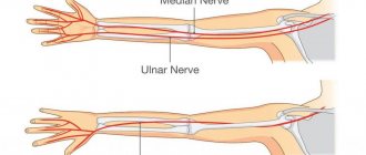
The definition of “acoustic neuroma” has developed historically, although it would be more correct to call it vestibular schwannoma . The tumor begins in the vestibular part of the vestibular-cochlear nerve (VIII pair of cranial nerves), which transmits information from the vestibular organs to the brain, and not from the cochlear part, which conducts auditory impulses. The VIII pair of cranial nerves passes together with the facial nerve (VII pair of cranial nerves) from the brain stem into the internal auditory canal (a small bony canal in the area of the temporal bone) to the organs of hearing and vestibular apparatus in the area of the temporal bone. Most often, tumor growth begins in the internal auditory canal; as the tumor increases in size, it spreads to neighboring areas and ultimately affects the cerebellar pontine angle.
Typical symptoms
Most often, the description of symptoms begins with the moment of hearing loss - with acute unilateral hearing loss and often with simultaneous noise in the ear (tinnitus). In many cases, after an injection that helps improve blood circulation in the area of the hearing loss, a short-term improvement is observed. Subsequently, symptoms of hearing loss reappear, with progressive deterioration of hearing. The most common complaints are dizziness and problems with the vestibular system, which are observed together with hearing loss and tinnitus. With the help of a gentle electro-physiological measurement method, which helps measure the auditory signal from its entry into the hearing organ to its perception by the brain, in the case of such symptoms, it is possible to identify vestibular schwannoma at an early stage and prescribe additional examination to identify the full picture of the disease. Large tumors can also put pressure on the cerebellum and brain stem, further leading to problems with walking and movement. Large tumors also have a negative impact on the functioning of neighboring cranial nerves. This can be expressed in a feeling of numbness of the face (group V cranial nerves), impaired facial mobility (group VII cranial nerves), and disorders of the swallowing reflex (group IX and X cranial nerves). The largest tumors prevent the free circulation of cerebrospinal fluid, which can lead to what is called obstructive hydrocephalus. The resulting intracranial pressure leads to headaches, nausea and loss of consciousness.
Therefore, in order to avoid unpleasant symptoms and serious consequences, it is necessary to undergo regular and timely examinations. Neuromas, for the most part, are treatable and today there are various methods of treatment, depending on the severity and size of the tumor.