What is it and what does it consist of
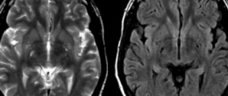
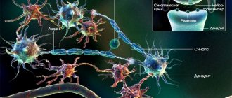
White matter of the brain is a collective concept that refers to a complex of nerve structures through which electrical and chemical impulses are transmitted. The nerve cell can be thought of as a trading post where travelers sell and buy goods, relax and discuss prices. However, for successful commercial activities, traders need roads, thanks to which they make long journeys from one point to another, delivering valuable cargo. It’s the same in the brain: the white substance ensures the delivery of nerve impulses.
The white matter of the nervous system serves as a springboard for the gray matter. The latter, unlike white, acts as a generator and collector of information. The white substance transmits the nerve impulse and is not responsible for its creation. On the other hand, there are opinions of many experts that white matter determines the speed and quality of brain functioning, namely the number of formed nerve pathways. Indeed, the development of the mental component of the mental sphere in children usually means the formation of white matter in the brain.
The white substance is opposed to the gray one. Gray matter is a collection of nerve cell bodies and their appendages (glial tissue, capillaries, partially short processes and early axons). The functions of gray matter include providing programs for higher nervous activity, such as thinking, memory, and perception. The contrast is not only functional, but also anatomical. If the gray matter is the cortex (the final layer of the brain), then the white substance is located between the cortex and the deep structures of the brain.
Speaking of structure, the substantia alba differs from the gray matter: the white matter of the brain consists of bundles of long processes - axons, covered with a myelin sheath. This layer, consisting of fat components, provides a person with an electrical impulse transmission speed of up to 100 m/sec on average. An axon that does not have myelinated fibers transmits information up to 10 m/sec. The white color of the substance is provided by myelin, and on a section the subcortical ball of the substance looks whitish-cream.
So, the white matter of the brain is represented by myelinated axons that connect different parts of the brain. Anatomically, the processes are divided into long ones, responsible for communication between distant parts of the brain, and short ones, connecting nearby structures (brain convolutions). They are located as follows:
- Short . They lie directly under the cortex of the brain and are called subcortical.
- Long or intracortical. This part of the white matter is located in the deep parts.
In addition, white matter is conventionally divided into 3 types, depending on anatomical features:
Associative connections . Fibers of this type of white matter provide a general connection between areas of the cortex, but located in the same hemisphere. For example, association fibers connect the area of general sensitivity (parietal cortex) with the frontal cortex.
Commissural fibers . These structures are represented by cerebral adhesions and articulate similar areas, but on different hemispheres. For example, the hearing area on the temporal cortex of one hemisphere with the same area in the other part of the brain. The largest structure here is the corpus callosum. In the physiological aspect, the structure ensures the interconnection of both hemispheres. The corpus callosum has not been fully studied.
Projection fields . This type of white matter connects the cerebral cortex with structures morphologically located below. Functionally divided into two subtypes:
- Efferent fibers. Along these pathways, the nerve impulse is sent from the cortical centers to the underlying structures;
- afferent. These fibers ensure the delivery of electrical signals from underlying structures (internal organs, tissues) to the brain.
There are phenomena where people who do not have this unifying structure (corpus callosum) have phenomenal memory. Experts say that this is due to the corpus callosum, which acts as a kind of barrier that limits the flow of electrical impulses. In the case where it is not present, the areas are connected to each other directly, without any collector system or filters.
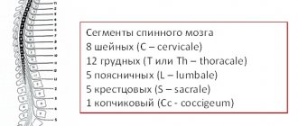
The white matter of the medulla oblongata is represented by short and long fibers. The latter include the pyramidal tracts running through the anterior clusters of the spinal cord. The fibers of the medulla oblongata form several tracts:
- Rubrospinal;
- Vestibulospinal;
- Reticulospinal tract.
Through these structures, information flows from the nuclei of the medulla oblongata, reticular and vestibular nuclei to the spinal cord.
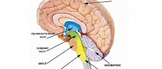
The white matter of the midbrain forms a cluster represented by the cerebral body, located deep in the cerebellum. Branching, the fibers of the body pierce all the convolutions of the coordinating center of the brain. The fibers of the white matter of the cerebellum form pathways leading to the cerebral cortex and neighboring brainstem structures.
Anatomical structures of the white matter of the cerebral hemispheres
Corpus callosum. The arch and its departments. Associative, commissural and projection pathways of the cerebral hemispheres. Lateral ventricles of the brain and their communication. Choroid plexuses of the ventricles. Basal ganglia and their significance.
The white matter of the hemisphere includes the internal capsule and fibers, which have different directions. These are fibers passing to the other hemisphere of the brain through its commissures (corpus callosum, anterior commissure, fornix commissure) and heading to the cortex and basal ganglia of the other side (commissural), as well as projection nerve fibers running from the cerebral hemisphere to its underlying parts and to the spinal cord and in the opposite direction from these formations.
The corpus callosum is formed by commissural fibers connecting both hemispheres. The free upper surface of the corpus callosum, facing the longitudinal fissure of the cerebrum, is covered with a thin plate of gray matter. The middle part of the corpus callosum - its trunk - bends downwards in front, forming the knee of the corpus callosum, which, thinning, passes into a beak, which continues downwards into the terminal (border) plate. The thickened posterior part of the corpus callosum ends freely in the form of a ridge. The fibers of the corpus callosum form its radiance in each hemisphere of the cerebrum. The genu corpus callosum fibers connect the cortex of the frontal lobes of the right and left hemispheres. Brainstem fibers connect the gray matter of the parietal and temporal lobes. The roller contains fibers connecting the cortex of the occipital lobes. The areas of the frontal, parietal and occipital lobes of each hemisphere are separated from the corpus callosum by the fissure of the same name.
Under the corpus callosum there is a thin white plate - the vault, consisting of two arched cords connected in its middle part by a transverse commissure of the vault. The body of the vault, gradually moving away in the anterior part from the corpus callosum, arches forward and downward and continues into the column of the vault. The lower part of each column of the fornix first approaches the terminal plate, and then the columns of the fornix diverge laterally and are directed downward and posteriorly, ending in the mastoid bodies.
Between the crura of the fornix at the back and the terminal plate at the front there is a transverse anterior (white) commissure, which, along with the corpus callosum, connects both hemispheres of the cerebrum.
Posteriorly, the body of the fornix continues into the flat peduncle of the fornix, fused with the lower surface of the corpus callosum. The crus of the fornix gradually moves laterally and downwards, separates from the corpus callosum, becomes even more dense and on one side fuses with the hippocampus, forming the hippocampal fimbria. The free side of the fimbria, facing the cavity of the lower horn of the lateral ventricle, ends in the hook, connecting the temporal lobe of the telencephalon with the diencephalon.
The area bounded above and in front by the corpus callosum, below by its beak, terminal plate and anterior commissure, and behind by the pedicle of the fornix, is occupied on each side by a sagittally located thin plate - the transparent septum. Between the plates of the transparent septum there is a narrow sagittal cavity of the same name, containing a transparent liquid. The lamina pellucidum is the medial wall of the anterior horn of the lateral ventricle.
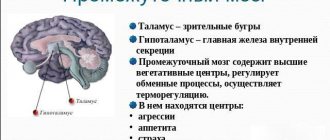
The internal capsule (capsula interna) is a thick, angled plate of white matter, bounded on the lateral side by the lentiform nucleus, and on the medial side by the head of the caudate nucleus (in front) and the thalamus (back). The internal capsule is formed by projection fibers connecting the cerebral cortex with other parts of the central nervous system. The fibers of the ascending pathways, diverging in different directions to the cerebral cortex, form the corona radiata. Downward, the fibers of the descending pathways of the internal capsule in the form of compact bundles are directed to the peduncle of the midbrain.
Lateral ventricle (ventriculus lateralis). The cavities of the cerebral hemispheres are the lateral ventricles (1st and 2nd), located in the thickness of the white matter under the corpus callosum. Each ventricle has four parts: the anterior horn lies in the frontal lobe, the central part in the parietal lobe, the posterior horn in the occipital lobe, and the inferior horn in the temporal lobe. The anterior horn of both ventricles is separated from the adjacent one by two plates of a transparent septum. The central part of the lateral one bends from above around the thalamus, forms an arc and passes posteriorly into the posterior horn, downwards into the inferior horn. The medial wall of the inferior horn is the hippocampus (a section of the ancient cortex), corresponding to the deep groove of the same name on the medial surface of the hemisphere. The fimbria stretches medially along the hippocampus, which is a continuation of the crus of the fornix. On the medial wall of the posterior horn of the lateral ventricle of the brain there is a protrusion - a bird's spur, corresponding to the calcarine groove on the medial surface of the hemisphere. The choroid plexus protrudes into the central part and lower horn of the lateral ventricle, which connects with the choroid plexus of the 3rd ventricle through the interventricular foramen.
The choroid plexus of the lateral ventricle is formed by protrusion into the ventricle through the vascular fissure of the pia mater of the brain with the blood vessels it contains.
The white matter of the telencephalon is represented by three types of pathways:
1. Associative (connect neurons of one hemisphere);
2. Commissural (connect neurons of two hemispheres);
3. Projection (connect neurons of the cerebral cortex with neurons of the underlying parts of the central nervous system).
The latter includes the main motor or pyramidal tract - a system of nerve fibers along which voluntary motor impulses from pyramidal neurocytes (Betz pyramidal cells) located in the horse of the precentral gyrus (layer V) are sent to the motor nuclei of the cranial nerves and the anterior horns of the spinal cord, and from them to the skeletal muscles.
The pyramid path is divided into two parts:
1. cortical-nuclear path going to the nuclei of the cranial nerve,
2. corticospinal tract (lateral and anterior).
The fibers of the cortical-nuclear tract pass to the opposite side to the motor nuclei of the cranial nerve. At the border of the medulla oblongata with the spinal cord, part of the fibers of the corticospinal tract passes to the opposite side. Thus, all pyramidal paths are crossed.
Extrapyramidal pathways are phylogenetically older than pyramidal ones. They start from the neurons of the cerebral cortex and end on the motor nuclei of the brain stem and the anterior horns of the gray matter of the spinal cord.
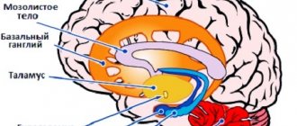
Basal ganglia and white matter of the telencephalon. In the thickness of the white matter of each cerebral hemisphere there are accumulations of gray matter, forming separately lying nuclei. These nuclei lie closer to the base of the brain and are called basal (subcortical, central). These include the striatum, which in lower vertebrates constitutes the predominant mass of the hemispheres, the fence and the amygdala.
The striatum (corpus striatum) in sections of the brain looks like alternating stripes of gray and white matter. Most medially and anteriorly is the caudate nucleus , located lateral and superior to the thalamus, being separated from it by the knee of the internal capsule. The nucleus has a head located in the frontal lobe, protruding into the anterior horn of the lateral ventricle and adjacent to the anterior perforated substance. The body of the caudate nucleus lies under the parietal lobe, limiting the central part of the lateral ventricle on the lateral side. The tail of the nucleus participates in the formation of the roof of the inferior horn of the lateral ventricle and reaches the amygdala, which lies in the anteromedial parts of the temporal lobe (posterior to the anterior perforated substance). The lenticular nucleus is located lateral to the caudate nucleus. A layer of white matter - the internal capsule - separates the lenticular nucleus from the caudate nucleus and from the thalamus. The lower surface of the anterior part of the lentiform nucleus is adjacent to the anterior perforated substance and is connected to the caudate nucleus. The medial part of the lenticular nucleus in a horizontal section of the brain narrows and is angled towards the knee of the internal capsule, located on the border of the thalamus and the head of the caudate nucleus. The convex lateral surface of the lenticular nucleus faces the base of the insular lobe of the cerebral hemisphere.
On the frontal section of the brain, the lenticular nucleus also has the shape of a triangle, the apex of which faces the medial side and the base faces the lateral side. Two parallel vertical layers of white matter divide the lenticular nucleus into three parts. The darker shell lies most laterally, the “globus pallidus” is located more medially, consisting of two plates: medial and lateral. The caudate nucleus and putamen belong to phylogenetically newer formations, while the globus pallidus belongs to older ones. The nuclei of the striatum form the striopallidal system, which, in turn, belongs to the extrapyramidal system involved in the control of movements and the regulation of muscle tone.
A thin vertically located fence , lying in the white matter of the hemisphere on the side of the shell, between it and the cortex of the insular lobe, is separated from the shell by the outer capsule, and from the insular cortex by the outermost capsule. The amygdala lies in the white matter of the temporal lobe of the hemisphere, approximately 1.5–2 cm posterior to the temporal pole.
White matter functions
Primarily, the white matter of the brain is responsible for coordinating information in the central nervous system. Thanks to the white substance, the brain is capable of “communication” between its own areas. In addition to the brain, the substantia alba is also located in the spinal cord, but its set of functions in the periphery is different. The white matter of the spinal column is responsible for the sensory and motor components of nervous activity. White matter acts as a conductor. The white substance also provides:
- Connection of similar structures of the hemispheres;
- connection of various parts of the cerebral cortex with other parts of the nervous system, in particular with the spinal cord.
Functions
Providing a safe environment for the functioning of the nuclei and other parts of the brain and conducting signals throughout the nervous system are the main tasks of white matter.
Constantly, uninterruptedly connecting all parts of the central nervous system is the main goal of the action of white matter. This ensures coordination of general life activities. A signal is transmitted through neural processes, which allows for a variety of human actions.
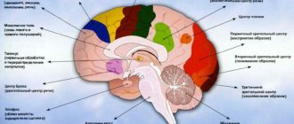
On the cerebral cortex, grooves and ridges that form convolutions can be clearly visible. The central sulcus divides the parietal and frontal lobes. On both sides of this groove are the temporal lobes. The furrows and convolutions separate the hemispheres, forming 4 lobes in each:
- Frontal lobes. They have undergone great changes in the process of evolution. They developed faster than others and have the largest mass. In them, the white matter must provide all motor processes. Here, thinking processes are started, the structure of speech and writing is regulated, and all complex forms of life support are controlled.
- Temporal lobes. They border on all other lobes. The functioning of the white matter in them is aimed at understanding speech and learning opportunities. Allows you to draw conclusions by receiving all kinds of information through hearing, sight, and smell.
- Parietal lobes. Responsible for pain, temperature, tactile sensitivity. They make possible the work of centers that have been brought to automaticity: eating, drinking, dressing. A three-dimensional understanding of the world around you and yourself in space is built.
- Occipital lobes. In this area, functions are aimed at remembering processed visual information. The form is evaluated.
Difference from gray matter
Gray matter differs from white matter not only functionally, but also anatomically. Location: gray matter is located on the surface of the cerebral hemispheres and is its upper layer. The white matter is located between the gray and deep structures of the brain.
Structure
gray matter consists of soma neurocytes and its appendages
white matter consists of myelinated fibers
Function
gray nuclei are responsible for specific specific functions of the nervous system
White matter is a conductor of nerve impulses
To further understand the differential differences, you can go to the article difference between white matter and gray matter.