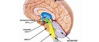
The structure and location of the frontal lobe of the brain
The frontal lobe is actually made up of two paired lobes and makes up two-thirds of the human brain. The frontal lobe is part of the cerebral cortex, and the paired lobes are known as the left and right frontal cortex. As its name suggests, the frontal lobe is located near the front of the head under the frontal bone of the skull.
All mammals have a frontal lobe, although it varies in size. Primates have the largest frontal lobes than other mammals.
The right and left hemispheres of the brain control opposite sides of the body. The frontal lobe is no exception. Thus, the left frontal lobe controls the muscles on the right side of the body. Likewise, the right frontal lobe controls the muscles on the left side of the body.
Structure and location
The frontal lobes are divided into two main areas: the motor cortex and the prefrontal cortex.
The area of the brain involved in language and speech, known as Broca's area, is located in the left frontal lobe. The prefrontal cortex is the front part of the frontal lobes and controls complex cognitive processes such as memory, planning, reasoning, and problem solving.
This area of the frontal lobe helps set and maintain goals, control negative impulses, organize events in a temporal order, and form individual personalities.
Functions of the frontal lobe of the brain
The brain is a complex organ with billions of cells called neurons that work together. The frontal lobe works alongside other areas of the brain and controls the functions of the brain as a whole. Memory formation, for example, depends on many areas of the brain.
What's more, the brain can "repair" itself to compensate for damage. This does not mean that the frontal lobe can recover from all injuries, but other areas of the brain can change in response to head trauma.
The frontal lobes play a key role in future planning, including self-management and decision making. Some functions of the frontal lobe include:
- Speech
: Broca's area is an area in the frontal lobe that helps verbalize thoughts. Damage to this area affects the ability to speak and understand speech. - Motor function
: The frontal lobe cortex helps coordinate voluntary movements, including walking and running. - Comparing objects
: The frontal lobe helps categorize objects and compare them. - Memory formation
: Almost every region of the brain plays an important role in memory, so the frontal lobe is not unique, but it plays a key role in the formation of long-term memories. - Personality formation
: The complex interplay of impulse control, memory, and other tasks helps shape the basic characteristics of a person. Damage to the frontal lobe can radically change personality. - Reward and motivation
: Most of the brain's dopamine-sensitive neurons are located in the frontal lobe. Dopamine is a brain chemical that helps maintain feelings of reward and motivation. - Management of attention
, including
selective attention
: When the frontal lobes cannot manage attention, attention deficit hyperactivity disorder (ADHD) may develop.
Frontal lobe
According to T.I. Yudin (1941), when the frontal lobes of the brain are removed, a person loses the ability to navigate new complex situations and make new plans for the future, which is somewhat reminiscent of symptoms quite typical of schizophrenia. T.I. Yudin believed that the frontal lobes contain mechanisms through which the future is planned. Modern neuropsychological studies show that damage to even both frontal lobes may not be accompanied by either aspontaneity or changes in temperament, but it does reduce the patient from his creative level.
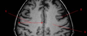
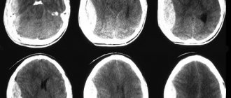
| | MRI allows you to diagnose changes in the size of the frontal lobe, and you can track brain activity in these areas using EEG |
In 1974, K. Ingvar and T. Franzen showed that patients with schizophrenia have a decrease in the volume of blood flow in the frontal lobes of the brain. Insufficient blood supply (hypofrontality) of the frontal lobes was so noticeable in patients with schizophrenia with severe mental disorders that it allowed us to talk about a special “hypofrontality phenomenon” in schizophrenia.
Frontal lobe changes in schizophrenia
- The phenomenon of hypofrontality (reduced blood flow in the frontal lobes of the brain)
- Reduction of volumes of the anterior cingulate, orbitofrontal, dorsolateral and ventromedial cortices
- Disruption of connections between the frontal lobes and deep brain structures
- Disruption of connections between the anterior parts of the right and left hemispheres
Using MRI slices in schizophrenia, decreased volumes of the anterior cingulate, orbitofrontal, dorsolateral and ventromedial cortices were found.
In other studies, on the contrary, hyperfrontality was identified, but such data were found significantly less than the results indicating a decrease in the functional activity of the frontal cortex, especially in schizophrenia with severe negative symptoms.
Recent studies have shown that in patients with schizophrenia, connections between the frontal lobes and much deeper brain structures are weakened. The cause of these changes is likely due to either increased activity of these brain structures or suppressed activity of the frontal cortex.
Many psychopathological syndromes that are observed in schizophrenia probably have their own pathoanatomical substrate, most likely manifesting itself at the level of the relationship between certain brain structures, for example, along the main pathways of the ascending dopaminergic tract: mesocortical, nigrostriatal, mesolimbic.
Some authors, noting anomalies in schizophrenia, associate them with a violation of cell migration from the cortical plate to the gray matter of the cortex during the process of brain maturation in ontogenesis.
This assumption is partly confirmed by research in the field of neuropsychiatry. Anosognosia and symptoms of depersonalization, which in some cases occur in schizophrenia, can be explained by predominant damage to the right hemisphere of the brain.
The clinical picture of damage to the left frontal lobe is characterized by aspontaneity, up to “paralysis of initiative,” a decrease in emotional experience regarding oneself and the world around (Bely B.I., 1987).
In addition to anosognosia, lesions of the right frontal lobe are characterized by fragmented perception with a tendency to interpret the entire proposed picture as a whole on the basis of randomly perceived individual episodes, uncriticality of one’s own mistakes, false recognitions, instability of attention, absent-mindedness, slipping into unrelated topics, difficulty switching attention, exhaustion, tendency to stereotypes. At the same time, the range of symptoms of damage to the right frontal lobe also includes other symptoms: confabulatory additions to what is perceived, mobility, verbosity, familiarity. The second group of symptoms, although found in schizophrenia, is recorded less frequently than the first. It is a fair assumption that the degree of anatomical damage to the right frontal lobe in schizophrenia is not as total as in tumors of this area of the brain, where both groups of symptoms are often noted in the clinical picture of the disease.
In the initial stages of the malignant variant of the course of schizophrenia, one can detect symptoms quite characteristic of damage to the basal-frontal regions: inadequacy of emotional reactions, verbosity, sloppiness, increased appetite (Sternberg E.Ya., 1967).
In schizophrenia, thinking is impaired, the ability to plan actions is weakened, and there is difficulty solving problems, which may indicate damage to the frontal lobe of the brain.
Symptoms of neurocognitive deficits and negative symptoms, as a rule, are more pronounced where there is damage to the frontal lobe of the brain, an indirect sign of which can be considered the expansion of the subarachnoid spaces around some areas of this lobe. According to our observations, the severity of delirium and lack of criticism of one’s condition are more noticeable in those patients who demonstrate changes in the frontal lobe of the brain when studied using modern neuroimaging methods.
The frontal lobes have been found to be involved in initiating actions. Response suppression, in turn, depends on interactions between the frontal cortex and the underlying areas of the brain that process sensations.
Some researchers believe that impaired functioning of the frontal lobe in schizophrenia is associated with a deficiency of dopamine, others speak of insufficient glutamatergic activity in this area of the brain.
It is interesting to note that with exacerbation of paranoid symptoms, there is an accumulation of dopa in the striatum. However, at present, the relationship between the concentration of dopa decarboxylase and dopamine synthesis remains unclear, since there is no direct relationship between them. At the same time, insufficient activation of the prefrontal cortex during the Wisconsin test is accompanied by the accumulation of dopa in the striatum. As a result of the above, it can be assumed that in schizophrenia, dysfunction of the frontal lobes is correlated with excessive activation of dopaminergic structures in the striatum.
In the frontal cortex, dopamine is considered a neurotransmitter that enhances the activity of neurons (D1 receptors), and increased activity of the glutamatergic system tends to enhance the activity of the dopaminergic system. Because of the above, a lack of glutamate in the frontal lobe of the brain may also contribute to increased negative symptoms and manifestations of neurocognitive deficits. A decrease in the severity of negative symptoms in schizophrenia as a result of taking atypical antipsychotics is also associated with changes in the activity of the serotonergic system of the brain.
In schizophrenia, neuropsychological studies indicate a disruption of the relationship between the anterior parts of the brain hemispheres.
Usually a normal person can control his actions (“model of subsequent message”), as if dosing their intensity; in schizophrenia, this process is disrupted.
Patients with mental automatism syndrome do not detect differences in time, manipulation of handling both small and larger objects. Let us recall that normally, when a person operates with smaller objects, his movements become slower.
Suppression of responses to sensations that a person induces through his actions occurs under the influence of signals coming from areas of the brain associated with the formation of actions.
Return to Contents
Consequences of damage to the frontal lobe of the brain
One of the most notorious head injuries occurred to railroad worker Phineas Gage. Gage survived an iron spike piercing his frontal lobe. Although Gage survived, he lost an eye and suffered a personality disorder. Gage changed dramatically, the once meek worker became aggressive and out of control.
It is not possible to accurately predict the outcome of any frontal lobe injury, and such injuries can develop very differently in each individual. In general, damage to the frontal lobe due to a blow to the head, stroke, tumor, and disease can cause symptoms such as:
- speech problems;
- personality change;
- poor coordination;
- difficulties with impulse control;
- planning problems.
Treatment of frontal lobe damage
Treatment for frontal lobe damage is aimed at eliminating the cause of the injury. Your doctor may prescribe medications for an infection, perform surgery, or prescribe medications to reduce your risk of stroke.
Depending on the cause of the injury, treatment is prescribed that may help. For example, if a frontal injury occurs after a stroke, it is important to adopt a healthy diet and physical activity to reduce the risk of future stroke.
The drugs may be useful for people who have problems with attention and motivation.
Treatment of frontal lobe injuries requires ongoing care. Recovery from injury is often a lengthy process. Progress can come suddenly and cannot be completely predicted. Recovery is closely related to supportive care and a healthy lifestyle.
Functions of the frontal lobes
The frontal lobes regulate motivational processes. They are also responsible for the perception and resolution of conflicts, as well as for constant attention, for the control of emotions and social behavior. They regulate emotional processing and control behavior based on context.
Functions of the premotor cortex
The primary function of the motor cortex is to control voluntary movement, including expressive language, writing, and eye movements. The primary motor cortex sends commands to neurons in the brainstem and spinal cord. These neurons are responsible for specific voluntary movements. Within the primary motor cortex of the two hemispheres there is a representation of the contralateral half of the body. That is, in each hemisphere there is a representation of the opposite side of the body. This area controls the programming of preparation and movement. The premotor cortex automates, harmonizes, and archives movement programs associated with previous experiences.
The primary motor cortex of the frontal lobes is involved in voluntary movement. It has nerve connections to the spinal cord that allow this area of the brain to control muscle movements. Movement in different areas of the body is controlled by the primary motor cortex, with each area connected to a specific area of the motor cortex. Body parts that require fine control of movement take up large areas of the motor cortex, while those that require simpler movements take up less space. For example, areas of the motor cortex that control movement of the face, tongue, and hands take up more space than areas associated with the hips and trunk. The premotor cortex of the frontal lobes has neural connections with the primary motor cortex, spinal cord, and brain stem. The premotor cortex allows you to plan and execute correct movements in response to external signals. This cortical region helps determine the specific direction of movement.
Functions of the prefrontal cortex
The prefrontal cortex is located in the front of the frontal lobe. It is considered the ultimate expression of human brain development. It is responsible for cognition, behavior and emotional activity. The prefrontal cortex receives information from the limbic system (involved in emotional control) and acts as an intermediary between cognition and feelings through executive functions. Executive functions are a set of cognitive skills needed to control and self-regulate behavior.
Functions of the dorsolateral prefrontal cortex
It is one of the most recently formed parts of the human brain. It makes connections with the other three areas of the brain and transforms information into thoughts, decisions, plans and actions.
It is responsible for cognitive abilities such as:
- attention;
- focus;
- braking;
- information maintenance and processing;
- programming upcoming actions;
- analysis of possible results;
- self-analysis of cognitive activity;
- analyzing the situation and developing an action plan;
- ability to adapt to new situations;
- organization of behavior in relation to a new goal.
Literature
- Collins A., Koechlin E. Reasoning, learning, and creativity: frontal lobe function and human decision-making //PLoS biology. – 2012. – T. 10. – No. 3. – P. e1001293.
- Chayer C., Freedman M. Frontal lobe functions // Current neurology and neuroscience reports. – 2001. – T. 1. – No. 6. – pp. 547-552.
- Kayser AS et al. Dopamine, corticostriatal connectivity, and intertemporal choice // Journal of Neuroscience. – 2012. – T. 32. – No. 27. – pp. 9402-9409.
- Panagiotaropoulos TI et al. Neuronal discharges and gamma oscillations explicitly reflect visual consciousness in the lateral prefrontal cortex // Neuron. – 2012. – T. 74. – No. 5. – pp. 924-935.
- Zelikowsky M. et al. Prefrontal microcircuit underlies contextual learning after hippocampal loss // Proceedings of the National Academy of Sciences. – 2013. – T. 110. – No. 24. – pp. 9938-9943.
- Flinker A. et al. Redefining the role of Broca's area in speech //Proceedings of the National Academy of Sciences. – 2015. – T. 112. – No. 9. – pp. 2871-2875.