Neuroinfections are a group of pathologies of an infectious nature, which are characterized by a severe course, damage to the anatomical structures of the nervous system, and a high mortality rate. Neuroinfection of the brain progresses when pathogenic microorganisms enter the body of an adult or child.
Neuroinfection can develop both primary and secondary. Primary neuroinfections are characterized by the fact that infectious agents initially affect the structures of the nervous system. The development of a secondary neuroinfection is said to occur if infectious agents were transferred by lymphogenous or hematogenous routes from other pathological foci.
Varieties
Neuroinfections in children and adults can occur in acute and chronic forms. The following diseases are classified as acute:
- myelitis;
- tetanus;
- encephalitis;
- rabies;
- meningitis;
- arachnoiditis.
Chronic types of neuroinfection of the brain:
- brucellosis;
- neurosyphilis;
- neurobrucellosis;
- neuroAIDS.
Neuroinfection according to the ICD does not have its own code, but the pathologies that are included in this group have a code. For example, the rabies code according to ICD-10 is A82.
Clinic
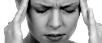
Symptoms of neuroinfection are usually very pronounced. It is important to immediately go to see a qualified doctor when the first signs appear, as delay can cost the patient’s life.
Any pathology belonging to the group of nephroinfections is accompanied by three syndromes, which are characterized by certain symptoms:
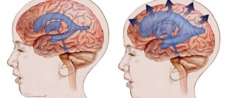
- liquor hypertension syndrome. It is characterized by intense headache, impaired consciousness, mood changes, tachycardia, tachypnea; intoxicating. An increase in body temperature to critical levels, nausea and vomiting, weakness, decreased ability to work, etc.; liquor syndrome.
Signs of neuroinfection
Diseases caused by neuroinfection are divided into acute, subacute and chronic. The most common conditions are:
- encephalitis, meningitis and myelitis and their forms are the most acute and serious diseases of the brain and spinal cord that require immediate hospitalization;
- radiculitis, neuritis;
- secondary neuroinfections (neurosyphilis, neurobrucellosis, tuberculosis, etc.).
Infection with a neuroinfection occurs through the bites of insect carriers (for example, encephalitis ticks) or infected animals, consumption of milk from such animals, such as complications after acute respiratory infections or ENT diseases of an infectious nature, through direct contact with infected blood and mucous membranes, from mother to child during childbirth.
Symptoms of neuroinfection have varying degrees of manifestation, depending on the severity of the condition:
- the acute form is characterized by: high temperature and general signs of intoxication, headaches, photophobia, convulsions, confusion, speech impairment, impaired coordination of movements, temporary hearing and vision disorders (hallucinations, decreased acuity, blindness or deafness), loss of consciousness and coma;
- Chronic infection is rarely accompanied by an increase in temperature above 37.2˚C, but headaches, frequent nausea, paresis of the limbs, sensory disturbances, depressed consciousness, drowsiness and lethargy, tachycardia with a decrease in blood pressure, and decreased labor and social activity are recorded.
It would not be amiss to once again emphasize that neuroinfection is dangerous in itself, but if suffered at an early age, it threatens severe complications, ranging from disturbances of sensitivity and motor activity, to mental retardation, disability and death.
Therapeutic measures
The consequences of neuroinfection can be extremely unfavorable if it is not detected and treated in a timely manner. Treatment of neuroinfection directly depends on which pathogen is identified during diagnosis. In case of bacterial infection, antibiotics are used. For viral neuroinfection, antiviral therapy is indicated, in particular interferon is prescribed. In addition, symptomatic treatment is carried out - diuretics, nootropics, neuroprotectors, vascular drugs, etc. are prescribed.
If a person has suffered a neuroinfection, then he may experience some changes in behavior, frequent mood changes, decreased mental activity, memory, etc.
Neuroinfection with SARS-CoV-2: what is currently known
Anosmia, encephalopathy, and stroke are the most common neurological syndromes associated with SARS-CoV-2 infection, although many other manifestations have been reported.
Analysis of biopsy samples from patients suggests that anosmia occurs primarily as a result of SARS-CoV-2 damage to non-neuronal cells of the olfactory epithelium and olfactory bulb, which leads to local inflammation and malfunction of neurons.
A significant proportion of patients admitted to intensive care units with a diagnosis of COVID-19 develop delirium; There is evidence that microvascular and inflammatory mechanisms underlie this phenomenon.
Autopsy data demonstrate activation of astrocytes and microglia in association with COVID-19 infection, particularly in the brainstem, where cytotoxic T cells also infiltrate.
SARS-CoV-2 can be detected in the brain by PCR diagnostics and immunohistochemistry, but current evidence suggests that the virus is primarily located in vascular and immune cells rather than directly infecting neurons.
Historically, serious neurological diseases such as Japanese encephalitis and polio directly affected the central nervous system. As the coronavirus pandemic progresses and reports of an increase in the number of patients with neurological diseases, the question has arisen, will this virus behave in a similar way?
Early reports of patients with COVID-19 who had clinical evidence of inflammation of the brain tissue raised the possibility of encephalitis caused by SARS-CoV-2. However, the fact that the virus was rarely found in the cerebrospinal fluid (CSF) suggests that immune-mediated damage predominates rather than virus replication directly in neurons. As the number of case reports has increased, it has become clear that anosmia, encephalopathy and stroke are the main neurological syndromes associated with COVID-19.
Anosmia and associated dysgeusia occur during SARS-CoV-2 infection, often in the absence of other symptoms. Herpes simplex virus type 1 infects the olfactory bulb and then the brain, leading to encephalitis. Animal models have shown that the same thing happens during infection with some coronaviruses, including SARS-CoV, which causes severe acute respiratory syndrome. Consequently, concerns have been raised that olfactory infection with SARS-CoV-2 may lead to CNS disease. However, a study published in July 2021 showed that the SARS-CoV-2 pathogen infects the supporting cells of the olfactory epithelium, rather than sensory neurons.
SARS-CoV-2 infection of supporting cells can lead to anosmia through several mechanisms. First, supporting cells of the olfactory epithelium regulate local water and electrolyte balance, and their damage can affect the conduction of signals from olfactory neurons to the brain. Second, infection of these cells and pericytes of the olfactory bulb may disrupt neuronal signaling due to local inflammation with the release of cytokines. Third, vascular damage and hypoperfusion in the olfactory bulb may contribute to its dysfunction. Finally, any of these changes may indirectly trigger the death of olfactory neurons. During observation of patients with COVID-19 and anosmia, hyperintensity and swelling of the olfactory bulb were identified, which is consistent with the presence of inflammation, which subsequently resolves, as do the symptoms in most patients.
Anosmia is the most common neurological symptom of mild coronavirus infection, but changes in higher mental functions are most important among hospitalized patients. Terms such as encephalopathy and delirium are used to describe these changes; different specialties prefer different terms, making data comparisons difficult.
In one study, 118 of 140 patients admitted to intensive care units (ICUs) with COVID-19 developed delirium associated with acute impairment of attention, consciousness, and cognition; 88 had evidence of corticospinal tract involvement. Delirium can develop in any patient in the ICU, but accumulating evidence suggests that these symptoms may be characteristic of coronavirus infection, especially since delirium, encephalopathy and other neuropsychiatric disorders are also observed in patients with milder forms of COVID-19. who are not treated in the ICU.
Analysis of colony-stimulating factor (CSF) in autopsy and research specimens is beginning to elucidate the mechanisms that may underlie cognitive impairment. Typically, pleocytosis is not observed in individuals with COVID-19-associated encephalopathy, but protein levels may be elevated due to oligoclonal IgG. Elevated levels of plasma and CSF cytokines, glial fibrillary acid proteins, and neurofilament light chain in COVID-19 are thought to reflect proinflammatory systemic and brain responses that include microglial activation and subsequent neuronal damage. Additional evidence for inflammatory mechanisms comes from imaging studies that have shown diffuse dilatation and white matter abnormalities such as microhemorrhages.
In addition to the inflammatory changes, coagulopathy, and endothelial vascular dysfunction that cause large vascular insults in COVID-19, microhemorrhages may also lead to small vessel occlusion, which may precipitate neurological and neuropsychiatric changes, as suggested by clinical and imaging studies . However, definitive data on the underlying mechanisms come from autopsy. One such study included 43 patients, most of whom died in ICUs, general wards, or nursing homes from pneumonia or sepsis associated with COVID-19. Six of them had acute ischemic brain damage. Activation of astrocytes was widespread in the brain, while activation of microglia was limited to the brainstem and cerebellum. Cytotoxic T cells have also been seen in the brainstem and meninges of many patients. RNA detection and immunohistochemistry results suggest that SARS-CoV-2 was widely distributed throughout the brain tissue, especially in the brainstem. However, no correlation was observed between the location of the virus and inflammation, or between the results of PCR diagnostics and immunohistochemical analysis of the virus. Without double immunostatization, it is difficult to say with certainty which cell types were infected, but electron microscopy results indicate infection of vascular endothelial cells rather than neurons. However, autopsies of 33 patients provided PCR and immunohistochemical data on SARS-CoV-2 in cells considered to be olfactory neurons and in anatomically related brain regions. However, some patients with encephalopathic changes respond to corticosteroids, highlighting the importance of immune-mediated mechanisms rather than direct viral effects.
Ultimately, if the retina is called the window to the brain, then to understanding SARS-CoV-2, the nose will probably be that. Just as SARS-CoV-2 causes olfactory impairment without infecting olfactory neurons, research data suggest that impairment of higher mental functions occurs predominantly without infection of CNS neurons (Figure 1). Although the virus can enter the brain, it appears to preferentially infect vascular and immune cells. Local inflammation upregulates astrocytes and microglia, possibly exacerbating the effects of circulating proinflammatory cytokines in severe systemic disease. Microvascular infarctions and hemorrhages, which are part of the systemic coagulopathy and vasodilation of COVID-19, also likely play an important role in the development of encephalopathy, delirium, and other neurological manifestations of SARS-CoV-2 infection.
Figure 1 | Up-to-date insight into the underlying mechanisms of neurological disease in COVID-19