Author: TainT
October 27, 2021 12:14
Community: Science
Tags: macro photography science neuroscience photo it's interesting
6281
22
The brain is a whole universe. What is there, what is being done and how it all works today we can see with the help of a microscope and macro photography. Let's lift the curtain and take a look into this wonderful world.
0
See all photos in the gallery
"Snail" of a newborn rat
0
This stunning image, which took eighth place in the 2021 Nikon Small World competition, shows us the main organ through which we hear: the snail. Hair cells are shown in green, the deviation of which triggers a nerve impulse that ultimately arrives in the auditory cortex. Red - nerve cells of the cochlea, “collecting” signals from hair cells.
Simplified case: receptors, pathways and neuronal circuits
Although reflex arcs are activated by external influences, they are nevertheless fully integrated into the execution of voluntary movements. For example, you turn your head from side to side as you read this article.
In this way, you ensure that the arbitrary task is completed, while your eyes provide focus. This is one of the important properties of sensorimotor adaptation, and it occurs without the participation of voluntary mechanisms. Oculomotor coordination is more complex than the normal reflex arc.
Voluntary actions subordinated to external goals are associated with the cortex in humans. Complex subcortical systems are also involved in the planning and execution of various actions. Spinal centers are capable of executing commands from higher structures using sensorimotor reflexes; they also send feedback signals to the brain.
At all levels of movement control, there are both endogenous and exogenous (sensory) inputs, both conscious and unconscious (Goodale and Milner, 1992). Thus, although there are a number of simple reflexes, such as the famous knee reflex .
Reflexes rarely work in isolation. Normally, they serve to perform tasks determined by the cortex.
Reflexes are innate mechanisms . They are formed during the development process; some of them subsequently disappear, such as the infant grasp reflex.
However, acquired involuntary processes are not reflexes. They are complex processes called automated skills and depend on practice.
So, reading this book, you can perfectly distinguish the letters a and b from each other, whereas for a person unfamiliar with the alphabet, this is much more difficult to do. When speaking and writing, the control of muscle contractions is very precise and largely unconscious. A person uses many automated skills to perform everyday tasks and needs.
Such skills are most often unconscious, do not require significant effort and do not require detailed conscious control. They should not be confused with reflexes. Automatic skills are initially under the control of the cortex, but after intensive practice they gradually come under the control of subcortical structures.
Each sensory nerve may contain several parallel channels, each carrying slightly different information. Thus, the visual tract has a channel for transmitting color, called small-cell, and a channel for transmitting the shape and size of an object, called large-cell. In the same way, somatosensory pathways combine channels for transmitting touch, pressure, pain and some others.
Most sensory fibers terminate in the thalamus , where they transmit signals to neurons that terminate in the cortex. Thalamic nuclei provide connections. Vision and touch are organized topographically, the sensory fields of primary receptors transform into fields of a higher level. Hearing is organized tonotopically; arrays of neurons correspond to a certain sound frequency.
Similarities of sensory pathways – touch, vision and hearing. All sensory systems begin with arrays of receptors, such as the touch receptor layers in the skin or the retina of the eye.
After initial processing in situ, sensory information is sent to the cortex for further processing. Note that all three of these sensory pathways terminate in the thalamic nuclei (green).
All these paths are also divided into two parts, one of which ends in the opposite hemisphere. This phenomenon is called crossover.
On their way from the periphery to the cortex, most sensory pathways cross the plane of symmetry of the body. The evolutionary significance of this phenomenon is still unclear, but nevertheless it is extremely common in both humans and other mammals.
Sensory organs interact with the nuclei of the thalamus. The thalamus is often called the hub of the brain.
However, it is quite possible that he is the most important part of it. Note that visual, auditory, and somatosensory information reaches the thalamus before reaching the cortex.
At the same time, signals also arrive from the cortex to the thalamus, which means that there is a continuous flow of information between the cortex and numerous thalamic nuclei. In some cases, the thalamus enhances the activity of the cortex, while in others it inhibits or blocks it.
Note the similarity of the cortical input and output of all three systems. JIKT – lateral geniculate body, RN – reticular nuclei, MCT – medial geniculate body, VB – ventrobasal complex of thalamic nuclei.
Although we are accustomed to thinking that signals travel in only one direction along various paths, in fact this is rarely true. Thus, signals from the retina to the thalamus go in one direction, while the outgoing signal from there is bidirectional. Up to 90% of neurons connecting the thalamus and area VI transmit the signal in the opposite direction - from area VI to the thalamus. In the auditory analyzer, the return flow of signals goes directly to the receptors.
Thus, in most signaling pathways there are feedback loops - such as in a neural network with two or more layers. Idelman and his co-authors attached particular importance to feedback, considering it one of the main properties of the brain. From this point of view, the brain appears to be a system of arrays and networks influencing each other.
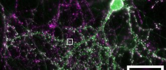
Dividing neuronal stem cells
0
The skin, liver, heart, kidneys, lungs and blood can form new cells to replace damaged ones. Until recently, experts believed that this ability to regenerate did not extend to the central nervous system, consisting of the brain and spinal cord. However, over the past five years, neuroscientists have discovered that the brain does change throughout life: new cells are formed to cope with emerging difficulties. This plasticity helps the brain recover from injury or disease, increasing its potential.
Neuron and its structure
You can often hear that a person’s mental abilities are guaranteed by the presence of gray matter. What is this substance and why is it gray? This is the color of the cerebral cortex, which consists of microscopic cells. These are neurons or nerve cells that ensure the functioning of our brain and control of the entire human body.
How does a nerve cell work?
A neuron, like any living cell, consists of a nucleus and a cell body called the soma. The size of the cell itself is microscopic - from 3 to 100 microns. However, this does not prevent the neuron from being a real repository of various information. Each nerve cell contains a complete set of genes - instructions for producing proteins. Some of the proteins are involved in the transmission of information, others create a protective shell around the cell itself, others are involved in memory processes, others provide mood changes, etc.
Even a small malfunction in one of the programs for the production of a certain protein can lead to serious consequences, illness, mental impairment, dementia, etc.
Each neuron is surrounded by a protective sheath of glial cells; they literally fill the entire intercellular space and make up 40% of the brain substance. Glia or a collection of glial cells performs very important functions: it protects neurons from unfavorable external influences, supplies nerve cells with nutrients and removes their waste products.
Glial cells guard the health and integrity of neurons, and therefore prevent many foreign chemicals from entering nerve cells. Including medications. Therefore, the effectiveness of various drugs designed to enhance brain activity is completely unpredictable, and they affect each person differently.
Dendrites and axons
Despite the complexity of the neuron, it itself does not play a significant role in the functioning of the brain. Our nervous activity, including mental activity, is the result of the interaction of many neurons exchanging signals. The reception and transmission of these signals, more precisely, weak electrical impulses, occurs with the help of nerve fibers.
The neuron has several short (about 1 mm) branched nerve fibers - dendrites, so named because of their resemblance to a tree. Dendrites are responsible for receiving signals from other nerve cells. And the axon acts as a signal transmitter. A neuron has only one fiber, but it can reach a length of up to 1.5 meters. Connecting with the help of axons and dendrites, nerve cells form entire neural networks. And the more complex the system of relationships, the more complex our mental activity.
Neuron operation
The most complex activity of our nervous system is based on the exchange of weak electrical impulses between neurons. But the problem is that initially the axon of one nerve cell and the dendrites of another are not connected; between them there is a space filled with intercellular substance. This is the so-called synaptic cleft, and the signal cannot cross it. Imagine that two people reach out to each other and just barely reach each other.
This problem is easily solved by a neuron. Under the influence of a weak electric current, an electrochemical reaction occurs and a protein molecule, a neurotransmitter, is formed. This molecule blocks the synaptic cleft, becoming a kind of bridge for the passage of the signal. Neurotransmitters also perform another function - they connect neurons, and the more often a signal passes along this nerve chain, the stronger this connection. Imagine a ford across a river. Walking along it, a person throws a stone into the water, and then each subsequent traveler does the same. The result is a strong, reliable transition.
This connection between neurons is called a synapse, and it plays an important role in brain activity. It is believed that even our memory is the result of the work of synapses. These connections provide a high speed of passage of nerve impulses - the signal along the chain of neurons moves at a speed of 360 km/h or 100 m/sec. You can calculate how long it takes for a signal from a finger that you accidentally pricked with a needle to reach your brain. There is an old riddle: “What is the fastest thing in the world?” Answer: "Thought." And this was very accurately noted.
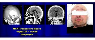
Immune cells heal brain after bleeding
0
In a new paper in Nature Communications, Houston neuroscientists show how immune cells called neutrophils can repair the brain after a hemorrhagic stroke. Neutrophils are known as the "infantry" in the body's war against infection. It turns out that they have another function: a new study shows that these immune cells may play a crucial role in protecting the brain from stroke, and can also be used in the treatment of intracerebral hemorrhages.
Functions of neurons
Considering that neurons ensure the functioning of all body systems, the functions of nerve cells must be very diverse. Moreover, all of them have not yet been fully clarified. Among the many different classifications of these functions, we will choose one that is the most understandable and closest to the problems of psychological science.
Information transfer function
This is the main function of neurons, with which others are associated, although no less significant. This same function is also the most studied. All external signals received by the organs enter the brain, where they are processed. And then, as a result of feedback in the form of impulses-commands, they are transferred along the efferent nerve fibers back to the sense organs, muscles, etc.
This constant circulation of information occurs not only at the level of the peripheral nervous system, but also in the brain. The connections between neurons exchanging information form incredibly complex neural networks. Just imagine: there are at least 30 billion neurons in the brain, and each of them can have up to 10 thousand connections. In the middle of the 20th century, cybernetics tried to create an electronic computer that worked on the principle of the human brain. But they failed - the processes occurring in the central nervous system turned out to be too complex.
Experience saving function
Neurons are responsible for what we call memory. More precisely, as neurophysiologists have found, the preservation of traces of signals passing through neural circuits is a kind of side effect of brain activity. The basis of memory is the same protein molecules - neurotransmitters, which arise as connecting bridges between nerve cells. Therefore, there is no special part of the brain responsible for storing information. And if, as a result of injury or illness, the destruction of nerve connections occurs, then the person may partially lose memory.
Integrative function
This ensures interaction between different parts of the brain. Instant “flares” of transmitted and received signals, foci of increased excitation in the cerebral cortex - this is the birth of images, feelings and thoughts. Complex neural connections that connect different parts of the cerebral cortex and penetrate into the subcortical zone are the product of our mental activity. And the more such connections arise, the better the memory and the more productive the thinking. That is, in essence, the more we think, the smarter we become.
Protein production function
The activity of nerve cells is not limited to information processes. Neurons are real protein factories. These are the same neurotransmitters that not only act as a “bridge” between neurons, but also play a huge role in regulating the functioning of our body as a whole. Currently, there are about 80 types of these protein compounds that perform various functions:
- Norepinephrine, sometimes called the rage or stress hormone. It tones the body, increases efficiency, makes the heart beat faster and prepares the body for immediate action to repel danger.
- Dopamine is the main tonic of our body. It is involved in the activation of all systems, including during awakening, during physical activity, and creates a positive emotional mood, even euphoria.
- Serotonin is also a “good mood” substance, although it does not affect physical activity.
- Glutamate is a transmitter necessary for memory function; without it, long-term storage of information is impossible.
- Acetylcholine controls the processes of sleep and awakening, and is also necessary for enhancing attention.
Neurotransmitters, or more precisely their quantity, affect the health of the body. And if there are any problems with the production of these protein molecules, then serious diseases can develop. For example, a lack of dopamine is one of the causes of Parkinson's disease, and if too much of this substance is produced, schizophrenia can develop. If not enough acetylcholine is produced, then very unpleasant Alzheimer's disease can occur, which is accompanied by dementia.
The formation of brain neurons begins even before a person is born, and throughout the entire period of growing up, the active formation and complication of neural connections occurs. For a long time it was believed that new nerve cells cannot appear in an adult, but the process of their death is inevitable. Therefore, mental development of the individual is possible only through the complication of neural connections. And even then, in old age, everyone is doomed to decline in mental abilities.
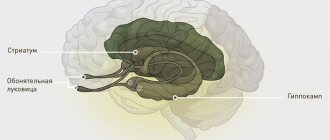
But recent studies have refuted this pessimistic forecast. Swiss scientists have proven that there is a part of the brain that is responsible for the birth of new neurons. This is the hippocampus; it produces up to 1,400 new nerve cells every day. And we can only more actively include them in the work of the brain, receive and comprehend new information, thereby creating new neural connections and complicating the neural network.
Cerebellar granule cells
0
Here is a snapshot of the cells of the granular layer of the cerebellum. Granule cells are small neurons, about 10 micrometers in diameter.
0
In the vast majority of cases, the cause of vision loss is the death of a large number of photoreceptor cells in the retina, which convert light into electrical nerve signals. Retinal cells that are insensitive to photons remain intact.
Classification of neurons
Nerve cells are diverse as such, so neurons can be classified based on their different parameters and attributes, namely:
- Body shape. In different parts of the brain there are neurocytes of different soma shapes: stellate;
- fusiform;
- pyramidal (Betz cells).
- unipolar: have one process;
- axo-somatic. In this case, the axon contacts the soma of the neighboring cell of the nervous tissue;
Types of neurons
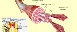
In order to carry out conscious movements, it is necessary that the impulse formed in the motor convolutions of the brain can reach the necessary muscles. Thus, the following types of neurons are distinguished: central motor neuron and peripheral motor neuron.
The first type of nerve cells originates from the anterior central gyrus, located in front of the largest groove of the brain - Roland's groove, namely from Betz's pyramidal cells. Next, the axons of the central neuron deepen into the hemispheres and pass through the internal capsule of the brain.
Peripheral motor neurocytes are formed by motor neurons of the anterior horns of the spinal cord. Their axons reach various formations, such as plexuses, spinal nerve clusters, and, most importantly, the performing muscles.