The main task of the human body is to maintain a constant internal environment (i.e. homeostasis), as well as adaptation (i.e. adaptation) to the environment. These tasks are performed by 3 body systems: 1. The immune system, which is responsible for protecting against foreign genetic information. 2. The humoral (endocrine) system is responsible for the slow regulation of the activity of individual organs and tissues of the body. 3. The nervous system, which appeared later than the previous two; is responsible for the rapid and precise regulation of the activity of individual organs and tissues.
Content
- Functions of the nervous system
- NS departments
- Reflex as the basic principle of the nervous system
Neurophysiology views the nervous system as part of a living system that specializes in the transmission, analysis and synthesis of information, and neuropsychology as the material substrate of complex forms of mental activity, formed on the basis of the integration of various parts of the brain into functional systems.
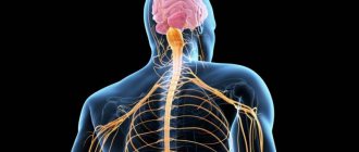
The nervous system (NS) is a set of anatomically and functionally interconnected nervous structures that provide regulation and coordination of the activities of the human body and its interaction with the environment.
The structural unit of the NS is a cell with a process ( neuron , or neurocyte). The nervous system is a collection of neurons that communicate with each other through synapses.
Changes in the excitability of the central nervous system
A further feature of the central nervous system is its extremely high sensitivity to changes occurring in the body. In response to certain changes, its excitability changes. The slightest changes in gas exchange and blood circulation affect the excitability of nerve cells.
The central nervous system consumes oxygen more than all other organs of our body: 100 g of a dog’s brain consumes 10 ml of oxygen per minute, while the same amount of liver consumes 10 times less, and the same amount of muscles consumes 22 times less oxygen. A decrease in oxygen supply can very quickly lead to loss of excitability and then to the death of nerve cells.
Brain activity also depends on normal blood circulation. It is enough to cause a disturbance in the blood circulation of the brain for a short period of time, as its excitability decreases or even completely disappears, and the person loses consciousness.
The excitability of the central nervous system is affected by some poisons that act primarily on the brain.
Strychnine is an exceptionally potent poison. Strychnine increases the excitability of the central nervous system. It is enough to inject the animal with a small dose of strychnine, and it begins to react violently even to mild irritations. If a small amount of a weak solution of strychnine is injected into the lymphatic sac of a frog, then knocking on the table on which it lies causes convulsions. The same picture can be observed in warm-blooded animals, in which, after the administration of a small dose of strychnine, convulsions appear in response to irritations, to which this animal did not react before the administration of strychnine.
In small doses, strychnine is sometimes used for medicinal purposes.
The cerebral hemispheres are affected by poisons called drugs. These include chloroform, ether, alcohol, etc. The first two are widely used in surgical practice as substances that cause anesthesia. These poisons initially cause an increase and then a sharp drop in the excitability of the nervous system and deep sleep. An important fact is that they act on the cerebral hemispheres and have almost no effect on the medulla oblongata, and this is very important for the body. In the medulla oblongata there are such important centers as the respiratory center, the center of cardiac activity, etc., suppression of the activity of which could lead to the death of the organism.
The excitability of the central nervous system changes especially sharply when it is damaged. If the spinal cord is cut, the activity of the nerve centers located below the damaged area is inhibited. This loss of excitability of the nerve centers located below the damaged area is called spinal shock. After some time, the shock passes, and the reflex activity of the spinal cord is restored. The duration of the shock varies from animal to animal: the higher the animal is on the zoological ladder, the stronger and longer the shock. In a frog the shock goes away within a few minutes, but in cats and dogs it takes days and weeks. Shock is the most severe and lasting in monkeys and humans.
Functions of the nervous system
The nervous system occupies a special position among other body systems. It ensures the relationship of the organism with the environment . Receptors respond to any signals from the external and internal environment, converting them into streams of nerve impulses that enter the central nervous system. Based on the analysis of the flow of nerve impulses encoding information, the brain forms an adequate response.
Together with the endocrine glands, the nervous system regulates the functioning of all organs . This regulation is carried out due to the fact that the spinal cord and brain are connected by nerves to all organs through bilateral connections. The central nervous system receives signals from the organs about their functional state, and the nervous system, in turn, sends signals to the organs, correcting their functions and ensuring all vital processes - movement, nutrition, excretion, etc. The nervous system ensures coordination of the activities of cells, tissues, organs , organ systems. In this case, the body functions as a single whole.
The nervous system is the material basis of mental processes : attention, memory, speech, thinking, etc., with the help of which a person not only learns about the environment, but can also actively change it.
Thus, several functions of the nervous system can be distinguished: 1. Connects the body with the environment (perception and transmission). 2. Provides interaction between tissues of organs and body systems and their regulation.
otdelymozga.jpg
Rice. ↑. Divisions of the brain. Source: https://mgtsrb.rf/pozvonochnik/stvolovoj-gemorragicheskij-insult.html
Detailed video lecture on the histology of the central nervous system: Go
Video: Central nervous system
The nervous system is functionally and structurally divided into the peripheral and central nervous systems (CNS).
The central nervous system consists of the brain and spinal cord.
Spinal cord
Brain
The brain is located inside the cranium, and the spinal cord is located in the spinal canal. The peripheral part of the nervous system consists of nerves, i.e. bundles of nerve fibers that extend beyond the brain and spinal cord and are directed to various organs of the body. It also includes nerve nodes, or ganglia - clusters of nerve cells outside the spinal cord and brain. The nervous system functions as a whole.
Functions of the nervous system: 1) formation of excitation; 2) transfer of excitation; 3) inhibition (cessation of excitation, reduction of its intensity, inhibition, limitation of the spread of excitation); 4) integration (combination of various excitation flows and changes in these flows); 5) perception of irritation from the external and internal environment of the body with the help of special nerve cells - receptors;
6) coding, i.e. transformation of chemical and physical irritation into nerve impulses; 7) trophic, or nutritional, function - the formation of biologically active substances (BAS).
A neuron is the basic structural and functional unit of the nervous system.
↑ Go to contents
Neuron
Definition of the concept
A neuron is the basic structural and functional unit of the nervous system.
A neuron is a specialized cell that is capable of receiving, conducting, and transmitting neural stimulation to process information in the nervous system. © 2016 Sazonov V.F. © 2021 kineziolog.su.
A neuron is a complex, excitable, secreting , highly differentiated nerve cell with processes that perceives nervous stimulation, processes it and transmits it to other cells. In addition to the excitatory effect, a neuron can also have an inhibitory or modulating effect on its target cells.
Neurons are part of bioregulation and chemoregulation .
Functionally, a neuron can be considered as one of the levels of organization of the nervous system, which connects several other levels with each other at once: on the one hand, molecular, synaptic and subcellular levels and, on the other hand, supracellular levels: local neural networks, nerve centers and large functional brain systems that organize behavior.
NS departments
Central nervous system and peripheral nervous system
According to topographical principles, the nervous system is divided into central and peripheral. The central nervous system (CNS) includes those parts that are contained in the cranial cavity and the spinal canal, i.e. brain and spinal cord. The spinal cord is a tube with a small canal in the middle, surrounded by neurons and their processes. The brain is an extension of the spinal cord. The topographic boundary with the spinal cord is a plane passing through the lower edge of the foramen magnum. The average brain weight is 1400 g with individual variations from 1100 to 2000 g.
The peripheral nervous system (PNS) includes all the nerve structures located outside of it. These are nodes and bundles of fibers that connect the central nervous system with sensory organs and various effectors (muscles, glands, etc.), i.e. ganglia and nerves. The peripheral nervous system connects the spinal cord and brain with receptors and effectors. It consists of 12 pairs of cranial and 31-33 pairs of spinal (spinal) nerves.
Somatic nervous system and autonomic nervous system
According to the functional classification, the nervous system is divided into somatic nervous system and autonomic nervous system. Both somatic and autonomic nervous systems include central and peripheral parts.
The somatic nervous system includes the parts of the nervous system that regulate the functioning of skeletal muscles. Responsible for connecting the body with the external environment, providing sensitivity and movement, causing contraction of skeletal muscles. It regulates primarily the functions of voluntary movement. Its neurons are located in the anterior horns of the spinal cord, and their axons through the anterior roots of the spinal cord are directed to the skeletal (striated) muscles.
The autonomic (autonomic) nervous system is a collection of nerves and ganglia through which the heart, blood vessels, internal organs, glands, etc. are regulated. Internal organs receive double innervation (supply of organs and tissues with nerves, which ensures their connection with the central nervous system) - from the sympathetic and parasympathetic divisions of the autonomic nervous system. These two departments have excitatory and inhibitory influences, determining the level of activity of the organs.
The vegetative nervous system provides metabolism, respiration, and excretion. Influencing the metabolic activity in various organs and tissues in accordance with the changing conditions of their functioning and the external environment, it carries out an adaptive-professional function.
The autonomic nervous system innervates smooth muscles; it is also called the visceral nervous system. The division of the peripheral nervous system into somatic and autonomic is quite arbitrary, since in the central nervous system there is a significant overlap of projections of both, and somatic and autonomic reactions are equal components of any behavioral reaction.
In the autonomic nervous system, there are 2 divisions that are functional antagonists : sympathetic and parasympathetic . They differ in the localization of centers in the brain and peripheral nodes, as well as the nature of the effect on internal organs.
Differences between sympathetic and parasympathetic nervous system
1. The fibers of the sympathetic nervous system emerge from the thoracic and lumbar spinal cord, where the first sympathetic neuron lies. They then converge on the sympathetic ganglia located along the spine, where the second sympathetic neuron is located. The fibers of the parasympathetic nervous system begin in the spinal cord above or below the place where the sympathetic nerves exit the cranial and sacral regions, and then converge in ganglia located not along the spinal column, but close to the innervated organ.
2. The peculiarities of the location of the ganglia of these two systems suggest a difference in the effect they have. The action of the sympathetic nervous system is more diffuse, while the parasympathetic nervous system is more specific, since it is associated only with changes in the organ next to which the ganglion is located.
3. These systems also differ in the mediators involved in synaptic transmission. The main transmitter for the sympathetic nervous system is adrenaline, and for the parasympathetic nervous system it is acetylcholine.
4. The results of the activity of these two systems are largely opposite. While the main function of the sympathetic nervous system is to mobilize the body for fight or flight, the parasympathetic nervous system primarily ensures the maintenance of homeostasis.
5. Activation of the sympathetic nervous system underlies the behavior of a person eager to fight. The stimulation of the parasympathetic nervous system ensures digestion in a person lying on the couch after a hearty lunch.
The sympathetic nervous system excites, and the parasympathetic nervous system inhibits the activity of the heart, the first weakens the motor activity of the intestines, the second strengthens it. At the same time, they can act at the same time: together they increase the motor activity of the salivary and gastric glands, although the composition of the secreted juice varies depending on the share of participation of each system.
Anatomy. Human nervous system
Nervous system
Irritability or sensitivity is a characteristic feature of all living organisms, meaning their ability to respond to signals or stimuli.
The signal is perceived by the receptor and transmitted via nerves and/or hormones to the effector , which carries out a specific reaction or response.
Animals have two interconnected systems for coordinating functions - nervous and humoral (see table).
| Nervous regulation | Humoral regulation |
| Electrical and chemical conduction (nerve impulses and neurotransmitters at synapses) | Chemical conduction (hormones) through the CS |
| Fast swipe and response | Slower conduction and delayed response (exception: adrenaline) |
| Mostly short term changes | Mostly long term changes |
| Specific signal propagation path | Non-specific signal pathway (with blood throughout the body) to a specific target |
| The response is often highly localized (eg, one muscle) | The response may be highly generalized (e.g. height) |
The nervous system consists of highly specialized cells with the following functions:
- perception of signals - receptors;
— conversion of signals into electrical impulses (transduction);
- conduction of impulses to other specialized cells - effectors, which, having received the signal, give a response;
Communication between receptors and effectors is carried out by neurons.
A neuron is a structural and functional unit of the nervous system.
A neuron is an electrically excitable cell that processes, stores, and transmits information using electrical and chemical signals. The neuron has a complex structure and narrow specialization. A nerve cell contains a nucleus, a cell body, and processes (axons and dendrites).
There are about 90-95 billion neurons in the human brain. Neurons can connect to each other to form biological neural networks.
Neurons are divided into receptor, effector and intercalary.
Neuron body: nucleus
(with a large number of nuclear pores) and
organelles
(ER, ribosomes, Golgi apparatus, microtubules), as well as from processes (dendrites and axons).
Neuroglia – a set of auxiliary cells of the NS; makes up 40% of the total volume of the central nervous system.
Neuron processes:
- Axon is a long extension of a neuron; conducts impulses from the cell body; covered by the myelin sheath (forms the white matter of the brain)
- Dendrites are short and highly branched processes of a neuron; conducts impulses to the cell body; do not have a shell
Important!
A neuron may have several dendrites and usually only one axon.
Important!
One neuron can have connections with many (up to 20 thousand) other neurons.
Types of neurons:
- sensitive - transmits excitation from the sensory organs to the spinal cord and brain
- motor – transmits excitation from the brain and spinal cord to muscles and internal organs
- intercalary – communicate between sensory and motor neurons in the spinal cord and brain
Nerve processes form nerve fibers.
Bundles of nerve fibers form nerves.
Nerves are sensory (formed by dendrites
), motor (formed
by axons
), mixed (most nerves).
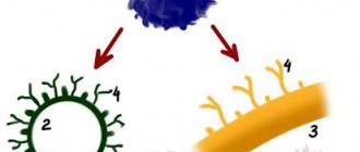
A synapse is a specialized functional contact between two excitable cells that serves to transmit excitation
In neurons, the synapse is between the axon of one cell and the dendrite of another; in this case, physical contact does not occur - they are separated by space - the synaptic cleft.
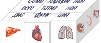
Nervous system:
- peripheral (nerves and nerve ganglia) – somatic and autonomic
- central (brain and spinal cord)
Depending on the nature of the innervation of the NS:
- Somatic – controls the activity of skeletal muscles, obeys the will of a person
- Vegetative (autonomous) – controls the activity of internal organs, glands, smooth muscles, does not obey the will of a person
The somatic nervous system is part of the human nervous system, which is a collection of sensory and motor nerve fibers that innervate muscles (in vertebrates - skeletal), skin, and joints.
It represents part of the peripheral nervous system, which is responsible for delivering motor (motor) and sensory (feeling) information to the central nervous system and back. This system consists of nerves attached to the skin, sensory organs and all the muscles of the skeleton.
Somatic NS:
- spinal nerves – 31 pairs; connected to the spinal cord; contain both motor and sensory neurons, therefore mixed;
- cranial nerves – 12 pairs; depart from the brain, innervate the receptors of the head (with the exception of the vagus nerve - innervates the heart, breathing, digestive tract); There are sensory, motor (motor) and mixed
A reflex is a quick automatic response to a stimulus, carried out without conscious control of the brain.
A reflex arc is the path traversed by nerve impulses from the receptor to the working organ.
From the receptor -
- in the central nervous system - along the sensitive pathway;
- from the central nervous system – to the working organ – along the motor pathway
Reflex arc:
- receptor (end of the dendrite of a sensory neuron) - perceives irritation
- sensitive (centripetal) nerve fiber - transmits excitation from the receptor to the central nervous system
- nerve center - a group of interneurons located at different levels of the central nervous system; transmits nerve impulses from sensory neurons to motor neurons
- motor (centrifugal) nerve fiber - transmits excitation from the central nervous system to the executive organ
Simple reflex arc:
two neurons - sensory and motor (example - knee reflex)
Complex reflex arc:
three neurons – sensitive, intercalary, motor (thanks to intercalary neurons, feedback occurs between the working organ and the central nervous system, which allows changes to be made in the work of the executive organs)
Autonomic (autonomous) nervous system - controls the activity of internal organs, glands, smooth muscles, and is not subject to human will.
Divided into sympathetic and parasympathetic .
Both consist of vegetative nuclei (clusters of neurons lying in the spinal cord and brain), vegetative ganglia (clusters of neurons outside the NS), nerve endings (in the walls of working organs)
The path from the center to the innervated organ consists of two neurons (in the somatic one - one).
| Sign | Sympathetic NS | Parasympathetic NS |
| Place of exit from the central nervous system | From the spinal cord - to the cervical, lumbar, thoracic regions | From the brain stem and the trunk of the sacral spinal cord |
| Location of the nerve ganglion | On both sides of the spinal cord, with the exception of the nerve plexuses (directly in these plexuses) | In or near innervated organs |
| Reflex arc mediators | In the prenodal fiber – acetylcholine; in the postnode - norepinephrine | Both fibers contain acetylcholine |
| Names of major nodes or nerves | Solar, pulmonary, cardiac plexuses, mesenteric node | Nervus vagus |
General effects of the sympathetic and parasympathetic nervous system on organs:
- Sympathetic NS - dilates the pupils, inhibits salivation, increases the frequency of contractions, dilates the blood vessels of the heart, dilates the bronchi, increases ventilation of the lungs, inhibits intestinal motility, inhibits the secretion of digestive juices, increases sweating, removes excess sugar in the urine; the overall effect is stimulating, increases the intensity of metabolism, reduces the sensitivity threshold; activates in times of danger, stress, controls reactions to stress
- Parasympathetic NS - constricts the pupils, stimulates lacrimation, reduces heart rate, maintains the tone of intestinal arterioles, skeletal muscles, lowers blood pressure, reduces ventilation, increases intestinal motility, dilates arterioles in the facial skin, increases the excretion of chlorides in the urine; the general effect is inhibitory, reduces or does not affect the intensity of metabolism, restores the sensitivity threshold; dominates at rest, controls functions in everyday conditions
Central nervous system (CNS) – ensures the interconnection of all parts of the nervous system and their coordinated work
In vertebrates, the central nervous system develops from the ectoderm (outer germ layer)
CNS – 3 shells:
- dura mater - outside;
- arachnoid membrane;
- pia mater ( pia mater ) - adjacent directly to the brain.
Brain
located in the brain part of the skull; contains
- white matter - pathways between the brain and the spinal cord, between parts of the brain
- gray matter - in the form of nuclei inside the white matter; cortex covering the cerebral hemispheres and cerebellum
The mass of the brain is 1400-1600 grams.
5 departments:
- the medulla oblongata is a continuation of the spinal cord; centers of digestion, respiration, cardiac activity, vomiting, coughing, sneezing, swallowing, salivation, conductive function
- hindbrain - consists of the pons and cerebellum; the pons
connects the cerebellum and medulla oblongata with the cerebral hemispheres;
the cerebellum
regulates motor acts (balance, coordination of movements, maintaining posture) - diencephalon – regulation of complex motor reflexes; coordination of the work of internal organs; implementation of humoral regulation;
- midbrain - maintaining muscle tone, indicative, sentry, defensive reflexes to visual and sound stimuli;
- forebrain (cerebral hemispheres) – the implementation of mental activity (memory, speech, thinking).
The diencephalon includes the thalamus, hypothalamus, epithalamus
The thalamus is the subcortical center of all types of sensitivity (except olfactory), regulates the external manifestation of emotions (facial expressions, gestures, changes in pulse, breathing)
Hypothalamus - centers of the autonomic nervous system, ensure the constancy of the internal environment, regulate metabolism, body temperature, feelings of thirst, hunger, satiety, sleep, wakefulness; The hypothalamus controls the pituitary gland
Epithalamus - participation in the work of the olfactory analyzer
The forebrain has two cerebral hemispheres: left and right
- Gray matter (cortex) is located on top of the hemispheres, white matter is inside
- White matter is the pathways of the hemispheres; among it are the nuclei of gray matter (subcortical structures)
The cerebral cortex is a layer of gray matter, 2-4 mm thick; has numerous folds and convolutions
Each hemisphere is divided by grooves into lobes:
- frontal - gustatory, olfactory, motor, musculocutaneous zones;
— parietal – motor, musculocutaneous zones;
- temporal - auditory zone;
- occipital - visual zone.
Important!
Each hemisphere is responsible for the opposite side of the body.
- The left hemisphere is analytical; responsible for abstract thinking, written and oral speech;
- The right hemisphere is synthetic; is responsible for imaginative thinking.
Spinal cord
located in the bony spinal canal; looks like a white cord, length 1m; there are deep longitudinal grooves on the front and back sides
At the very center of the spinal cord is the central canal, filled with cerebrospinal fluid.
The canal is surrounded by gray matter
(has the appearance of a butterfly), which is surrounded by
white matter.
- In the white matter - ascending (axons of spinal cord neurons) and descending pathways (axons of brain neurons)
- The gray matter resembles the outline of a butterfly and has three types of horns.
— front horns -
they contain motor neurons (motoneurons) - their axons innervate skeletal muscles
- hind horns -
contain interneurons – connect sensory and motor neurons
- lateral horns -
contain vegetative neurons - their axons go to the periphery to the vegetative nodes
Spinal cord – 31 segments; 1 pair of mixed spinal nerves, each having a pair of roots, departs from each segment:
— anterior (axons of motor neurons);
- posterior (axons of sensory neurons.
Functions of the spinal cord:
- reflex - implementation of simple reflexes (vasomotor, respiratory, defecation, urination, sexual);
- conductive - conducts nerve impulses from and to the brain.
Damage to the spinal cord leads to disruption of conduction functions, resulting in paralysis.
Reflex as the basic principle of the nervous system
I. M. Sechenov in 1863 in his work “Reflexes of the Brain” he developed the idea that the reflex is the basic principle of the functioning of not only the spinal cord, but also the brain.
A reflex is the body’s response to irritation with the participation of the central nervous system. Reflexes are divided into: 1) unconditioned reflexes: innate (hereditary) reactions of the body to stimuli carried out with the participation of the spinal cord or brain stem; 2) conditioned reflexes: temporary reactions of the body acquired on the basis of unconditioned reflexes, carried out with the obligatory participation of the cerebral cortex, which form the basis of higher nervous activity.
Each reflex has its own reflex arc - this is the path along which excitation passes from the receptor to the effector (executive organ).
The reflex arc is represented by a chain of neurons that provide the perception of irritation, the transformation of the energy of irritation into a nerve impulse, the conduction of a nerve impulse to the nerve centers, the processing of incoming information and the implementation of a response.
Any reflex arc consists of 5 components
1. A receptor is a specialized cell designed to perceive a stimulus (sound, light, chemical, etc.). 2. Afferent pathway, which is represented by afferent neurons. 3. Part of the central nervous system, represented by the spinal cord or brain; 4. The efferent pathway consists of the axons of efferent neurons extending beyond the CNS. 5. An effector is a working organ (muscle, gland, etc.).
The simplest reflex arc includes 2 neurons and is called monosynaptic (based on the number of synapses). A more complex one is represented by 3 neurons and is called three-neuron or disynaptic. However, most reflex arcs include a large number of interneurons and are called polysynaptic.
Reflex arcs can pass through the spinal cord only (for example, withdrawing a hand when touching a hot object) or through the brain only (for example, closing the eyelids when a stream of air is directed at the face), or through both the spinal cord and the brain.
Reflex arcs are closed into reflex rings using feedback connections. The concept of feedback and its functional role was indicated by Bell in 1826. He wrote that two-way connections are established between the muscle and the central nervous system. With the help of feedback, signals about the functional state of the effector are sent to the central nervous system.
The morphological basis of feedback is the receptors located in the effector and the afferent neurons associated with them. Thanks to feedback afferent connections, fine regulation of the effector’s work and an adequate response of the body to environmental changes are carried out.
Excitation speed
Any reflex takes place over a certain period of time: some happen faster, others slower. The time that elapses from the beginning of stimulation of the receptors to the beginning of the response is called reflex time. The reflex time consists of the time that is necessary to cause excitation in the receptors and conduct the resulting impulse into the central nervous system, then for the excitation to pass through the central nervous system and to spread along the centrifugal nerves, then for the transition to the working organ and, finally, for the hidden period of excitation of this organ. Thus, the reflex time, as we see, represents the sum of many terms.
Special studies and measurements have shown that the speed of excitation is not the same in different parts of the reflex arc. Excitation passes most slowly through the central nervous system, where excitation is transferred from one neuron to another. Therefore, it is customary to talk about central, or synaptic, delay. Slow conduction in the central nervous system is called delay because it seems as if the excitation, having reached the synapse, encounters some kind of obstacle and is therefore delayed.