Author Zybina A.M.
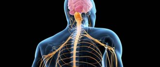
The nervous system integrates the entire organism into a single orchestra, carries out its interaction with the environment, voluntary movements (together with the muscular system), and all manifestations of mental activity. All functions of the nervous system are performed by a network of neurons connected to each other through synapses. Their viability is supported by glial cells.
The nervous system is divided according to its anatomical location into central (CNS) and peripheral (PNS). The central nervous system consists of the brain and spinal cord. PNS - made up of nerves (a bundle of nerve cell processes) and nerve ganglia, or ganglia (a collection of neuron bodies) located outside the nervous system.
Based on their functions, the nervous system is divided into somatic (animal, SomNS) and vegetative (autonomous, ANS) divisions. The SomNS controls voluntary contractions of skeletal muscles. The ANS controls the activity of internal organs. It is divided into two divisions: sympathetic (SNS) and parasympathetic (PNS). Both the SomNS and the ANS have both central and peripheral sections.
Structure of the central nervous system
The CNS consists of the brain and spinal cord, each of which has white and gray matter. White matter consists of pathways, myelinated and unmyelinated axons. Myelin is white, which gives the corresponding shade to the tissue. Gray matter consists of neuron cell bodies. It can be located in the nervous system in the form of a tube (spinal cord); nuclei, or ganglia (clusters of neuron bodies in the thickness of the white matter), as well as the cortex (gray matter on the surface of the white matter).
The spinal cord is located in the spinal canal and its mass is 40 g. On its lateral surface, the dorsal roots, carrying afferent (sensitive, to the brain) information, enter from the back, and the anterior roots, carrying efferent (motor, from the brain) information, exit from the front. The section of the spinal cord corresponding to each pair of roots is called a segment. The segments are named according to where the roots exit the spine. The spinal cord has 8 cervical, 12 thoracic, 5 lumbar, 5 sacral and 1 coccygeal segments. In general, the number of spinal cord segments corresponds to the number of vertebrae. Exceptions are the cervical region, where there are 8 segments per 7 vertebrae; and coccygeal, where there is 1 segment for 3-4 vertebrae (Fig. 1).
Rice. 1. Structure and location of spinal cord segments.
In a cross section of the spinal cord, there is gray matter in the center surrounded by white matter. The gray matter has the shape of a butterfly, in the center of which is the spinal foramen, filled with cerebrospinal fluid (CSF). The butterfly consists of approximately 13 million neurons and has anterior and posterior horns (Fig. 2b, 3). The middle horns are also well defined in the middle sections of the spinal cord. Sensitive (sensory) information to interneurons (interneurons) enters the dorsal horns along the dorsal root. The anterior horns contain motoneurons (motor neurons) that send motor information to the muscles; it is their axons that form the anterior root. The middle horns contain neurons of the central sections of the ANS.
The spinal cord works on a reflex principle. A reflex is a stereotypical response of the body to any (external or internal) influence. The simplest reflex is monosynaptic. To implement it, two neurons are enough. An example of such a reflex is the knee reflex. When the receptor is irritated, the impulse is transmitted along the dendrite to the body of the neuron located in the nerve ganglion near the spinal cord. The axon of this neuron enters the spinal cord through the dorsal roots and forms a synapse with the motor neuron in the anterior horn. The axon of the motor neuron exits through the anterior roots and goes to the effector organ, where it changes the activity of the organ itself (Fig. 50a). The polysynaptic reflex includes an additional link in the form of one or more interneurons between the ganglion and motor neurons. Interneurons can additionally process information, compare it with other stimuli and the internal state of the body, making a decision about how to respond to the stimulus.
Rice. 2. Reflex arc (a) and histological section (b) of the spinal cord.
Rice. 3. Diagram of the structure of a section of the spinal cord.
The white matter of the spinal cord includes pathways. It is divided by the butterfly into anterior, posterior and lateral funiculi (Fig. 3).
In the posterior cords there are ascending tracts through which information is transmitted from the PNS to the spinal cord and further to the brain. In the anterior horns of the spinal cord there are descending tracts through which information goes from the brain to the spinal cord, and from the latter to the PNS. In the lateral horns, the ascending tracts are located posteriorly, and the descending tracts are located anteriorly.
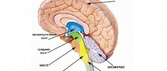
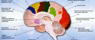
The brain is located in the skull and consists of 5 sections. Its average mass is 1.5 kg and it contains up to 100 billion neurons. There are 12 pairs of cranial nerves (cranial nerves) that arise from the brain.
The medulla oblongata is the junction of the spinal cord and the brain. Its length is approximately 25 mm. In the lower part of the medulla oblongata one can still distinguish a butterfly; in the upper parts the bodies of neurons are collected into nuclei. The IX-XII pairs of the cranial nerves (Fig. 5) depart from the medulla oblongata and the corresponding nuclei lie in it. These nerves control movement and sensation in the throat, tongue, and neck. The medulla oblongata contains the largest center of the parasympathetic nervous system, which, through the X nerve (vagus, vagus nerve), controls the activity of all internal organs. The medulla oblongata contains centers for regulating breathing and vital reflexes, such as sneezing and coughing. Here is the olive kernel, which is responsible for balance. All tracts from the spinal cord to the brain pass through the medulla oblongata.
The hindbrain consists of the pons and cerebellum. The pons serves as a continuation of the medulla oblongata. It contains a lot of white matter that connects the cerebellum to the rest of the brain. This white substance forms a ridge on the underside of the bridge, making it easy to distinguish. The pons, together with the medulla oblongata, form the bottom of the 4th ventricle of the brain (a continuation and expansion of the spinal canal). V-VIII ChMN depart from the bridge. Here lie the auditory and vestibular nuclei, nuclei that innervate sensitivity and facial muscles (including facial muscles). The pons contains the locus coeruleus, which is responsible for regulating sleep.
Rice. 4. Main parts of the brain.
Rice. 5. Cranial nerves. I-olfactory, II-visual, III-oculomotor, IV-trochlear, V-trigeminal, VI-abducens, VII-facial, VIII-vestibular-cochlear, IX-glossopharyngeal, X-vagus, XI-accessory, XII-hyoid.
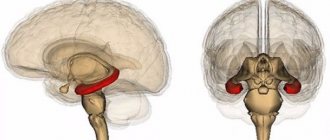
The cerebellum is well developed in humans due to upright posture and fine motor skills of the hands. This part of the brain is responsible for maintaining posture, balance, motor learning, and some motor reflexes. The cerebellum has a cortical structure. The cerebellar cortex consists of three layers and is divided into two hemispheres by the vermis. Under the cortex there is white matter, among which there are 3 pairs of cerebellar nuclei. To carry out its functions, it receives information from the vestibular apparatus, olive and other parts of the human motor system.
Rice. 6. External structure (a) and histological section (b) of the cerebellar cortex.
The midbrain consists of the cerebral peduncles and the roof (Fig. 7). The aqueduct of Sylvius passes through the center of the spinal cord and connects the third and fourth ventricles. The third and fourth pairs of cranial nerves depart from the midbrain. These nerves control the movements of the eyeballs. The third nerve contains parasympathetic fibers that control pupil width. The midbrain contains elements of the motor system: the red nucleus and the substantia nigra. On the roof of the brain is the quadrigeminal region. The colliculus receives visual information, and the inferior colliculus receives auditory information. This is necessary for the implementation of the orientation reflex.
Rice. 7. Appearance (a) and section (b) of the midbrain.
The medulla oblongata, pons, and midbrain together form the brainstem. The reticular formation runs through the entire trunk, regulating the overall level of brain activity.
The diencephalon consists of the thalamus, hypothalamus, pituitary gland and pineal gland. The third ventricle of the brain is located here. It serves as the origin of the second cranial nerve. The pituitary gland is the gland through which the nervous system controls the humoral one. The pineal gland is also a gland that regulates circadian rhythms. The thalamus filters information entering the cortex and removes unimportant repetitive sensory stimuli (heartbeat, gastrointestinal function, nose in the field of view, touch of clothing, etc.) In addition, the thalamus contains nuclei of the limbic system (forms mood), motor and associative kernels. The hypothalamus controls the activity of the pituitary gland and also regulates the internal state of the body. It contains the centers of hunger, thirst, sexual behavior, pleasure, displeasure, etc. Thus, the main function of the hypothalamus is to maintain homeostasis of the entire organism.
The telencephalon (forebrain) consists of the cerebral cortex and basal ganglia (nuclei). The first and second ventricles of the brain are located symmetrically under the cortex. Its area is about 220 cm2, it forms grooves and convolutions (Fig. 8). It consists of 6 layers. The hemispheres are connected to each other by the corpus callosum - a ridge of white matter. The cerebral cortex processes sensory information, the formation of voluntary movements, memory and higher nervous activity. The first cranial nerve approaches the olfactory bulbs. The basal ganglia are nuclei of gray matter located in the thickness of the white matter. They play an important role in voluntary movements, motor learning and the formation of emotions.
Rice. 8. Structure (a) and histological sections (b, c) of the cerebral cortex.
Propagation of nerve impulses
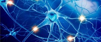
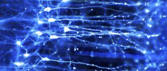
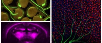
As a result of the evolution of the nervous system of humans and other animals, complex information networks arose, the processes of which are based on chemical reactions. The most important element of the nervous system are specialized cells called neurons.
.
Neurons consist of a compact cell body containing a nucleus and other organelles. Several branched processes extend from this body. Most of these projections, called dendrites
, serve as contact points for receiving signals from other neurons.
One process, usually the longest, is called an axon
and transmits signals to other neurons. The end of an axon can branch multiple times, and each of these smaller branches is able to connect to the next neuron.
The outer layer of the axon contains a complex structure formed by many molecules that act as channels through which ions can flow both into and out of the cell. One end of these molecules, deflecting, attaches to the target atom. Energy from other parts of the cell is then used to push that atom out of the cell, while the process in the opposite direction brings another molecule into the cell. The most important is the molecular pump, which removes sodium ions from the cell and introduces potassium ions into it (sodium-potassium pump).
When a cell is at rest and not conducting nerve impulses, the sodium-potassium pump moves potassium ions into the cell and removes sodium ions out (imagine a cell containing fresh water and surrounded by salt water). Because of this imbalance, the potential difference across the axon membrane reaches 70 millivolts (approximately 5% of the voltage of a regular AA battery).
However, when the state of the cell changes and the axon is stimulated by an electrical impulse, the equilibrium on the membrane is disturbed, and the sodium-potassium pump begins to work in the opposite direction for a short time. Positively charged sodium ions enter the axon, and potassium ions are pumped out. For a moment, the internal environment of the axon acquires a positive charge. In this case, the channels of the sodium-potassium pump are deformed, blocking further sodium influx, and potassium ions continue to flow out, and the original potential difference is restored. Meanwhile, sodium ions spread inside the axon, changing the membrane at the bottom of the axon. At the same time, the state of the pumps located below changes, promoting further propagation of the impulse. A sudden change in voltage caused by the rapid movement of sodium and potassium ions is called an action potential
. When an action potential passes through a certain point on the axon, the pumps turn on and restore the resting state.
The action potential travels quite slowly—no more than a fraction of an inch per second. In order to increase the speed of impulse transmission (since, after all, it is not good for a signal sent from the brain to take a minute to reach the hand), the axons are surrounded by a sheath of myelin, which prevents the influx and outflow of potassium and sodium. The myelin sheath is not continuous - at certain intervals there are breaks in it, and the nerve impulse jumps from one “window” to another, due to this the speed of impulse transmission increases.
When the impulse reaches the end of the main part of the axon body, it must be transmitted either to the next underlying neuron or, in the case of neurons in the brain, through numerous branches to many other neurons. For such transmission, a completely different process is used than for transmitting an impulse along the axon. Each neuron is separated from its neighbor by a small gap called a synapse
.
The action potential cannot jump across this gap, so some other way must be found to transmit the impulse to the next neuron. At the end of each process there are tiny sacs called ( presynaptic
)
vesicles
, each of which contains special compounds -
neurotransmitters
. When an action potential occurs, these vesicles release neurotransmitter molecules that cross the synapse and bind to specific molecular receptors on the membrane of underlying neurons. When a neurotransmitter attaches, the balance on the neuron membrane is disrupted. Now we will consider whether a new action potential arises with such an imbalance (neuroscientists continue to search for the answer to this important question to this day).
After neurotransmitters transmit a nerve impulse from one neuron to the next, they can simply diffuse out, undergo chemical breakdown, or return back to their vesicles (this process is awkwardly called reuptake
). At the end of the 20th century, an astonishing scientific discovery was made - it turns out that drugs that affect the release and reuptake of neurotransmitters can radically change a person’s mental state. Prozac* and similar antidepressants block the reuptake of the neurotransmitter serotonin. It appears that Parkinson's disease is associated with a deficiency of the neurotransmitter dopamine in the brain. Researchers studying borderline states in psychiatry are trying to understand how these compounds affect human reasoning.
There is still no answer to the fundamental question of what causes a neuron to initiate an action potential - in the professional language of neurophysiologists, the mechanism of “firing” of a neuron is unclear. Particularly interesting in this regard are neurons in the brain, which can receive neurotransmitters sent by a thousand neighbors. Almost nothing is known about the processing and integration of these impulses, although many research groups are working on this problem. We only know that the neuron carries out the process of integrating incoming impulses and makes a decision whether or not to initiate an action potential and transmit the impulse further. This fundamental process controls the functioning of the entire brain. It is not surprising that this greatest mystery of nature remains, at least today, a mystery for science!
Autonomic nervous system
The ANS includes two divisions, the sympathetic (SNS) and the parasympathetic (PNS). The SNS is activated during a stressful situation. It increases the heart rate, constricts blood vessels, pupils, increases blood flow to the muscles and outflow from the gastrointestinal tract. The center of the SNS is located in the thoracic and lumbar regions of the spinal cord (Fig. 9). The PNS has the opposite effect. It is activated in a calm environment and leads to a rush of blood to the gastrointestinal tract, outflow from the muscles, a decrease in heart rate, pupil dilation, etc. PNS centers are located in the medulla oblongata, some nuclei of the cranial nerve and the sacral spinal cord.
The main difference between the autonomic reflex arc and the somatic one is the presence of another synaptic switch in the ganglion after the spinal cord. Thus, the autonomic reflex begins from the receptor, then the sensitive neuron from the ganglion transmits information to the neuron of the middle horns of the spinal cord (or another center of the ANS). The axon of the autonomic neuron exits through the anterior roots and goes to the ganglion, where it forms a synapse with the ganglion neuron, the process of which goes directly to the effector organ. The nerve fiber running from the spinal cord to the ganglion is called preganglionic. The nerve fiber running from the ganglion to the organ is called postganglionic. The ganglia of the SNS are located next to the spinal cord, so the preganglionic fiber is short and the postganglionic fiber is long. The ganglia of the PNS are located near or in the wall of the organ, so their preganglionic fiber is long and the postganglionic fiber is short. The effector neurotransmitter of the sympathetic nervous system is norepinephrine, and the parasympathetic nervous system is acetylcholine.
Rice. 9. Effects of the SNS and PNS.
Rice.
10. Comparison of the reflex arc of the somatic and autonomic reflex. # Human anatomy