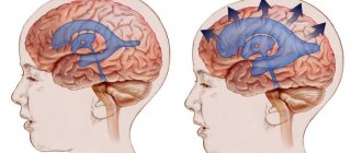
About the importance and circulation of cerebrospinal fluid
It is known that when a person is suspected of meningitis, doctors take cerebrospinal fluid, that is, cerebrospinal fluid, for analysis by performing a puncture. This fluid is contained in the central nervous system in small quantities - only half a glass or a small glass for an adult, but its functions are very important.
Thus, cerebrospinal fluid maintains a certain level of pressure, performs a nutritional and trophic function, and also mechanically protects the brain from shock, playing the role of a “shock-absorbing cushion.”
It circulates in the subarachnoid spaces and washes the spinal roots. The cerebrospinal fluid is located inside the blood-brain barrier, so many substances cannot penetrate there.
The cerebrospinal fluid begins from the basal cistern at the level of the base of the spinal cord . The spinal cord itself seems to “hang”, immersed in the cerebrospinal fluid, and inside it there is a central canal, which also contains cerebrospinal fluid.
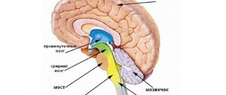
This central canal continues when the spinal cord enters the foramen magnum, but since the brain in its phylogenetic development slightly “rotated” and twisted, then inside the brain there are extended extensions of the central canal and its lateral branches, which are called the lateral ventricles .
In the image below, the lateral ventricles are shown in green, and the unpaired structures, which include the cerebral aqueduct, or aqueductus cerebri, are shown in purple. Let us remember that the word aqueductus is Latin.
All anatomical structures, according to the established rules of the BNA, or international anatomical classification, must be Latin. But they are often duplicated in Greek. So, “cerebrum” is the brain translated from Latin. Hence the terms “cerebral”, “cerebrovascular” and so on. And translated from Greek “brain” is “encephalon”, that is, “what is inside the head”. And terms such as “encephalitis” and “encephalopathy” are also applicable and exist on a par with the Latin names.
Unpaired liquor structures
The central structures are the unpaired ventricles of the brain, namely the IV and III ventricles, if you rise up from the spinal cord. The floor of the fourth ventricle is nothing more than the famous “diamond-shaped fossa”, in which the nuclei of the cranial nerves lie.
Gradually, the roof of the fourth ventricle narrows, turning into a narrow canal, which then opens into the cavity of the third ventricle, ventriculi tercii. This narrow cerebral canal, which connects the unpaired ventricles, is the aqueductus cerebri, or cerebral aqueduct. It lies directly under the quadrigeminalis, and is about 2-2.5 cm long in an adult. If you draw its cross section, the aqueductus cerebri will look like a triangle, an ellipse, or a rhombic cross-section. All of these are variants of the norm.
The diagram below shows the projection of the aqueduct, when viewed from above, and its relationship between the third and fourth ventricles.
Illustrations
- Cross section of the midbrain. Number 2 shows the cerebral aqueduct
- Cross section of the midbrain at the level of the inferior colliculus.
- Cross section of the midbrain at the level of the superior colliculus.
- Midsagittal section of the brain.
- Projection of the ventricles of the brain onto its surface
- Brain plumbing
- Brain plumbing
More about plumbing
Since this central narrow duct leaves the region of the trunk and pons, running in the structures of the midbrain, sometimes, taking into account its topography, it is called the aqueduct of the midbrain, aqueductus cerebri mesencephali.
Further, after flowing into the third ventricle, the unpaired or middle part of the liquor ends, giving way to the lateral ventricles of the brain. On a sagittal section, the topography of the aqueduct is clearly visible: ventrally it passes over the cerebellar peduncles, leaving above the lamina tecti, the plate of the roof of the midbrain.
What is the role of the plumbing? It performs a trophic function for the surrounding brain structures, and the following formations are located around the Sylvian aqueduct:
- the nuclei and network of the reticular formation, which we described in detail in the corresponding article;
- separately lying nuclei of the oculomotor and trochlear nerves, that is, the III and IV pairs of cranial nerves;
- A little more ventral in the thickness of the gray matter are the Yakubovich nuclei, which carry out the autonomic innervation of the eye: they compress and dilate the pupil, more precisely its sphincter, or the orbicularis muscle, depending on the amount of light entering the retina. The pupil itself is an opening and does not contain any structures.
Also, the role of the water supply is very important in maintaining normal intracranial pressure. When the aqueductus cerebri is compressed by a tumor, or a space-occupying formation of another nature, for example, a traumatic or parasitic cyst, intracranial hypertension syndrome may occur, which is manifested by bursting headaches, nausea, vomiting, and progressive decrease in vision.
Silviev water supply
As mentioned earlier, in the brain there is a canal that connects the third and fourth ventricles to each other. This is the Sylvian aqueduct, which is an integral part of the central canal. The cross-section of a water pipeline may look like a triangle, rhombus or ellipse. Its length does not exceed two centimeters.
Why was the Sylvian aqueduct invented and created by nature? Its function is trophic, that is, it is to deliver nutrients to brain cells. Without food they may die. In addition, the brain is located around it. The table of his departments shows this clearly. These are the nuclei of the reticular formation, the oculomotor nerve. Thanks to the Sylvian aqueduct, cerebrospinal fluid circulates in the brain, creating pressure. In total it contains just over one hundred milliliters.
Liquor is indispensable for creating the phenomenon of depreciation and balance. In addition, it serves to form a hydrostatic membrane and ensures a position of the nerve roots that reduces the tension of the blood vessels. Liquor is also indispensable for providing tissues with nutrition. With its help, nutritional cells are delivered to them. And after the process of processing them, the liquor removes waste substances. The cellular immune systems included in its composition protect against microbes.
The plumbing is extremely important for maintaining normal intracranial pressure. If the fluid is lost, the pressure will drop, which will immediately affect it in the form of unbearable headache, nausea, vomiting, and decreased vision. If there is a loss of cerebrospinal fluid, then an urgent magnetic resonance imaging (MRI) procedure is necessary.
Thus, the role of the aqueduct of Sylvius in the brain is irreplaceable. It is thanks to him that a person feels good, his intracranial pressure remains normal, and brain cells can feed normally and, therefore, function.
Classification of hydrocephalus
Etiological classification Depending on the causes of occurrence, hydrocephalus is divided into congenital (a consequence of developmental defects or intrauterine damage to the nervous system) and acquired (as a result of traumatic brain injury, inflammatory processes, tumors, cerebrovascular pathology, etc.). Particular attention is paid to the etiological factors of hydrocephalus in children, which differ from those in adults. These include:
- congenital pathology without meningomyelocele
- congenital pathology in combination with meningomyelocele
- perinatal intracranial hemorrhages
- brain tumors
- inflammatory processes
- traumatic brain injury
- cerebrovascular pathology.
Congenital pathology leading to hydrocephalus:
- Arnold-Chiari malformation 1 due to obstruction of the flow of cerebrospinal fluid from the fourth ventricle;
- Arnold-Chiari malformation 2 with or without meningomyelocele;
- primary stenosis of the cerebral aqueduct (occurs in utero, but is clinically realized more often in adults);
- secondary stenosis of the cerebral aqueduct due to gliosis as a result of intrauterine infection or hemorrhage of the germinal matrix;
- Dandy-Walker malformation (atresia of the foramen of Luschka or Mozhandie);
- rare genetic hereditary abnormalities associated with the X chromosome.
Acquired pathology causing hydrocephalus:
- infectious (the most common cause of communicating hydrocephalus);
- posthemorrhagic (the second most common cause of reported hydrocephalus):
- after subarachnoid hemorrhage
- after intraventricular hemorrhage (20-50% of such patients develop progressive hydrocephalus);
- secondary in case of volumetric intracranial processes:
- non-tumor (arteriovenous malformations, cysts)
- tumor (as a result of obstruction of the cerebrospinal fluid ducts - tumors of the cerebellum, quadrigeminal cistern. III or lateral ventricle;
- as a result of overproduction of cerebrospinal fluid - plexus papilloma, plexus carcinoma, plexus meningioma
- in combination with spinal cord tumors
- postoperative (about 20% of children require shunt surgery after removal of tumors of the posterior cranial fossa).
Morphological classification
There are open (communicating), closed (occlusive) and ex vacuo hydrocephalus. Open hydrocephalus involves free communication of the cerebrospinal fluid spaces: its development is associated with a violation of the relationship between the processes of production and resorption of cerebrospinal fluid. There are hyperproductive, aresorptive and mixed forms, in which production processes prevail over resorption processes. Closed hydrocephalus occurs as a result of separation of the cerebrospinal fluid ducts at various levels. With occlusion, an expansion of one lateral ventricle of the brain is formed at the level of the interventricular foramen, at the level of the third ventricle - both lateral ventricles, at the level of the cerebral aqueduct and the fourth ventricle - the lateral and third ventricle (triventricular form), at the level of the occipital cistern - expansion of the entire ventricular system of the brain . Hydrocephalus ex vacua is a consequence of a decrease in the volume of brain parenchyma as a result of atrophy, with aging of the body (as a physiological norm) or with pathological conditions of the central nervous system, accompanied by atrophic changes (for example, Creutzfeldt-Jakob disease, Alzheimer's disease). Hydrocephalus ex vacua is not true hydrocephalus, caused by impaired cerebrospinal fluid dynamics, but a consequence of the filling of “free” intracranial spaces with cerebrospinal fluid. Morphologically, hydrocephalus is also divided according to the location of the expanded cerebrospinal fluid spaces in relation to the brain tissue: internal (intraventricular), external (subarachnoid) and mixed.
Functional classification
Depending on the level of cerebrospinal fluid pressure, hypertensive and normotensive hydrocephalus are distinguished. In functional and clinical aspects, hydrocephalus is divided into progressive (increasing), stabilized (not undergoing changes over time) and regressive (decreasing). Progressive hydrocephalus in clinical terms is decompensated or subcompensated: it is this that is the object of conservative or surgical treatment. Stabilized and regressive hydrocephalus is always compensated and usually does not require treatment.
Excerpt describing the Brain Plumbing
“Le duc d'Oldenbourg supporte son malheur avec une force de caractere et une resignation admirable, [The Duke of Oldenburg bears his misfortune with remarkable willpower and submission to fate," said Boris, respectfully entering into the conversation. He said this because, while passing from St. Petersburg, he had the honor of introducing himself to the Duke. Prince Nikolai Andreich looked at the young man as if he wanted to say something to him about this, but decided against it, considering him too young for that. “I read our protest about the Oldenburg case and was surprised at the poor wording of this note,” said Count Rostopchin, in the careless tone of a man judging a case well known to him. Pierre looked at Rostopchin with naive surprise, not understanding why he was bothered by the poor edition of the note. – Doesn’t it matter how the note is written, Count? - he said, - if its content is strong. “Mon cher, avec nos 500 mille hommes de troupes, il serait facile d'avoir un beau style, [My dear, with our 500 thousand troops it seems easy to express ourselves in a good style,” said Count Rostopchin. Pierre understood why Count Rostopchin was worried about the edition of the note. “It seems that the scribblers are pretty busy,” said the old prince: “they write everything there in St. Petersburg, not just notes, but they write new laws all the time.” My Andryusha wrote a whole lot of laws for Russia there. Nowadays they write everything! - And he laughed unnaturally. The conversation fell silent for a minute; The old general drew attention to himself by clearing his throat.
Pathogenesis
Normally, cerebrospinal fluid (CSF) is produced by the choroid plexus of interconnected ventricles of the brain. The largest amount of it is formed in the lateral ventricles, from where the cerebrospinal fluid enters the third ventricle, and from it through the Sylvian aqueduct into the fourth ventricle. Then the cerebrospinal fluid enters the subarachnoid (subarachnoid) space, which extends over the entire surface of the brain, and in the caudal direction passes the region of the craniovertebral junction and then surrounds the spinal cord along its entire length. The cerebrospinal fluid located in the subarachnoid space is constantly absorbed by the arachnoid (arachnoid) membrane of the spinal cord and brain and enters the blood. The above etiological factors that disrupt the production, movement and absorption of cerebrospinal fluid lead to its excessive accumulation and the occurrence of hydrocephalus.
Human cerebral plumbing[ | ]
In humans, it is a canal about 15 mm long that connects the cavity of the third ventricle of the brain with the fourth in the brain [1]. The dorsal wall is formed by the plate of the quadrigeminal midbrain, the ventral wall is formed by the tegmentum of the cerebral peduncles. It is formed in ontogenesis from the cavity of the third medullary vesicle[2].
Around the Sylvian aqueduct is the central gray matter (substantia grisea centralis) [2], anatomically related to the tegmentum of the midbrain. This gray matter sends its ascending projections to the raphe nuclei and locus coeruleus, as well as to the somatosensory and viscerosensory nuclei of the thalamus. It also has descending projections into the spinal cord. The ascending nerve fibers of the spinothalamic tract, which conduct sensations of pain and temperature, make an intermediate “stop” in the periaqueductal gray matter on their way to the thalamus. This part of the spinothalamic tract is called the spinomesencephalic tract. In turn, the thalamic nuclei that perceive pain and temperature sensations send their descending feedback fibers to the spinal cord also through the central gray matter. In the central gray matter, in the area of the bottom of the aqueduct, the nuclei of two pairs of cranial nerves are located. at the level of the superior colliculi of the midbrain quadrigeminal, closer to the midline lies the paired nucleus of the oculomotor nerve (III pair of cranial nerves). More ventral to it lies the accessory nucleus of the oculomotor nerve (n. oculomotorius accessorius) - the Yakubovich nucleus, the Westval-Edinger nucleus. Somewhat higher and anterior to the nucleus of the oculomotor nerve lies one of the nuclei of the reticular formation - the intermediate nucleus (nucleus interstitialis, nucleus of Cajal) [2].
Diagnosis of the disease
When the first symptoms appear, you should immediately consult a neurologist. After collecting complaints and asking about the possible causes of the pathology, instrumental research methods will be prescribed.
Computed tomography is used to determine normal/pathological processes in brain structures. With its help, benign and malignant neoplasms, changes in the contours of the ventricles and subarachnoid space are identified.
MRI is used to determine the type of hydrocephalus.
Cisternography is performed to determine the direction of the flow of cerebrospinal fluid and the location of its accumulation.
Angiography of cerebral vessels - the presence of occlusion in the arteries.
General information
Hydrocephalus literally means “dropsy of the head.” In modern neurology, this is a common clinical syndrome that can be observed in many diseases, congenital anomalies or post-traumatic conditions of the brain. The occurrence of hydrocephalus is associated with certain disorders in the cerebrospinal fluid system of the brain. People of any age are susceptible to hydrocephalus. Hydrocephalus can occur in newborns, be congenital, develop in children and adults, and accompany atrophic processes occurring in the brain in old people. However, it is most often encountered in pediatric practice.
Hydrocephalus
Causes
Three pathological mechanisms lead to the accumulation of an excess amount of cerebrospinal fluid in the cerebrospinal fluid system of the brain: the production of an excess amount of cerebrospinal fluid, a violation of its absorption, or a disorder of the cerebrospinal fluid circulation. Hydrocephalus may be based on one of these mechanisms or a combination of them. The causes that cause disturbances in the functioning of the cerebrospinal fluid system can act during fetal development and cause congenital hydrocephalus or affect the brain after birth and cause the appearance of so-called acquired hydrocephalus. Causes of hydrocephalus include:
1. Congenital hydrocephalus
:
- malformations of the cerebrospinal fluid system (atresia of the foramina of Magendie and Luschka
- defects in the structure of the subarachnoid space
- stenosis of the Sylvian aqueduct, Dandy-Walker syndrome, etc.)
- craniovertebral anomalies (Chiari malformation, congenital basilar impression)
- intrauterine infections (toxoplasmosis, congenital syphilis, cytomegaly, rubella), birth trauma.
Symptoms of hydrocephalus
Hydrocephalus in adults
The accumulation of excess cerebrospinal fluid in a limited space of the cranium leads to increased intracranial pressure, which causes the most typical symptoms of hydrocephalus. In adults and older children, these include: intense headache that cannot be relieved by analgesics, nausea, vomiting, and a feeling of pressure on the eyeballs. These symptoms may occur acutely or increase gradually, being transient at the onset of the disease. Atrophic hydrocephalus often occurs without signs of increased intracranial pressure and is detected only with additional examination of the patient.
In most cases, hydrocephalus is accompanied by neurological symptoms, which are caused both by compression of brain structures by expanded cerebrospinal fluid spaces and by the underlying disease that causes the development of hydrocephalus. The most common symptoms of hydrocephalus are vestibular and visual disturbances. The first includes vestibular ataxia, manifested by dizziness, unsteady gait, noise in the ears and head, and nystagmus. On the visual side, there may be a significant decrease in visual acuity, loss of certain areas of the visual fields, congestive optic discs; with prolonged hydrocephalus, atrophy of the optic nerves may develop.
Hydrocephalus can occur with disturbances in the motor and sensory sphere: paresis and paralysis, increased tendon reflexes and muscle tone, decreased or complete loss of all types of sensitivity, and the formation of spastic contractures of the limbs. Occlusive hydrocephalus, caused by impaired circulation of cerebrospinal fluid in the posterior cranial fossa, is characterized by symptoms of cerebellar ataxia: impaired coordination and gait, large-scale disproportionate movements, changes in handwriting, etc.
In some cases, hydrocephalus is accompanied by mental disorders, which in adults are more often manifested by disorders of the emotional-volitional sphere: emotional instability, neurasthenia, causeless euphoria with a rapid transition to a state of indifference and apathy. With a sharp increase in intracranial pressure, aggressive behavior is possible.
Idiopathic stenosis of the cerebral aqueduct
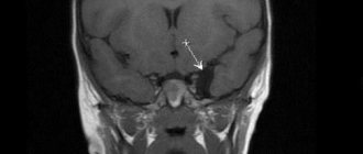
A 21-year-old woman was admitted with complaints of drowsiness and confusion. Over the past 5 months, she noted a gradual deterioration in her condition: increased headaches, nausea and vomiting.
Fundoscopy showed bilateral papilledema as a clinical symptom of intracranial hypertension. Computed tomography and magnetic resonance imaging (MRI) revealed supratentorial hydrocephalus caused by cerebral aqueductal stenosis. Sagittal MRI images performed with contrast agent showed acute insufficiency of fluid movement at the level of the cerebral aqueduct.
Important Borderline Personality Disorder
The patient was diagnosed with idiopathic aqueductal stenosis, most common with a combination of obstructive hydrocephalus in adults. There are unproven causes of aqueductal stenosis, such as tumor, infectious ventriculitis, or intraventricular hemorrhage.
Endoscopic ventriculotomy (Figure D) exposed the floor of the 3rd ventricle in the subarachnoid space of the brainstem. This procedure can treat idiopathic aqueductal stenosis and restore cerebrospinal fluid circulation without the need for external ventriculotomy or ventriculoperitoneal shunt.
All treatment measures were completed without complications, such as meningitis, severe hemorrhage, injury to neurological structures, impaired circulation of cerebrospinal fluid, and pneumocephalus. The patient had a good response to treatment with elimination of the neurological defect.
After 5 years, there were no symptoms of the disease and MRI showed no recurrence of hydrocephalus.
https://youtube.com/watch?v=deQhPDtXlIQVideo can't be loaded because JavaScript is disabled: Endoscopic Ventriculostomy of the Third Ventricle (https://youtube.com/watch?v=deQhPDtXlIQ)
In this material we wrote on the topic: “brain plumbing.”
← Do you want to know how to cover a roof with ondulin over an old roof? Watch the video! - ondulin how to cover a roofHow to build a frame house with your own hands - instructions in 4 steps - how to build a frame house with your own hands →