In Ozhegov's dictionary
SYMPATHETIC, -aya, -oe (special). 1. Reflectively arising in another, symmetrically or nearby place. Sympathetic pain. 2. Causing moral relief, satisfaction in the patient and usually neutral in its medical properties. Sympathetic means. * Sympathetic ink is colorless ink that becomes visible after special processing of the written material. The sympathetic nervous system is a division of the nervous system involved in regulating blood circulation, metabolism and other body functions.
Ciliary node (ganglion ciliare)
Flat, about 2 mm long and thick, located near the superior orbital fissure in the thickness of the fatty tissue at the lateral semicircle of the optic nerve. This node is formed by a cluster of cell bodies of second neurons of the parasympathetic part of the autonomic nervous system. Preganglionic parasympathetic fibers that come to this node as part of the oculomotor nerve end in synapses on the cells of the ciliary ganglion. Postganglionic nerve fibers, consisting of three to five short ciliary nerves, emerge from the anterior part of the ciliary ganglion, go to the back of the eyeball and penetrate it. These fibers innervate the ciliary muscle and the pupillary sphincter. Fibers that conduct general sensitivity (branches of the nasociliary nerve) transit through the ciliary ganglion, forming a long (sensitive) root of the ciliary ganglion. Sympathetic postganglionic fibers (from the internal carotid plexus) also transit through the node.
- The parasympathetic part of the facial nerve consists of the superior salivary nucleus, pterygopalatine, submandibular, hypoglossal nodes and parasympathetic nerve fibers. The axons of the cells of the superior salivary nucleus, which lies in the tegmentum of the pons, in the form of preganglionic parasympathetic fibers pass as part of the facial (intermediate) nerve. In the area of the genu of the facial nerve, part of the parasympathetic fibers is separated in the form of the large stony nerve (n. petrosus major) and exits the facial canal. The greater petrosal nerve lies in the groove of the same name in the pyramid of the temporal bone, then pierces the fibrous cartilage that fills the torn hole at the base of the skull, and enters the pterygoid canal. In this canal, the greater petrosal nerve, together with the sympathetic deep petrosal nerve, forms the nerve of the pterygoid canal, which exits into the pterygopalatine fossa and goes to the pterygopalatine ganglion.
Important How does a stroke affect life expectancy?
In the dictionary D.N. Ushakova
SYMPATHETIC, sympathetic, sympathetic. 1. adj. to sympathy" title='what is sympathy, the meaning of the word sympathy in Ushakov's dictionary'>sympathy in 1 · meaning, reflectedly arising in another, symmetrically and nearby place (med.). Sympathetic pain in the teeth (near or opposite those located from the diseased tooth). Sympathetic ophthalmia (painful sensation in the eye under the influence of pain in the other). 2. One that causes moral satisfaction in the patient, regardless of medical properties (about medicines, medicinal products; medical). A sympathetic remedy. Sympathetic drugs. 3. Based on sympathy (see sympathy" title='what is sympathy, the meaning of the word sympathy in Ushakov's dictionary'>sympathy in 2 · meaning; · book · outdated ). “There is no deep, sympathetic attachment, but only habit.” Goncharov. 4. The same as cute” title='what is cute, the meaning of the word cute in Ushakov's dictionary'>pretty (obsolete). “I see something sympathetic in your gaze.” Dostoevsky. “You will meet sympathetic people there, without any pretensions.” A. Turgenev. • Sympathetic nervous system (anat., physiol.) - a section of the autonomic nervous system that regulates the processes of plant life (digestion, respiration, blood circulation, etc.). Sympathetic ink is a colorless ink that becomes visible only when it is heated or wetted with a certain chemical composition.
Parasympathetic and peripheral
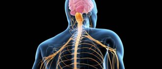
The parasympathetic nervous system should not be confused with the peripheral nervous system (PNS). As mentioned earlier, the brain and spinal cord make up the CNS. The PNS consists of all parts of the nervous system that are not the brain and spinal cord; in other words, all nerves and ganglia that are not in the brain and spinal cord are part of the PNS.
PSNS nerves originate in the brain or spinal cord, but most of the PSNS are not found in these areas and affect other areas of the body, so the PSNS is considered part of the PNS. But not all PNS are PSNS. The PNS also includes the sympathetic nervous system and the somatic nervous system (SoNS), which controls voluntary movements of the body.
To summarize the different sections of the nervous system: The parasympathetic nervous system controls the "rest and digest" actions, and the sympathetic nervous system controls the "fight or flight" actions. These systems make up the autonomic nervous system, which controls unconscious bodily actions. The somatic nervous system controls all voluntary movements of the body, such as walking or catching a ball. Parts of all these systems make up the peripheral nervous system, which is all parts of the nervous system, not including the brain and spinal cord. The brain and spinal cord make up the central nervous system.
- Sympathetic Nervous System (SNS) – Controls fight-or-flight actions such as increased heart rate and increased blood pressure.
- Autonomic Nervous System (ANS) – Controls the largely unconscious actions of the internal organs and consists of the parasympathetic and sympathetic nervous systems.
- Somatic Nervous System (SoNS) – Controls the voluntary movements of the skeletal muscle body.
- Peripheral Nervous System (PNS) – Parts of the nervous system that are not the brain and spinal cord, such as nerves and ganglia found throughout the body.
In the dictionary Complete accentuated paradigm according to A. A. Zaliznya
sympathetic, sympathetic, sympathetic, sympathetic, sympathetic, sympathetic, sympathetic, sympathetic, sympathetic, sympathetic, sympathetic, sympathetic, sympathetic, sympathetic, sympathetic, sympathetic, sympathetic, sympathetic, sympathetic, sympathetic, sympathetic ́sympathetic, sympathetic, sympathetic, sympathetic, sympathetic, sympathetic, sympathetic, sympathetic, sympathetic, sympathetic, sympathetic, sympathetic, sympathetic
Central department
This part of the autonomic nervous system represents various structures of the brain. It turns out that it is scattered throughout the entire brain. In the central section, segmental and suprasegmental structures are distinguished. All formations belonging to the suprasegmental department are united under the name hypothalamic-limbic-reticular complex.
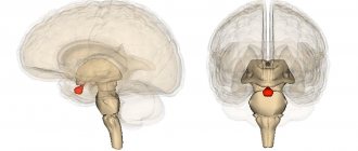
Hypothalamus
The hypothalamus is a structure of the brain located in the lower part, at the base. This cannot be said to be an area with clear anatomical boundaries. The hypothalamus smoothly passes into the brain tissue of other parts of the brain.
In general, the hypothalamus consists of a cluster of groups of nerve cells, nuclei. A total of 32 pairs of nuclei were studied. Nerve impulses are formed in the hypothalamus, which reach other brain structures through various pathways. These impulses control blood circulation, breathing, and digestion. The hypothalamus contains centers for regulating water-salt metabolism, body temperature, sweating, hunger and satiety, emotions, and sexual desire.
In addition to nerve impulses, substances with a hormone-like structure are formed in the hypothalamus: releasing factors. With the help of these substances, the activity of the mammary glands (lactation), adrenal glands, gonads, uterus, thyroid gland, growth, fat breakdown, and the degree of skin color (pigmentation) is regulated. All this is possible thanks to the close connection of the hypothalamus with the pituitary gland, the main endocrine organ of the human body.
Thus, the hypothalamus is functionally connected with all parts of the nervous and endocrine systems.
Conventionally, two zones are distinguished in the hypothalamus: trophotropic and ergotropic. The activity of the trophotropic zone is aimed at maintaining the constancy of the internal environment. It is associated with a period of rest, supports the processes of synthesis and utilization of metabolic products. It exerts its main influences through the parasympathetic division of the autonomic nervous system. Stimulation of this area of the hypothalamus is accompanied by increased sweating, salivation, slowing of heart rate, decreased blood pressure, vasodilation, and increased intestinal motility. The trophotropic zone is located in the anterior parts of the hypothalamus. The ergotropic zone is responsible for the body’s adaptability to changing conditions, ensures adaptation and is realized through the sympathetic division of the autonomic nervous system. At the same time, blood pressure increases, heartbeat and breathing accelerate, pupils dilate, blood sugar increases, intestinal motility decreases, and urination and bowel movements are inhibited. The ergotropic zone occupies the posterior parts of the hypothalamus.
Limbic system
This structure includes part of the temporal lobe cortex, hippocampus, amygdala, olfactory bulb, olfactory tract, olfactory tubercle, reticular formation, cingulate gyrus, fornix, and papillary bodies. The limbic system is involved in the formation of emotions, memory, thinking, ensures eating and sexual behavior, and regulates the sleep-wake cycle.
To realize all these influences, the participation of many nerve cells is necessary. The functioning system is very complex. In order for a certain model of human behavior to be formed, it is necessary to integrate many sensations from the periphery, transmitting excitation simultaneously to various structures of the brain, as if circulating nerve impulses. For example, in order for a child to remember the names of the seasons, repeated activation of structures such as the hippocampus, fornix, and papillary bodies is necessary.
Reticular formation
This part of the autonomic nervous system is called the reticular system because, like a network, it interweaves all the structures of the brain. This diffuse location allows it to participate in the regulation of all processes in the body. The reticular formation keeps the cerebral cortex in good shape, in constant readiness. This ensures instant activation of the desired areas of the cerebral cortex. This is especially important for the processes of perception, memory, attention and learning.
Individual structures of the reticular formation are responsible for specific functions in the body. For example, there is a respiratory center, which is located in the medulla oblongata. If it is affected for any reason, then independent breathing becomes impossible. By analogy, there are centers of cardiac activity, swallowing, vomiting, coughing, and so on. The functioning of the reticular formation is also based on the presence of numerous connections between nerve cells.
In general, all structures of the central part of the autonomic nervous system are interconnected through multineuron connections. Only their coordinated activity allows the vital functions of the autonomic nervous system to be realized.
Segmental structures
This part of the central part of the visceral nervous system has a clear division into sympathetic and parasympathetic structures. Sympathetic structures are located in the thoracolumbar spinal cord, and parasympathetic structures are located in the brain and sacral spinal cord.
Sympathetic department
Sympathetic centers are localized in the lateral horns in the following segments of the spinal cord: C8, all thoracic (12), L1, L2. Neurons in this area are involved in the innervation of smooth muscles of internal organs, internal muscles of the eye (regulation of pupil size), glands (lacrimal, salivary, sweat, bronchial, digestive), blood and lymphatic vessels.
Parasympathetic Division
Contains the following structures in the brain:
- accessory nucleus of the oculomotor nerve (nucleus of Yakubovich and Perlia): control of pupil size;
- lacrimal nucleus: accordingly, regulates tear secretion;
- superior and inferior salivary nuclei: provide saliva production;
- dorsal nucleus of the vagus nerve: provides parasympathetic influences on internal organs (bronchi, heart, stomach, intestines, liver, pancreas).
The sacral section is represented by neurons of the lateral horns of segments S2-S4: they regulate urination and defecation, blood flow to the vessels of the genital organs.
In the dictionary Dictionary of foreign words
and I
1. In combination: sympathetic nervous system (physiol.) - part of the autonomic nervous system, involved in the regulation of the activity of internal organs, activating processes associated with the breakdown of energy necessary for the interaction of the organism with the external environment.||Cf. PARASYMPATHETIC" title='PARASYMPATHETIC, PARASYMPATHIC is what is PARASYMPATHETIC, PARASYMPATHETIC interpretation'>PARASYMPATHETIC.
2. In combination: sympathetic ink is a colorless liquid used in secret correspondence. | What is written in sympathetic ink becomes visible only after being heated or moistened with a certain chemical composition.
Share the meaning of the word:
Anatomy of the Human Parasympathetic Nervous System - Information:
The parasympathetic part of the autonomic nervous system historically develops as a suprasegmental department, and therefore its centers are located not only in the spinal cord, but also in the brain.
Parasympathetic centers
The central part of the parasympathetic division consists of the head, or cranial, division and the spinal, or sacral, division. Some authors believe that the parasympathetic centers are located in the spinal cord not only in the region of the sacral segments, but also in other parts of it, in particular in the lumbar-thoracic region between the anterior and posterior horn, in the so-called intermediary zone. The centers give rise to efferent fibers of the anterior roots, causing vasodilation, delayed sweating and inhibition of contraction of involuntary hair muscles in the trunk and limbs.
The cranial section, in turn, consists of centers located in the midbrain (mesencephalic part), and in the rhombencephalon - in the pons and medulla oblongata (bulbar part).
- The mesencephalic part is represented by the nucleus accessorius n. oculomotorii and the median unpaired nucleus, due to which the muscles of the eye are innervated - m. sphincter pupillae and m. ciliaris.
- The boulevard part is represented by the nucleus saliva tonus superior n. facialis (more precisely, n. intermedius), nucleus salivatorius inferior n. glossopharyngei and nucleus dorsalis n. Vagi.
Sacral department. The parasympathetic centers lie in the spinal cord, in the substantia intermedialateralis of the lateral horn at the level of the II-IV sacral segments.
Peripheral division of the parasympathetic part
The peripheral part of the cranial section of the parasympathetic system is represented by:
- preganglionic fibers running as part of the III, VII, IX and X pairs of cranial nerves (possibly also as part of I and XI);
- terminal nodes located near organs, namely: ganglia ciliare, pterygopalatinum, submandibulare, oticum, and
- postganglionic fibers; postganglionic fibers either have an independent course, such as nn. ciliares breves, extending from the ganglion ciliare, or are part of any nerves, such as, for example, postganglionic fibers, extending from the ganglion oticum and coming as part of n. auriculotemporalis.
Some authors indicate that parasympathetic fibers also emerge from other segments of the spinal cord and go through the anterior roots, heading to the walls of the trunk and limbs. The peripheral part of the sacral section of the parasympathetic system is represented by fibers that, as part of the anterior roots of the II-IV sacral nerves and then as part of their anterior branches, forming the plexus sacralis (animal plexus), enter the small pelvis. Here they are separated from the plexus and in the form of nn. splanchnici pelvini are directed to the plexus hypogastricus inferior, innervating together with the latter the pelvic viscera: the rectum with the colon sigmoideum, the bladder, the external and internal genital organs. Irritation nn. splanchnici pelvini causes contraction of the rectum and bladder (m. detrusor vesicae) with weakening of their sphincters.
The fibers of the sympathetic hypogastric plexus delay the emptying of these organs; they also stimulate uterine contraction, while nn. splanchnici pelvini slow it down. Nn. splanchnici pelvini also contain vasodilator fibers (nn. erigentes) for the corpora cavernosa penis et clitoridis, which cause erection. Parasympathetic fibers extending from the sacral spinal cord go to the pelvic plexuses not only as part of the nn. erigentes and nn. splanchnici pelvini, but also as part of the nervus pudendus (preganglionic fibers). The pudendal nerve is a complex nerve that, in addition to animal fibers, also contains autonomic (sympathetic and parasympathetic) fibers that enter the inferior hypogastric plexus. Sympathetic fibers arising from the nodes of the sacral sympathetic trunk as postganglionic fibers join the pudendal nerve in the pelvic cavity and pass through the inferior hypogastric plexus to the pelvic organs.
The parasympathetic nervous system also includes the so-called intramural nervous system . In the walls of a number of cavitary organs there are nerve plexuses containing small nodes (terminal) with ganglion cells and non-myelinated fibers - the ganglion-reticular, or intramural, system.
The intramural system is especially pronounced in the digestive tract, where it is represented by several plexuses.
- The myenteric plexus, plexus myentericus, is between the longitudinal and circular muscles of the digestive tube.
- Submucosal plexus, plexus submucosus, located in the submucosa.
The latter passes into a plexus of glands and villi. To the periphery of these plexuses there is a diffuse nerve network. Nerve fibers from the sympathetic and parasympathetic systems approach the plexuses. In the intramural plexuses, a switch occurs from prenodal fibers of the parasympathetic system to postnodal fibers. Intramural plexuses, like extraorgan plexuses of the body cavities, are mixed in composition. Recently, cells of a sympathetic nature have been discovered in the intramural plexuses of the digestive tract.