Types of damage
Damage to the nerves of the hand is represented by the following groups.
- Complete rupture of a nerve is called neurotmesis. Surgical treatment is indicated. Recovery occurs over months or years depending on the severity of the damage.
- Partial rupture accompanied by various disorders - neuropraxia - occurs with closed injuries. A condition with preserved nerve sensitivity, when the lack of conduction is temporary.
- Neuropathy is a disorder resulting from a fracture, bruise or cut in the hand area.
- A pinched nerve occurs when the patient cannot bend the wrist. The little finger is completely immobilized, the ring finger is partially immobilized, the thumb moves with difficulty. The pain radiates to the little finger.
Mechanisms of functional restoration in case of damage to peripheral nerves and plexuses
Simultaneously with the process of denervation that occurs when the nerve is damaged, restoration processes begin, which can proceed in three directions.
(1) Nerve regeneration: the proximal stump forms axonal outgrowths (influxes of axoplasm, or “growth flasks”), which begin to move distally and grow into the endoneurial tubes (of course, only in cases where the latter have retained their integrity). The myelin sheath of the newly formed fiber is formed from strands of lemmocytes. The rate of axonal regeneration is approximately 1.5–2 mm per day. Individual nerve conductors have different abilities for regeneration: among the peripheral nerves, the function of the radial and musculocutaneous nerves is especially well restored, and the ulnar and peroneal nerves have the worst regenerative abilities. To achieve good repair, growing axons must connect to the distal nerve stump before it develops significant peri- and endoneurial scarring. In cases where a connective tissue scar is formed along the sprouting fiber, some of the axons do not spread in the distal direction, but randomly deviate to the sides, forming a traumatic neuroma. With complete anatomical damage to the nerve trunk, an amputation neuroma forms at the central end 2–3 weeks after the injury. Regeneration of the nerve trunk can occur heterogeneously: some of the motor fibers grow into the sensory membranes, and the same fibers grow into bundles innervating opposite parts of the limb.
(2) In cases where not all, but only part of the nerve fibers in the nerve trunk are affected, restoration of muscle function is possible due to the branching of the surviving axons and their “capture” of those muscle fibers that were innervated by the dead axons; in this case, the motor units of the muscle are enlarged. Due to this mechanism, a muscle can maintain its performance in cases of loss of up to 50% of the axons that innervated it (and for muscles that do not develop significant efforts - even up to 90%), however, it takes about a year to complete the process of compensatory innervation restructuring.
(3) In some cases (usually with a nerve trunk injury such as a bruise), restoration or improvement of functions is associated with the reversibility of certain pathomorphological processes: with the disappearance of reactive inflammatory phenomena, with the resorption of minor hemorrhages, etc. In mild injuries, nerve conduction, even after complete its loss is restored within the first days or weeks.
The main factors that determine the speed and degree of spontaneous recovery of impaired functions in peripheral neuropathies and plexopathies (and, consequently, the volume and direction of therapeutic interventions) include the following:
- degree of damage to the nerve conductor;
- level of damage;
- the nature of the damaging agent.
Rehabilitation specialists most often determine the degree of nerve damage in 3 categories according to the H. Seddon classification. Sometimes the classification of S. Sunderland is also used, which distinguishes 5 degrees of nerve damage; this classification is based on H.Seddon's classification, detailing it. According to H. Seddon’s classification, all local damage to nerve trunks is divided, depending on the safety of the axon and connective tissue structures, into three groups:
- neurapraxia;
- axonotmesis;
- neurotmesis.
Neurapraxia is a nerve injury that does not lead to axon death. Often observed with nerve compression (for example, “Saturday night palsy” due to compression of the radial nerve), with mild nerve injury. Clinically characterized by a decrease in vibration, proprioceptive, and sometimes tactile sensitivity. Pain sensitivity is less affected. Motor disturbances and paresthesia are often observed. The nerve impulse conduction block observed due to local damage to the myelin sheath is transient and regresses as the myelin is restored. Restoration of motor and sensory functions can last up to 6 months.
Axonotmesis (axonotmesis, English) is damage to the nerve, leading to the death of the axon while the epineurium, perineurium, endoneurium and Schwann cells are preserved. It is often observed with closed fractures or dislocations of the bones of the extremities, as well as with compression of the nerve trunks. The motor, sensory and sudomotor functions of the nerve are impaired. Functional restoration occurs due to axon regeneration. The speed and degree of recovery depends on the level of damage, age (in young people regeneration occurs faster) and the general condition of the patient. In cases where axon growth is slow, scarring of the endoneurial tube into which the axon grows may occur and repair does not occur. For the same reason, an unfavorable prognosis occurs in cases where the nerve trunk defect is of significant length. Under favorable conditions, gradual neurotization of the distal part of the damaged nerve occurs, which continues for many months, sometimes a year or more. There is a restoration of lost functions, but not always complete.
Neurotmesis (neurotmesis, English) is a rupture of a nerve with the intersection of the axon and connective tissue sheaths of the nerve. Due to the fact that the endoneurial tubes are damaged, it becomes impossible for axons to grow into them; axonal regeneration leads to the formation of a traumatic neuroma. The prognosis for recovery is unfavorable.
This classification is based on microscopic changes in the nerve trunk. It is almost impossible to discern the degree of damage macroscopically. Diagnosis is based on dynamic clinical and electrophysiological observation. In this regard, for closed injuries of the nerve trunks, a different classification is often used, based on the identification of the following 4 forms of damage to the nerve trunk:
‒ concussion
- bruise
‒ compression
- traction
A concussion is not accompanied by morphological changes in the nerve; nerve dysfunction is short-term (no more than 1–2 weeks) and completely reversible.
Nerve contusion is characterized by the occurrence of small hemorrhages, areas of crushing of nerve fibers and bundles, which leads to complete or partial disruption of conductivity, long-term and persistent loss of functions.
When a nerve is compressed, the degree of conduction disturbance depends primarily on the duration of the compression: with timely removal of the substrates compressing the nerve (hematoma, foreign body, bone fragment, etc.), a rapid and complete restoration of conductivity can be observed, whereas with prolonged compression in the nerve trunk degenerative changes develop. Failure to restore function within 2–3 months is a criterion for complete anatomical interruption of the nerve.
Traction (for example, traction of the branches of the brachial plexus during reduction of a dislocated shoulder) is usually accompanied by partial dysfunction, but restoration of conduction along the nerve takes a long time (within several months).
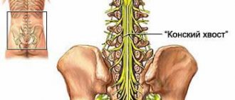
The more proximal the damage to the nerve trunk or plexus (i.e., the greater the distance from the site of damage to the peripheral endings), the worse the prognosis for restoration of function, since the longer it takes for the nerve fiber to grow and the greater the likelihood of irreversible development in the endoneurial tube of the peripheral segment of the nerve. scar changes. So, for example, according to S.I. Karchikyan, with wounds of the sciatic nerve in the upper third of the thigh, the first movements of the foot and fingers appear only 15–20 months or later after the application of the nerve suture, and with injuries of the same nerve in the lower third of the thigh - 10–15 months after surgery. The worst prognosis is observed for injuries at the radicular level, since the roots of the spinal nerves do not regenerate and cannot be restored surgically. Damage to the roots (usually a separation of the root at the cervical level), in contrast to damage to the plexus, is characterized by the following symptoms:
- intense burning pain radiating along the corresponding dermatome;
- paralysis of paravertebral muscles innervated by the posterior branches of the spinal nerves;
- paralysis of the scapula muscles due to dysfunction of the short nerves of the shoulder girdle (pterygoid scapula);
- Horner's syndrome (with damage to the C8 roots);
- trophic disorders and rapidly progressing muscle atrophy with severe secondary contractures.
Peripheral neuropathies and plexopathies can have very different etiologies. In peacetime, the most common form of peripheral nerve damage is tunnel neuropathies, accounting for about 30–40% of all diseases of the peripheral nervous system. Tunnel neuropathy is a local lesion of the nerve trunk caused by its compression and ischemia in the anatomical canals (tunnels) or due to external mechanical influence. Factors predisposing to the development of tunnel neuropathies include the genetically determined narrowness of the natural nerve receptacles, acquired narrowness of these receptacles due to edema and connective tissue hyperplasia in various diseases (for example, diabetes mellitus, hypothyroidism, collagenosis), prolonged overstrain of the musculo-ligamentous apparatus in persons of certain professions, consequences herbs, muscular-tonic and neurodystrophic disorders in reflex syndromes of spinal osteochondrosis, iatrogenic traumatic effects (incorrect application of a plaster cast, hemostatic tourniquet). Nerve dysfunction occurs due to both demyelination and axonal damage (deterioration of neurotrophic control due to failure of axonal transport). Tunnel nerve lesions are manifested primarily by pain, sensory and autonomic disorders. Motor disorders develop in only one third of patients and consist, as a rule, of decreased muscle strength, muscle wasting, and the development of contractures. The prognosis for functional restoration with early treatment is usually favorable, but this restoration can take quite a long time, up to several months. In addition, the prognosis depends on the underlying disease against which the neuropathy developed, and on whether occupational overload of the limb persists. In 30–40% of cases, tunnel neuropathies recur.
In second place in frequency are traumatic neuropathies. Among the causes of traumatic neuropathies, the most prognostically favorable are incised wounds, in which timely surgical intervention provides a good outcome.
Traction and gunshot injuries have a worse prognosis, since in them the central segment of the nerve and the neuron of the spinal centers are often altered, which significantly complicates nerve regeneration. Destruction of the nerve trunk over a long distance can also be observed due to electrical trauma or chemical damage (accidental injection of various medicinal substances into the nerve). A very unfavorable condition accompanying nerve damage is circulatory disturbance in the limb (bleeding or prolonged application of a hemostatic tourniquet, thrombosis of the main artery), which can lead to the development of an atrophying sclerosing process in the muscles, tendons, joint capsules, skin and subcutaneous tissue with the formation of contractures. Secondary changes in the joints and tendons, which develop as a result of stretching of the ligaments and joint capsules during passive hanging of the limbs in the case of flaccid paralysis or paresis, can also prevent the restoration of movements. For neuro- and plexopathies that have developed against the background of somatic diseases, due to immune, neoplastic, infectious, toxic lesions and effects, the prognosis depends on the nature of the course of the underlying disease or process.
Literature:
- Diagnosis of peripheral nerve damage S. M. Russell publishing house: Binom Year: 2009 Pages: 251
- Neurology and neurosurgery. Volume 1. 4th edition
- Author: Gusev E.I., Konovalov A.N., Skvortsova V.I.
- https://www.medicport.ru/doctors/stati_dlya_vrachej/materialy/lechenii_zabolevanij_perifericheskih_nervnyh_stvolov_i_spletenij/
- https://vse-zabolevaniya.ru/bolezni-nejrohirurgii/porazhenija-nervnoj-sistemy.html
- https://aupam.narod.ru/pages/medizina/reab_ruk_rbsdn_t2/page_28.htm
Diagnostics
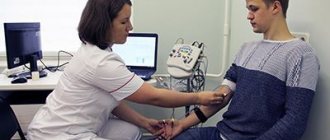
To make a diagnosis, examination using palpation and a series of tests is important.
- Discriminative two-point test - the sensitivity of the branches is checked in turn and the reaction is compared.
- The sensory function of the radial nerve is checked by a discrimination test at two points and pricking the folds of the thumb.
- Motor branches are tested by joint extension.
- The sensitivity of the ulnar nerve is determined on the little finger; to control motor capabilities, the patient spreads his fingers with force.
- Additional tests to analyze ulnar nerve function include ring finger flexion and thumb adduction.
- Motor function of the median nerve is tested by resisting flexion of the wrist and index finger.
- A visual test of the sensitivity of the median nerve is a discrimination test with an attachment in the palm.
When is surgery indicated?
- Impaired sensitivity and movement function.
- Tumors.
- Painful neuromas.
- Compression by scars.
- Damage due to trauma.
- Pain syndrome.
Taking into account the nature of the injury, the method of surgical treatment is selected:
- Excision of scar formations – neurolysis;
- Connecting the nerve sheath and applying a special suture;
- Carrying out plastic surgery of nervous tissue.
During surgical treatment, microsurgical techniques are used to make the comparison as accurate as possible.
Symptoms
Depending on the type of nerves affected (motor, sensory, autonomic) and which parts of them are involved in the pathological process, the symptoms of neuropathies are somewhat different. However, manifestations rarely occur in isolation; they are mostly combined: patients, as a rule, experience motor disturbances, changes in tissue sensitivity, and other symptoms. If only the myelin sheaths of the nerves are affected, the tissue changes are reversible when the cause is eliminated, but if the axon, the actual process of the nerve cell, is involved, then the damage may be irreversible.
Severe damage in the area of peripheral nerve fibers manifests itself in gradual muscle atrophy and progressive weakness, changes in tendon reflexes, they are usually reduced. Paresis (decrease in muscle strength and decrease in range of motion) in the area of the affected nerve fibers and sensitivity disorders are also observed - decrease or increase, loss of certain types of sensitivity (temperature, pain, vibration) or a crawling sensation, tingling, numbness. Also typical is a change in skin color and dryness, marbling of the skin, and sweating.
For many types of neuropathy, severe pain is typical, resulting from ischemic damage to the nerves.
Rehabilitation
The recovery period takes at least six months. First, touch is restored, then sensitivity when touching two points. For recovery, it is important to recognize objects by touch.
Principles of successful rehabilitation:
- Early intervention;
- Reducing the risk of complications;
- Ensuring healing;
- Restoring the functions of the nerve of the hand;
- Using a multi-stakeholder approach.
Restoration of the hand nerve is carried out at the clinic of the Central Clinical Hospital of the Russian Academy of Sciences, in the department of hand microsurgery. Everything is available here for effective treatment of a nerve rupture in the arm - experienced, highly qualified surgeons work, the most modern microsurgical equipment is used, and the staff is caring.
Registration for a consultation is carried out on the website. You can get the information you are interested in and find out the price of treatment by calling the number provided.