Causes
“Blue” heart defects, open and penetrating traumatic brain injury, lung pathology, as well as bacterial endocarditis and the presence of immunodeficiency (including those caused by HIV) can increase the risk of developing a brain abscess. The main pathogens are Escherichia coli, staphylococci, toxoplasma, anaerobic bacteria, streptococci, pneumococci, fungi and meningococci. 25% contain a sterile abscess. The infection enters the brain through contact or hematogenous means, also known as metastatic.
Brain abscess
Brain abscess is manifested by general infectious, cerebral and local (focal) symptoms. The latter characterize the localization of the abscess. General infectious symptoms: fever (sometimes intermittent), chills, blood leukocytosis, increased ESR, signs of a chronic infectious process (pallor, weakness, weight loss).
General cerebral symptoms appear due to increased intracranial pressure caused by an abscess. The most consistent symptom is headache, often with vomiting. In the fundus there are congestive discs or optic neuritis. Bradycardia up to 40-50 beats per minute and mental disorders are periodically detected. Noteworthy is the patient's lethargy and apathy, and the slowness of his thinking. Stupefaction and drowsiness gradually develop; in severe cases without treatment - coma. As a consequence of intracranial hypertension, general epileptic seizures may occur.
Focal symptoms depend on the location of the abscess in the frontal, temporal lobes, and cerebellum. Abscesses located deep in the hemispheres outside the motor zone may occur without conduction symptoms. Otogenic abscesses sometimes form not on the side of otitis media, but on the opposite side, giving the corresponding clinic. Along with focal symptoms, symptoms associated with swelling and compression and dislocation of brain tissue may be observed. When the abscess is close to the membranes and with a cerebellar abscess, meningeal symptoms are detected.
In the cerebrospinal fluid, pleocytosis (25-300 cells), consisting of lymphocytes and polynuclear cells, increased protein levels (0.75-3 g/l) and increased pressure are detected. However, often the cerebrospinal fluid is normal.
Course The onset of the disease is usually acute, with rapid manifestation of hypertensive and focal symptoms against a background of rising temperature. In other cases, the onset of the disease is less defined, and then the clinical picture resembles the course of a general infection or meningitis. Rarely, the initial stage of an abscess occurs latently with minimal symptoms and low fever. After the initial manifestations, after 5-30 days, the disease enters the latent stage, corresponding to the encystation of the abscess. This stage is asymptomatic or manifests itself with moderately severe symptoms of intracranial hypertension - frequent headache, vomiting, mental retardation. The latent stage can last from several days to several years. Subsequently, under the influence of an external factor (infection), and more often without obvious reasons, general cerebral and focal symptoms begin to progress rapidly. An extremely severe complication of an abscess, possible at any stage, is its rupture into the ventricular system or subarachnoid space, which usually ends in death.
Types of disease
Most often in the clinic there are contact abscesses caused by otitis, mastoiditis, suppuration in the nasal cavities, meninges, orbit or skull bones.
- Otogenic. Occurs in most cases. An abscess is more often complicated by chronic purulent otitis than by an acute process. In the case of otitis media, the infection enters the brain from the temporal bone through the cavernous sinuses and the roof of the tympanic cavity into the middle fossa of the skull, resulting in inflammation of the brain in the temporal lobe of the brain. An infection of otogenic origin can also penetrate into the posterior cranial fossa, passing through the labyrinth and then the sigmoid sinus, which is why a cerebellar abscess occurs.
- Rhinogenic. Often concentrated in the frontal parts of the brain. First, it is caused by local pachymeningitis, after which it passes into the stage of adhesive limited meningitis and ultimately this process ends with the formation of limited purulent encephalitis in the brain tissue. It is very rare that rhinogenic and otogenic types of disease manifest themselves hematogenously as a result of sinuses, venous thrombosis or septic arteritis. In this case, the abscesses are concentrated deep in the brain.
- Traumatic. Often occur during open skull trauma. If the dura mater is damaged, the infection enters the brain tissue through the perivascular gaps. If a foreign body enters the brain, the infection enters along with it. Nowadays, traumatic abscesses reach 15% of all possible types of this disease.
- Metastatic. Often occurs as a result of lung diseases such as pleural empyema, pneumonia (pneumonia) or bronchiectasis. Such abscesses are accompanied by the penetration of infection into the brain by septic embolism. About 30% of metastatic abscess diseases are multiple and form in the deep parts of the white matter of the brain.
Aftermath and recovery
After the operation, patients are under the supervision of qualified specialists. Hospitalization continues until specialists are confident that the patient has fully recovered. Qualified treatment is carried out at the Medsi clinic. Neurosurgeons completely eliminate the consequences of a brain abscess and develop an individual rehabilitation program. Highly qualified specialists make it possible to minimize possible complications both during and after surgery for a brain abscess.
Rehabilitation includes physical therapy. It restores impaired functions and helps the patient return to a full life in society. The exercise therapy complex is selected by a rehabilitation specialist. Exercises after surgery are supervised by a professional.
Diagnostics
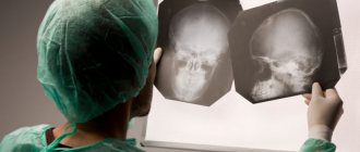
Diagnosis of the disease is made using MRI or computed tomography, both with and without contrast enhancement. Identifying the source of infection is essential for further treatment of the abscess and its successful completion. In most cases, the purulent process occurs in teeth affected by caries, the air sinuses of the nose, and the middle ear.
Causes and course of the disease
A brain abscess mainly forms in the frontal lobe of the brain, involving various areas from the affected sinus.
Much less common are abscesses located on the opposite side (contralateral). The microbial flora of the abscess is diverse and does not always coincide with the flora inhabiting the paranasal sinuses. Most often, the infection spreads by contact from the frontal sinus. But cases have been identified when, due to thrombosis of the cavernous sinus (sinus of the dura mater), the infection spreads through the blood (hematogenously).
Within 3 days from the onset of the disease, local inflammation develops around the blood vessels, which, due to the development of cerebral edema and tissue necrosis (death), leads to encephalitis (inflammation of the brain). From 4 to 9 days of the disease, maximum swelling is observed in the brain tissue, which is accompanied by an increase in the area of necrosis and the formation of pus (pus formation).
At the same time, a reticular network is formed (neurons that are connected by nerve fibers and form a kind of network), which signals the formation of a capsule. On days 10-13, the capsule becomes denser and its necrotic center is separated from the surrounding medulla. In the last stage, which is observed on the 14th day or slightly later, the well-formed capsule has the following layers:
- Necrotic;
- Zone of inflammatory fibroblasts (connective tissue) and cells;
- Collagen capsule;
- The area of newly formed vessels;
- The area of reactive gliosis (replacement of brain cells with denser tissue under the influence of the inflammatory process), which is accompanied by edema.
Treatment
Therapy for the disease must be comprehensive. Most often, surgical intervention is used in combination with adequate combination antibacterial therapy. Antibacterial therapy can last several weeks. Subsequently, a long period of rehabilitation is required. Depending on the resulting neurological defect, one should resort to neuroprotective therapy, massage, exercise therapy, and sometimes, when symptomatic epilepsy develops, the selection of antiepileptic drugs is necessary.
Rhinogenic intracranial complications
In the majority of patients, the primary manifestation of all types of intracranial complications is a sharp exacerbation of the general intoxication syndrome. This is manifested by a sharp increase in body temperature to 39.5-41.0 ° C, chills, severe weakness, malaise, increased heart rate and breathing, and confusion. Also, many rhinogenic brain lesions are characterized by general cerebral symptoms: pronounced diffuse headache, dizziness, nausea, vomiting that does not bring relief, mood lability, generalized convulsions and disturbances of consciousness - somnolence, stupor or coma.
The leading manifestations of intracranial hypertension syndrome are headache localized in the frontoparietal region, a feeling of internal pressure on the eyes. There is also nausea and vomiting, which worsens in the morning, severe vestibular disorders, blurred vision, excessive irritability, and insomnia. Meningeal syndrome is manifested by photophobia, hyperacusis, increased sensitivity to odors, stiff neck, forced “cocked” posture, constant vomiting and unbearable headache. Some patients develop psychomotor agitation, delirium followed by loss of consciousness.
Focal symptoms depend on the location of the pathological process. Damage to the frontal lobe often manifests itself as motor dysfunction: unsteadiness of walking, increased muscle tone of the limbs, paresis and paralysis. Further, epileptic seizures, speech disorders, mental disorders of the “frontal psyche” type and the emergence of primitive reflexes are observed. When the parietal lobe is involved in the pathological process, hyperesthesia, dysgraphia, dyslexia, and geographic agnosia are noted. Damage to the temporal lobe is accompanied by cortical deafness, tinnitus and auditory hallucinations, amnesia, and partial seizures. Localization of the pathological focus in the occipital lobe leads to visual agnosia, visual hallucinations, hemianopsia or complete loss of vision.
Optochiasmal syndrome is predominantly manifested by visual disturbances. Patients exhibit progressive deterioration of vision, the occurrence of central scotomas, deterioration of color perception and concentric narrowing of the visual fields. The headache is most pronounced in the frontal and occipital regions and has a burning, pressing character. Drooping eyelids, strabismus, diplopia, exophthalmos, hyposmia, and olfactory hallucinations are found somewhat less frequently.