Causes and mechanisms of development
Most often, a bladder infection is caused by bacteria that are usually found in the intestines. Cystitis or inflammation of the bladder is one of the common problems of the urinary system in children. Sometimes doctors use the more general term urinary tract infection (UTI) if it is impossible to accurately determine that only the bladder is affected, but there are signs of urethritis and pyelonephritis.
Bladder infections affect approximately 3 in 100 children each year. Infants under 12 months of age are more likely to get UTIs than older children. During the first few months of life, UTIs are more common in boys than girls. By age 1, girls are more likely to develop a UTI than boys, and girls continue to be at higher risk throughout childhood and adolescence.
Symptoms of the disease
The work of all organs and systems in the body is regulated by the nervous system, the bladder is no exception.
The disease is manifested by the following symptoms:
- frequent false urge to urinate;
- urinary incontinence of varying severity, caused by an unbearable urge to urinate;
- frequent urination;
- infrequent urination.
Symptoms of the disease can appear at any time of the day, at rest, during sleep, during various physical activities, tension in the anterior abdominal wall when coughing or laughing.
| Approximate norms of urination depending on age | |||
| Age | Volume of urine per day in milliliters | Number of urinations per day | Volume of urine per urination in milliliters |
| 0 - 6 months | 300 — 500 | 20 — 25 | 20 — 35 |
| 6 – 12 months | 300 — 600 | 15 — 16 | 25 — 45 |
| 1 – 3 years | 760 — 820 | 10 — 12 | 60 — 90 |
| 35 years | 900 — 1070 | 7 — 9 | 70 — 90 |
| 5 – 7 years | 1070 — 1300 | 7 — 9 | 100 — 150 |
| 7 – 9 years | 1240 — 1520 | 7 — 8 | 145 — 190 |
| 9 – 11 years | 1520 — 1670 | 6 — 7 | 220 — 260 |
| 11 – 13 years old | 1600 — 1900 | 6 — 7 | 250 — 270 |
| 14 years and older (adults) | 1500 — 1900 | 7 — 8 | 200 – 300 |
Which children are more likely to develop a bladder infection?
Girls are much more likely to develop bladder infections than boys, except in the first year of life. Among boys under 1 year of age, those who have not had their foreskin removed (circumcised) have a higher risk of bladder infection. However, most uncircumcised boys do not develop a bladder infection.
Typically, any medical condition or habit of holding urine in a child's bladder for too long can lead to infection. Other factors that may increase your chance of developing cystitis include:
- overactive bladder, a treatable condition that often goes away as the child gets older;
- incomplete emptying of the bladder;
- waiting too long to urinate;
- constipation—fewer than two bowel movements per week or hard stool that is painful or difficult to pass;
- Vesicoureteral reflux (VUR) is the backflow of some urine from the bladder towards the kidneys during urination;
- A narrowing of the urethra is a problem that restricts the normal flow of urine, such as a stone or a duct that is too narrow (stricture). In some cases, it may be due to a birth defect;
- violations of hygiene rules, defects in caring for a small child;
- family history of cystitis.
Among teenage girls, those who are sexually active are more likely to develop bladder infections. Due to their anatomy, girls are much more likely to develop a bladder infection than boys.
Girls have a shorter urethra than boys, so bacteria have a short distance to travel to reach the bladder and cause an infection.
In girls, the urethra is located closer to the anus, a source of bacteria that can cause a bladder infection.
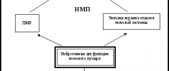
Pathophysiology of neurogenic bladder dysfunction
The lower urinary tract - the bladder and urethra - is a complex anatomical and physiological complex united by the unity of the functions of accumulation, retention and excretion of urine, while each of these three functions can normally be adequately controlled.
Urinary tract dysfunctions in patients with verified neurological diseases are classified as neurogenic dysfunctions. In the absence of neurological pathology, disorders are considered idiopathic.
From a neurological point of view, the lower urinary tract has a complex mixed innervation united by multi-level reflexes. According to the most common concept, a simplified diagram of the innervation of the lower urinary tract is as follows.
Scheme of innervation of the bladder
1 - center of voluntary control of urination of the cerebral cortex; 2 - stem urinary control center (bridge); 3 - lumbar and extraspinal sympathetic ganglia; 4 - parasympathetic ganglia and nerve pathways; 5 - bladder; 6 - sphincter apparatus of the bladder.
The adaptive function of the bladder is controlled by the sympathetic nervous system. Extraspinal sympathetic ganglia provide the function of urine storage by maintaining a relaxed state of the detrusor and tonic contraction of the internal sphincter. A constant inhibitory effect on filling signals coming from the bladder receptors is normally provided by the stem (pontine) urinary control center. Reaching the threshold of its adaptation is accompanied by the appearance of an urge. At the same time, further urinary retention is controlled by cortical centers, which provide voluntary contraction of the external urethral sphincter through somatic nerves. Sensory innervation and, ultimately, bladder emptying are provided primarily by parasympathetic ganglia and nerve pathways at the level of the sacral spinal cord. For adequate urination, simultaneous friendly (synergistic) contraction of the detrusor and relaxation of the bladder sphincters are necessary.
From the point of view of pathogenesis and the possibility of assessing the level of denervation, the following classification of neurogenic bladder dysfunctions seems successful:
- Uninhibited cortical bladder
- Reflex spinal bladder
- Unadapted bladder a - spinal
- b - ganglionic
- a - spinal
Uninhibited cortical bladder - characterized by functional weakness of cortical centers, which are unable to provide an inhibitory effect on spinal structures, while voluntary control over urinary continence when urge occurs is absent or weakened. In patients with an uninhibited bladder, a common manifestation is the presence of an uncontrollable (imperative) urge to urinate, which leads to urge urinary incontinence. An uninhibited bladder in children is often associated with intrauterine hypoxia, birth trauma, or delayed development and maturation of regulatory centers. In adults, loss of voluntary control over the lower urinary tract is caused by microcirculatory disorders, trauma and inflammatory damage to the frontal lobes of the cerebral cortex. Reflex spinal bladder - occurs in patients with damage to the spinal cord above the lumbosacral centers. In the absence of the inhibitory influence of the brain, urination occurs involuntarily with a small volume of bladder filling, similar to a spinal reflex. A number of such patients urinate when a feeling of heaviness appears in the lower abdomen by voluntary irritation of the skin of the thighs or by stimulating a reflex contraction of the detrusor by pressing on the bladder area (Creed, Valsalva maneuvers). In some patients, suprasacral denervation may be accompanied by persistent spasm of the external urethral sphincter or the appearance of its reflex contractions simultaneously with contraction of the detrusor (sphincter-detrusor dyssynergia). In these cases, patients develop chronic urinary retention. Unadapted bladder - characterized by a decrease in the adaptive (storage) function of the bladder due to damage to the sympathetic centers of the spinal cord or extraspinal sympathetic ganglia. In patients with an unadapted bladder, the urge is usually absent or sharply weakened, which is associated with the inability to fill the bladder to a threshold level. With ganglion lesions, incontinence in patients can be combined with a small amount of residual urine, which is due to partial preservation of sphincter function, since complete destruction of all ganglia, as a rule, does not occur. Reflex bladder - characterized by the absence or significant weakening of the urge to urinate due to loss of sensitivity due to damage to the spinal or ganglion parasympathetic centers, as well as underdevelopment or death of the peripheral nerve endings of the bladder (intramural forms). In such patients, the equivalent of urge can be achieved only with maximum filling of the bladder. Mixed bladder manifests itself as chronic urinary retention followed by dilatation of the upper urinary tract and increasing renal failure. Common symptoms in patients are the development of overflow urinary incontinence (ischuria paradoxa) and constipation. Patients with a reflex bladder require continuous urinary diversion or intermittent catheterization. Shriveled bladder - develops as a result of the progression of degenerative-sclerotic changes in the detrusor in conditions of its denervation, infection and/or prolonged drainage of the bladder.
Involvement of the area of Lieto's triangle in the process can lead to disruption of the outflow of urine from the upper urinary tract, which requires surgical measures. According to the above etiopathogenetic aspects, the term neurogenic bladder should be understood as dysfunction of the bladder and its sphincters, which develop as a result of neurological diseases and damage to the central and peripheral nervous system. The complexity, the presence of additional nerve pathways, the dominant positions of the autonomic system and a large number of control centers at different levels determine the multiplicity of options and paradoxical manifestations of neurogenic bladder dysfunction. Clinicians are well aware of situations that contradict or do not fit into the pathogenetic classification presented above.
Classification of the disease in children
- Complicated (with the addition of peritonitis and other ailments) or uncomplicated.
- Acute (accompanied by inflammation of the sub- and mucous layers, in some cases – bleeding of the walls of the bladder) or chronic (morphological pathologies affect the muscle layer).
- Focal or total (according to the degree of prevalence).
- Primary (without abnormalities in the structure and dysfunction of the bladder) or secondary (occurs due to residual urine due to improper anatomy and functionality of the bladder). Source: K.V. Mitrofanov Cystitis in children // Mother and Child in Kuzbass, 2005, No. 1(20), pp. 3-9
Causes of inflammation:
- infection coming from the kidneys, urethra, pelvic and more distant organs;
- dysfunction of the bladder;
- incorrect structure of the organ;
- incomplete and/or irregular bowel movements;
- coli;
- Pseudomonas aeruginosa, Klebsiella, Proteus (rarer pathogens);
- viral infection (indirect effect - leads to improper microcirculation of urine, which becomes a good background for the disease);
- ureaplasma, chlamydia, mycoplasma (usually the cause of infection is chlamydia in parents, as well as lack of hygiene, visiting public baths, etc.);
- phimosis (relevant for boys);
- vesicoureteral reflux;
- fungus (for immunodeficiency).
Additional risk factors for cystitis:
- urolithiasis disease;
- foreign objects entering the bladder;
- therapy with drugs toxic to the kidneys, including cytostatics in oncology;
- invasive examinations on the urological profile;
- dysbacteriosis;
- infection with worms;
- various infectious diseases of the intestinal tract;
- a wide range of gynecological diseases;
- inflammatory and purulent processes;
- dysfunction of the endocrine system;
- radiation;
- hypothermia;
- failure to comply with basic personal hygiene standards; Source: https://www.ncbi.nlm.nih.gov/pubmed/26075187 Hanna-Wakim R, Ghanem ST, El Helou MW, Khafaja SA, Shaker RA, Hassan SA, Saad RK, Hedari CP, Khinkarly RW, Hajar FM, Bakhash M, El Karah D, Akel IS, Rajab MA, Khoury M, Dbaibo GS Epidemiology and characteristics of urinary tract infections in children and adolescents // Front Cell Infect Microbiol. 2015 May 26;5:45. doi: 10.3389/fcimb.2015.00045. eCollection 2015
- etc.
Clinical and urodynamic classification of neurogenic urinary tract dysfunction
In 1992, Wayne proposed a simplified classification of neurogenic bladder dysfunctions in terms of urodynamic disorders:
- Overactive bladder.
- Sphincter-detrusor dyssynergia.
- Detrusor areflexia.
Overactive bladder is a chronic condition characterized by symptoms of frequency, urgency, and nocturia, with or without urgency urinary incontinence. It should be understood that the diagnosis of overactive bladder can be made if these symptoms are not caused by bladder diseases or metabolic disorders. Therefore, the diagnosis of any form of neurogenic dysfunction can be verified only on the basis of a comprehensive neurourological examination, including a mandatory urodynamic study (uroflowmetry, cystometry, profilometry, pressure-flow study, etc.). The most important urodynamic criterion for diagnosing hyperactivity is the appearance of involuntary contractions of the detrusor with a pressure amplitude of more than 5 cm of water. Art. during cystometry. Thus, the term “overactive bladder” should be considered as a diagnosis, and overactive detrusor as its main urodynamic manifestation.
In patients with spinal cord injury (reflex spinal bladder), when cystometry is performed, so-called terminal detrusor overactivity is often encountered, which is characterized by contraction of the detrusor and the occurrence of urination in the complete absence of urge. Detrusor overactivity occurs in 77% of patients with neurogenic bladder dysfunction. This is due to the fact that in most neurological diseases, the spinal link of the voiding reflex - its lumbosacral centers - is usually not completely destroyed. Experimental studies have shown that under conditions of the “decapitated” reflex in the bladder, unmyelinated C-fibers are activated and the content of nerve growth factor in the detrusor increases. The unity of histochemical changes in the bladder wall in patients with neurogenic and idiopathic detrusor overactivity suggests that the formation of overactive bladder syndrome is always a consequence of its complete or partial denervation, which developed for an established or unidentified reason. Sphincter-detrusor dyssynergia is characterized by involuntary contractions of the external urethral sphincter and/or paraurethral muscles during detrusor contraction. Violation of concomitant relaxation of the sphincter and contraction of the detrusor leads to the development of functional bladder outlet obstruction and the appearance of residual urine in patients. Sphincter-detrusor dyssynergia usually occurs in patients with suprasacral spinal cord injuries. Patients with sphincter-detrusor dyssynergia account for 33–55% of all patients with neurogenic bladder dysfunction. To define intermittent urination due to involuntary contraction of the sphincter and periurethral muscles in patients without neurological diseases, the term dysfunctional urination has been proposed. Detrusor areflexia is characterized by a decrease or complete absence of the ability of the detrusor to contract. The main manifestation of areflexia is the complete absence of the urge to urinate in patients. Most often, detrusor areflexia occurs in patients with damage to the sacral and infrasacral centers (meningomyelocele, radical surgery and injuries with damage to the pelvic organs). In this case, 50% of patients have a spastic state of the urethral sphincter. Patients with detrusor areflexia require constant urological supervision due to the high risk of urinary infection and the frequent occurrence of complications from the upper urinary tract. A number of patients with damage to the sacral centers develop obstruction due to sphincter spasm, which is characterized by persistent contraction of the urethral sphincters. In a number of these patients, urodynamic stress incontinence occurs when the bladder is full.
In modern clinical practice, the term stress incontinence is used to define urinary incontinence due to stress (from the English stress - tension), for example, coughing, laughing, sneezing. Involuntary leakage of urine due to stress incontinence is usually not associated with neurological disorders. The main role in the development of stress incontinence belongs to disturbances in the anatomy of the ligamentous apparatus and the function of the pelvic floor muscles and damage to the urethral sphincters.
How does cystitis manifest in a child: main symptoms
The child has ways to protect himself from infection in the urinary tract. For example, urine leaves your baby's kidneys through the ureters and into the bladder. Bacteria that enter the urinary tract are expelled during urination. This one-way flow of urine prevents bacteria from infecting the urinary tract. Sometimes the body's defenses weaken, and bacteria ascend into the bladder, causing inflammation. If your child has symptoms of cystitis or has a fever for no clear reason, contact your doctor within 24 hours.
Cystitis often occurs in children, especially in girls (which is due to the structural features of the urinary tract). Immediate treatment of cystitis in a child can prevent kidney damage (pyelonephritis). It can develop due to an infection that spreads up one or both ureters. Kidney infections are very painful, can be dangerous and cause serious health problems, so it is best to start treatment as early as possible.
Signs of the disease in infants and children under one year of age:
- decreased appetite or complete refusal to eat;
- restlessness that increases when the child urinates;
- crying, nervous excitement or, conversely, lethargy and weakness;
- increased body temperature, up to fever;
- spasm of the urethral sphincter from the outside, urinary retention due to a developed reflex;
- change in urine: cloudiness, sediment, flakes, repulsive odor.
In older children, the symptoms are quite bright and pronounced. The main symptoms are pain in the lower abdomen, back or side, as well as an urgent need to urinate or frequent trips to the toilet. Some toddlers who are already potty trained or toileting lose control of their bladder and may wet the bed. Parents may also see drops of blood in the urine and/or pink colored urine.
It is important to know how cystitis manifests itself in young children. Babies may have more general symptoms such as fussiness, decreased appetite, crying, fussiness on the potty, or fever. Other symptoms of cystitis include:
- burning or pain when emptying the bladder;
- foul-smelling or cloudy urine;
- a sharp urge to urinate, and then the release of only a few drops of urine;
- high temperature;
- nausea or vomiting;
- diarrhea.
How to determine acute cystitis?
The disease manifests itself quickly, with a rapid increase in symptoms:
- the urge to empty your bladder every 20 minutes or more often;
- pain above the pubis, radiating to the perineum and intensifying when palpating the abdomen and even when the bladder is not full;
- difficulty passing urine, accompanied by stinging and pain;
- urinary incontinence;
- a few drops of blood after urinating;
- urge that does not lead to urination.
Chronic form of the disease
A secondary disease, which is characterized by a similar clinical picture during periods of exacerbation. Children experience discomfort in the pubic area, suffer from incontinence and noticeably increased frequency of urination. Source: Pigareva, N.A. Tsap Chronic cystitis in children: modern view on etiology, pathogenesis, clinical picture, diagnosis and treatment (literature review) // Russian Bulletin, 2012, vol. II, no. 2, pp. 32-37
Pediatric urology PEDUROLOGY.RU
Urinary disorders, manifested by frequent urination, urinary incontinence, including urgency, are common complaints in childhood. However, parents usually pay attention to these symptoms in children over 4-5 years old, considering such manifestations to be a feature of younger age. But that's not true.
Not only the problem of social maladaptation of a child with “wet panties” syndrome, but also severe infectious complications from the urinary system, as well as vesicoureteral reflux, which may accompany this condition, determine the relevance of the problem. Due to the fact that not only highly specialized specialists (urologists), but also general practitioners are involved in the process of diagnosing and treating urinary disorders, in 1997 P. Abrams and A. J. Wein introduced the term “Overactive Bladder” to facilitate diagnosis and treatment.
Bladder overactivity is a symptom complex characterized by 1) pollakiuria (frequent urination > 8 times during the day); 2) urgency (an unexpected irresistible (imperative) urge to urinate; 3) urgent urinary incontinence (an episode of involuntary loss of urine immediately following an episode of urgency); 4) enuresis (bedwetting); Pollakiuria is combined with a decrease in the effective volume of the bladder below standard values. The most important sign of pollakiuria, as a pathological symptom, is the absence of residual urine, which is established retrospectively during an instrumental examination (catheterization of the bladder or ultrasound examination after urination). Urgency is diagnosed when the patient complains of periodically appearing, irresistible desires to urinate. If a child cannot suppress the urge to urinate, urge urinary incontinence occurs. Enuresis is urinary incontinence at night. If all of the above symptoms are present, we can speak of a complete syndrome of urinary dysfunction.
AT WHAT AGE SHOULD OVERACTIVE BLADDER IN CHILDREN BE CONSIDERED A DISEASE? By the age of 4, most children develop a “mature type of urination”, characterized by: complete retention of urine day and night; · the ability to delay and interrupt, if necessary, the act of urination; · the ability to empty the bladder without a previous urge to urinate; · behavioral reactions accompanying the act of urination - the desire for privacy when the urge appears, etc. Normally, children 3-4 years old empty their bladder 5-8 times while awake; The average effective bladder volume ranges from 120 to 150 ml.
The volume of urination can be calculated using the formula: (30xage) +30 ml. Thus, we can talk about the formation of an OAB symptom complex in cases where in a child 4 years of age and older, the number of urinations and the effective volume of the bladder go beyond the normative indicators, and urination is uncontrollable to one degree or another. However, we must not forget that pathology begins at an early age, and therefore it is very important to know, even in children in the first months of life, how often the child urinates, what the portions of urine are (in infants, the study should be carried out according to international standards within 4 hours). The diagnosis of OAB is established using a comprehensive staged examination, including at the prehospital stage: -physical examination; -laboratory tests; -keeping a diary of urination and bowel movements. At the first stage of the examination, the child should be examined by a psychoneurologist, since even during the initial examination behavioral problems, hyperactivity and attention deficit syndrome, and delayed psychomotor development can be identified. During a physical examination, the lumbosacral spine is examined to identify malformations and phenomena of spinal dysmorphism: (hyperpigmentation, subcutaneous lipoma, buttock asymmetry, hypertrichosis), which is an indication for R-graphy of the lumbosacral spine or MRI, which will reveal bone markers of myelodysplasia, the presence of additional formations and will allow assessing the state of liquorodynamics. When collecting anamnesis, it is necessary to determine the presence of constipation and/or stool. It is mandatory to study perineal sensitivity and reflexes, as well as the tone of the anal sphincter. When examining the lower extremities, it is necessary to pay attention to the symmetry of the extremities and gait. An examination of the external genitalia is carried out to identify concomitant pathologies: cicatricial phimosis, epispadias, meatal stenosis, vulvitis, vaginitis. It is fundamentally important to confirm or exclude the presence of infectious complications. Clinical laboratory tests (general blood count, general urinalysis), microbiological research methods (bacteriological culture of urine to determine the sensitivity of microorganisms to antibacterial drugs) are mandatory. A method for identifying an overactive bladder at the first stage of the examination is keeping a diary of urination and defecation, since The pelvic organs are innervated by the same nerve structures, and these disorders are often combined. According to the terminology of the International Society for Urinary Continence in Children, a “urination and defecation diary” involves recording the number of urinations, the time of onset of the urge, the volume of urine excreted per urination, symptoms of incontinence and urgency, urine production at night, taking into account the volume of fluid taken and recording acts of defecation . Urination diary Frequency of urination: ─ increased (> 7/day) ─ decreased (< 3/day) Volume of urine excreted: ─ portions Volume of fluid taken ─ N = 25-30 ml/kg/day (for diabetes 75-100 ml/kg day) ─ distribution over time Cases of urinary incontinence: ─ frequency ─ intensity Urgency: yes/no Constipation/stooling: yes/no The study should be carried out within 14 days, or at least within 4-5 days, determining the time and volume of urination. In infants, a 4-hour test is performed with ultrasound monitoring of bladder filling and mandatory recording of the volume of residual urine after each urination. Urine portions can also be determined by weighing diapers. The absence of changes in urine tests in the presence of pollakiuria should alert the doctor, and changes in the nature of urination are indications for further examination and treatment. Data on the nature of urination, assessed by the rhythm of urination, are analyzed using a table for assessing urinary urgency syndrome proposed by E.L. Vishnevsky in 1997, in which the main symptoms of urination disorders are assessed by a certain number of points, depending on the severity of the disorders. Urinary Urgency Assessment Chart.
| sign | state | points |
| Urinary urgency | No Not every day Every day 1-2 times Every day several times | 01 2 3 |
| Urgent incontinence | No Not every day Every day 1-2 times Every day several times | 0 1 2 3 |
| Involuntary urination during night sleep (enuresis) | No Not every day Several times a month Several times a week Every night 1 time Every night several times | 0 1 5 10 15 20 |
| Diuresis from 18 to 6 hours (in relation to daily) | Up to 40% 41-50% 51-60% 61-70% More than 71% | 0 1 2 3 4 |
| leukocyturia | No In urine analysis according to Nechiporenko In general urine analysis | 0 1 2 |
| Sign | Number of urinations per day | points |
| Urination rhythm | 5-8 9-10 11-12 13-14 15-16 17-18 19-20 20 or more | 0 1 2 3 4 5 6 7 |
Depending on the severity of urination disorders, 3 groups are distinguished: Group 1 - mild degree of urination disorders, total score 2-10. Group 2 - average degree of disorder, total score 11-20. Group 3 - severe urinary disorders, total score 21-30. Considering the high incidence and infectious complications and the development of vesicoureteral reflux with an overactive bladder, the damaging effect on the renal parenchyma and, as a consequence, the development of renal failure, a multidisciplinary approach to treatment tactics is necessary. Since the nature of OAB is polyetiological, treatment should be comprehensive, not symptomatic and aimed at restoring the act of urination as a whole.
Treatment of childhood enuresis.
The main methods of treatment are urotherapy, pharmacotherapy and isotherapy. The starting method in the treatment of OAB is urotherapy – a system of cognitive methods. Urotherapy means the elimination of behavioral disorders in a child, cognitive correlation, urination training in combination with the principle of biofeedback. The next stage of pathogenetically based treatment of OAB is pharmacotherapy (use, in particular, oxybutynin). But in children under 5 years of age, M-anticholinergics are not approved for use (and it is at this age that urinary disorders begin), and, in addition, 17-30% of children have intolerance to oxybutynin. Therefore, physiotherapy is of great importance in the treatment of OAB, both in the form of monotherapy (in children under 5 years of age) and in combination with pharmacotherapy. It is recommended to use thermal procedures (in particular ozokerite applications), sinusoidally simulated currents, as well as the use of modern types of energy (interference currents and laser therapy).
If you would like more information, please contact us or consult your doctor for specific diagnostic and treatment recommendations.
For many questions, an online consultation with a urologist is possible - https://pedurology.ru/index.php/on-line1
Diagnosis of pathology in children
If your child has symptoms of cystitis, contact your pediatrician immediately. Your doctor will order urine tests to check for bacteria. You need to collect urine in several ways:
- Older children can pee in a sterile cup.
- For younger children who are not toilet trained, a sterile polyethylene urine bag is used on the genitals to collect urine.
- For children who wear diapers, a tube (catheter) may be inserted into the urethra and bladder to collect a sample.
In the laboratory, they look at the sample under a microscope to determine if there are germs in the urine. Urine can also be cultured—a lab technician places the urine in a dish to see what bacteria grow in it. This can help your doctor pinpoint the germs that are causing your cystitis so he knows what medications to prescribe to kill them.
If your child has had multiple urinary tract infections or is diagnosed with chronic cystitis, the doctor may refer you to a nephrologist (kidney specialist) and do one or more of these imaging tests to look for problems in the urinary tract:
An ultrasound uses sound waves to identify any obstructions or other problems in the kidneys and urinary tract.
A voiding cystourethrogram, where fluid is injected into the bladder through a tube, is used to show any problems in the urethra or bladder when the baby urinates.
Nuclear scans use fluids containing small amounts of radioactive material to see how well the kidneys are working.
A CT scan, or computed tomography scan, is a high-powered X-ray that takes detailed pictures of the bladder and kidneys.
MRI, or magnetic resonance imaging, uses powerful magnets and radio waves to produce images of the bladder and kidneys.
Vesicoureteral reflux - symptoms and treatment
Timely detection of vesicoureteral reflux, quality treatment and monitoring can prevent serious complications with devastating consequences for the organ and the body as a whole.
A carefully collected medical history (including family history) forms the basis of diagnosis. You need to find out:
- the nature of urination (in children - restlessness, straining, increased frequency, decreased frequency, pain, two-component urination, incontinence, urinary incontinence, etc.);
- facts of unmotivated episodes of fever, attacks of urinary tract infections;
- the presence of unexplained pain in the abdomen, lower back, sacrum;
- heredity in terms of diseases of the urinary system in close and distant relatives;
- during pregnancy.
Physical examination is not the least important in making a diagnosis. At the same time, attention is paid to the physical development of the child, the color of the skin and mucous membranes, elasticity and moisture. Their pathological changes may be a sign of a latent inflammatory process in the urinary system, including chronic renal failure. The presence of edema of various localizations makes it possible to suspect a decrease in the water-excreting function of the kidneys, which accompanies PMR. Inspection and palpation of the abdomen and lumbar region are necessary, assessing pain at the costovertebral, costal-lumbar, and ureteral points. Mandatory for any type of objective examination is the measurement of blood pressure, respiratory rate, and heart rate. The patient or his legal representative must record the rhythm of spontaneous urination for 2-3 days. This will help reveal a picture of the functional state of the bladder.
A general blood test indirectly indicates any renal disease (anemia, leukocytosis, accelerated ESR). A biochemical blood test with changes in normal values of creatinine, urea, uric acid, and electrolytes indicates impaired tubular and glomerular functions. The Rehberg test, or calculation of the glomerular filtration rate (cleansing the blood of metabolic products), calculated using the Schwartz formula, clarifies the degree of impairment in the kidneys.
A general urinalysis, in addition to the inflammatory changes characteristic of PMR, can show persistent isolated proteinuria (an increase in the amount of protein in the urine), which reflects the presence of a scarring process in the kidneys due to reflux nephropathy. The fact that urine concentration processes are disrupted when obtaining the results of diagnostic tests (Zimnitsky test) indicates a decrease in tubular functions. Regular urine culture for established reflux (at least once every six months) helps to clarify the etiology and stage of the inflammatory process.
Of the instrumental research methods, the most accessible is ultrasound of the urinary system , which detects indirect signs of reflux:
- expansion of the pyelocaliceal system;
- changes in the structure of the mucous membrane of the pelvis and bladder with concomitant inflammation;
- suspicion of other malformations of the urinary and urinary organs, since reflux is often combined with these.
Dopplerography (duplex scanning of renal blood flow) allows you to determine the state of intrarenal hemodynamics, which visualizes the onset of reflux nephropathy earlier than other methods.
The primary radiopaque diagnostic method is voiding cystourethrography (MCUG) , which allows one to confirm or exclude suspected vesicoureteral reflux, as well as to determine its degree.
With radioisotope cystography (RC), the exposure to radiation is much less than with MCCG, but the anatomical details are reflected somewhat weaker.
Clinically, a special place is occupied by intermittent vesicoureteral reflux, which has not been proven by repeated x-ray methods, but has a characteristic clinical picture: recurrent pyelonephritis, periodic leukocyturia (increased number of leukocytes in the urine), indirect ultrasound and x-ray signs. In this case, the patient is managed conservatively, or the issue of surgical intervention is decided after visualization of the gaping ureteral orifices during cystoscopy [5].
All patients with established stage ΙIΙ-IV VUR are indicated for excretory urography . The study is carried out by intravenous administration of a radiocontrast agent followed by taking X-ray photographs. This method allows you to visualize the upper urinary tract and assess its functionality.
Sometimes a urodynamic study :
- to assess functional-obstructive or functional-non-obstructive neurogenic disorders of the bladder and sphincters;
- to determine functional disorders of the lower urinary tract (uroflowmetry, profilometry of the vesicoureteral junction, etc.) [8].
This testing is especially important for patients in whom secondary reflux is suspected (spina bifida or boys with posterior urethral valves). But in most cases, in patients with neurogenic dysfunctions of urination, diagnosis and assessment of the condition in the long term should be limited to non-invasive methods (ultrasound of the bladder, rhythm of spontaneous urination).
Static and dynamic nephroscintigraphy (administration and recording of the removal of a radioisotope) with subsequent assessment on a scintigram of the layer-by-layer structure and size of the kidneys with already established PMR is carried out regularly. Radioisotope renography evaluates kidney function and the rate of urine excretion from the body on the right and left separately; the frequency of studies is determined individually. Magnetic resonance imaging , which allows detailed assessment of the parenchyma, the presence of scars and abnormalities, is an infrequent alternative to excretory urography.
Equally important is prenatal (prenatal) diagnosis of reflux. The presence of unilateral or bilateral dilatation of the fetal pelvis, sometimes in combination with dilation of the ureter, unstable sizes and variability within the micturition cycle, and urinary disorders of the fetus are harbingers of the formation of VUR in the postnatal (extrauterine) period [3].
Observation of specialists is mandatory: therapist/pediatrician, nephrologist, urologist, dentist, otolaryngologist, and, if indicated, neurologist, ophthalmologist, orthopedist. The goal of surveillance should be to prevent infections and permanent damage to the renal parenchyma, as well as its late complications. For this purpose, antibiotic prophylaxis, renoprotective therapy and/or surgical correction of reflux are prescribed.
Treatment methods
After determining the diagnosis, the doctor determines how to treat cystitis in children. For the treatment of acute and chronic cystitis in childhood, clinical recommendations have been developed that define a set of actions and medications for each case.
Mostly antibiotics are used. These medications kill bacteria. Children usually take them for 3 to 10 days (most often 7-10 days). Your doctor may do another urine test after your child finishes taking the medicine to see if the infection has cleared up. Always take the full course of medications, even if your child gets better. Stopping too early can make germs resistant to antibiotics and cause cystitis to recur.
Most UTIs go away in about a week. In some children, symptoms persist for several weeks. Contact your doctor if your child's symptoms do not begin to improve after 3 days of starting antibiotics or if they get worse.
If the child's condition is serious, hospitalization is possible, where doctors will decide how cystitis is treated depending on age and severity. Intravenous antibiotics and pain medications may be indicated if the baby is less than 2 months old or has vomiting or abdominal pain. The duration of treatment depends on:
- how serious the infection is;
- whether the child’s symptoms and signs of cystitis disappear;
- whether the child has recurrent bladder infections, vesicoureteral reflux, or another urinary tract problem.
Important! Only a doctor can help a child and completely cure him. Self-medication can lead to certain complications and aggravate the course of the disease.
I'll tell you about these diseases.
Bladder dysfunction is a whole group of conditions, usually manifested by daytime complaints.
Urinary infections - manifested by an increased number of bacteria and leukocytes in urine tests
Chronic kidney disease is also accompanied by retardation in physical development or even loss of body weight, increased blood pressure, edema, abnormalities in urine tests - increased levels of protein and red blood cells.
The doctor can easily rule out this condition based on your complaints, examination results, and fairly simple urine and blood tests.
Congenital posterior urethral valve is a congenital anomaly in the form of folds of the mucous membrane at the outlet of the bladder, which prevent its emptying.
It is manifested by incomplete emptying of the bladder - even after urination you can feel a full bladder. It also manifests itself as a weak or intermittent stream, difficulty starting urination, and repeated urinary infections.
Ectopic urethra in girls is an incorrect location of the exit of the urethra. The main complaint is constant leakage of urine.
Severe constipation.
Links to my notes on this topic:
Risk situations for functional constipation.
Fecal incontinence in children: what parents need to know.
And an excellent video about the causes of constipation - in English, but everything is clear there without translation...
Sickle cell anemia is a very rare blood disease in Russia.
Convulsions - manifest themselves in stereotypical attacks - certain movements, stopping gaze, instant loss of muscle tone.
Diabetes mellitus - manifests itself in increased thirst, frequent urination, rapid weight loss, and sugar in the urine.
Diabetes insipidus is a rare condition that is manifested by thirst and a large volume of very low-density urine excreted per day.
Spinal dysraphism is a group of diseases of the spinal cord and spine that can manifest as disturbances in the functioning of the pelvic organs, including the bladder.
Obstructive sleep apnea —short-term episodes of lack of oxygen to the brain can cause a child to wet themselves.
By the way, at the beginning of September, my colleague, a wonderful ENT and phoniatrist Anna Kolesnikova, talked about this problem on her Instagram https://www.instagram.com/lor_kolesnikova/
View this post on Instagram
A post shared by ENT, phoniatrist Anna Kolesnikova (@lor_kolesnikova)
Pinworms usually manifest themselves as itching in the anus in the evening and at night.
Primary polydipsia is a condition when a child simply drinks too much. It occurs for various reasons and is often psychogenic.
Prevention
Drinking enough fluids, practicing good hygiene and diaper use, wearing loose-fitting clothing, and treating cystitis-related health problems can help prevent UTIs in a child or teen.
It is important to avoid constipation. Hard stool can put pressure on the urinary tract and block the flow of urine, allowing bacteria to grow. Helping your child to have bowel movements regularly can help prevent constipation.
If your child has vesicoureteral reflux, a urinary tract structure, or an anatomical problem, see a pediatric urologist or other specialist. Treating these conditions can help prevent recurrent bladder infections.
Diabetes and other health problems can increase the risk of a bladder infection. Ask your pediatrician how to reduce your risk of developing a bladder infection.
Sources:
- K.V. Mitrofanov. Cystitis in children // Mother and Child in Kuzbass, 2005, No. 1(20), pp. 3-9.
- https://www.ncbi.nlm.nih.gov/pubmed/26075187 Hanna-Wakim R, Ghanem ST, El Helou MW, Khafaja SA, Shaker RA, Hassan SA, Saad RK, Hedari CP, Khinkarly RW, Hajar FM, Bakhash M, El Karah D, Akel IS, Rajab MA, Khoury M, Dbaibo GS. Epidemiology and characteristics of urinary tract infections in children and adolescents // Front Cell Infect Microbiol. 2015 May 26;5:45. doi: 10.3389/fcimb.2015.00045. eCollection 2015.
- A.E. Pigareva, N.A. DAC. Chronic cystitis in children: modern view on etiology, pathogenesis, clinical picture, diagnosis and treatment (literature review) // Russian Bulletin, 2012, vol. II, no. 2, pp. 32-37.
- Sh.Sh. Mametova. Modern aspects of diagnosis and treatment of cystitis in children // Bulletin of KAZNMU, 2012, No. 1, pp. 255-257.
The information in this article is provided for reference purposes and does not replace advice from a qualified professional. Don't self-medicate! At the first signs of illness, you should consult a doctor.
Overactive bladder syndrome in children. Clinical case
Among all urinary disorders in children, overactive bladder (OAB) occupies a significant place. The term “overactive bladder,” introduced by the International Continence Society (ICS), has been used in pediatric urology since 2004 [1, 2]. According to the ICS, the guidelines of the European Association of Urology (EAU), the guidelines of the German Society of Child and Adolescent Psychiatry, Psychotherapy and Psychosomatics (Deutsche Gesellschaft für Kinder und Jugendpsychiatrie, Psychosomatik und Psychotherapie, DGKJP), the main clinical signs of OAB are pollakiuria, imperative urge to urinate, urge urinary incontinence of varying severity and enuresis. Overactive bladder in children is characterized by a large polymorphism of clinical symptoms and their severity due to age-related characteristics of the rate of maturation of the sympathetic and parasympathetic parts of the autonomic nervous system (ANS), instability of brain metabolism, as well as the inherent tendency of the child’s body to develop generalized reactions in response to local irritation [3].
The formation of OAB is caused by pathology of the hypothalamic-pituitary system, accompanied by a delay in the maturation of the neurohumoral regulation of the act of urination, detrusor ischemia and impaired bioenergetics [4]. The ANS plays an important, if not decisive, role in maintaining homeostasis, i.e., the constancy of the internal environment of the body [5]. When the regulatory influence of the ANS is disrupted, not only urination disorders develop, but also combined disorders of the visceral organs [6]. However, there is a problem of general underestimation of the role of autonomic maladaptation in the formation of children's health. Today, for a practicing doctor, any deviations from the ANS, as a rule, are regarded as pathological with an invariable diagnosis of “vegetative dystonia syndrome (vegetative vascular dystonia, SVD)”, the prescription of drug therapy, exemption from physical activity and referral to specialist doctors - a cardiologist, neurologist, gastroenterologist, nephrologist, urologist. At the same time, certain signs of vegetative instability have a fairly wide range of interpretation, due to the obligatory nature of stress factors, the tolerance of the compensatory-adaptive apparatus to them, the inherited type of response, and the training property of living conditions. And if the role of environmental factors is currently recognized, then the patterns and dynamics of transformation of adaptive-compensatory reactions in response to their influence remain beyond the attention of the doctor [7]. Despite the fact that there is no nosological unit of SVD as such, in certain territories of Russia the volume of the diagnosis “vegetative dystonia syndrome” is 20–30% of the total volume of registered morbidity data, while it is coded by doctors and statisticians of outpatient clinics as somatic diagnosis [8].
The influence of mental processes on the functions of the urinary system has long been known. When exposed to nervous tension, among other things, vascular tone changes, as a result of which the blood supply to the kidneys and bladder deteriorates, which causes a change in the composition of the blood and urine, affecting the entire body. Increased tone also means increased excitability of sensors in the wall of the bladder and, at the same time, a reduced threshold of excitation [9, 10]. The role of a child’s microsocial environment in shaping his health is invaluable. It has been proven that if the number of events leading to serious changes in an individual's life increases more than twice the average, then the probability of disease will be 80%. The child has such a close relationship with his parents that almost all important changes in their lives put the child at risk of developing the disease. The child's condition may be an indicator of the relationship of his parents; painful manifestations in the child may be the only expression of family disorganization (M. Basquen, 1983). All changes in family relationships that impede the development of the child’s individuality, preventing him from openly expressing his emotions, make him vulnerable to emotional stress (H. Jaklewicz, 1988). At the same time, the presence of strong social support has a beneficial effect on resistance to disease. Disturbed intrafamily contacts at an early age, especially between mother and child, subsequently increase the risk of developing psychosomatic diseases (G. Ammon, 1978).
In this multifactorial model of overactive bladder syndrome, psychological factors can act as provoking factors and determine the tactics of therapeutic measures.
Clinical case
First visit 12/15/2015
Patient K., 14 years old, complained of frequent urination in small portions and imperative urges. The duration of the disease before seeking medical help is about three months. Associated complaints included headaches, fatigue, irritability, and sleep disturbances. Before visiting the urologist, I did not contact other specialists or receive treatment.
Anamnesis morbi: the first symptoms of urinary disorder appeared at the end of September 2015, after starting school at a new school. Neither mother nor child associated the onset of the disease with any factor. After leading questions, the teenager burst into tears and said that at school “everyone was annoying him”, that there was a conflict with peers in the form of a discrepancy between the teenager’s behavior and the requirements of classmates and teachers. Pollakiuria and urgency were noted only during the daytime, usually at school. Examined at the place of residence - general urine analysis, Nechiporenko test - without pathology. Ultrasound of the kidneys and bladder before and after miction - no structural changes were detected echographically, urodynamics were not disturbed, there was no residual urine. Analysis of the outpatient pathology card did not reveal any urine tests throughout life.
Anamnesis vitae: from the 2nd pregnancy (the first was a miscarriage), which occurred against the background of the threat of miscarriage, fetoplacental insufficiency (FPI), chronic intrauterine fetal hypoxia (CIHF). First birth, by caesarean section, weight 2950 g, 50 cm, Apgar score 7–8 points. Breastfeeding for up to three months, then artificial feeding. During the first three years of the child’s life, there were no injuries or operations, but hospitalizations for obstructive syndrome were recorded.
Family history: complete family, only child in the family. The father is often on long business trips and has no emotional closeness with the child. Mom is timid, constantly apologizes during the appointment, and has bright spotty hyperemia on her face and neck. For the first 6 years of a child’s life, there were quarrels and scandals in the family.
Past diseases: perinatal encephalopathy, frequent acetonemic conditions, obstructive bronchitis.
Heredity: not burdened with kidney disease. My father has a stomach ulcer, my mother has unstable blood pressure.
Objective examination: the child’s posture attracts attention - crossed arms and legs, lowered head, stiffness, as well as hyperhidrosis of the hands, poor posture. No pathology was detected in the genitourinary organs. The area of the back, sacrum and coccyx are not visually changed. The bulbocavernosus reflex was preserved. Stool with tendency to constipation.
The comprehensive examination included methods common to mother and child: assessment of complaints, structured history taking, identification of symptoms of autonomic dysfunction using the A. M. Wein questionnaire (1998), clinical assessment of the condition of the lower urinary tract (diary of spontaneous urination, assessment of urinary urgency syndrome using qualimetric tables of E. L. Vishnevsky, 2001). The child underwent a urodynamic study - uroflowmetry (UROCAP, Canada) and cardiointervalography (CIG). Additionally, the mother used the International Lower Urinary Tract Symptom Scale (IPSS), which includes an assessment of quality of life in relation to symptoms of lower urinary tract dysfunction (QOL).
Examination results: according to the results of A. M. Vein’s questionnaire, the child and mother had severe vegetative disorders (35 and 56 points, respectively, with the norm being up to 15 points), the initial vegetative status of both was characterized by sympathicotonia. Cardiointervalography and clinoorthostatic test (COT) of the child showed a severe variant of maladaptation of autonomic regulation (initial autonomic tone (VT) - sympathicotonia, autonomic reactivity (VR) - hypersympathicotonia, COP - reduced response to orthostasis). According to the clinical assessment of urination, the child urinates 6–8–15 times a day, freely, painlessly, with a tense stream, with a feeling of complete emptying of the bladder. The average effective volume of the bladder is 100 ml, the maximum is 350 ml (in the morning). Urgent urge every day, 2-3 times a day, periodically - false urge to urinate. Holds urine day and night. Urinary disorders are provoked and aggravated by psycho-emotional stress. Testing using the table showed that the child has urinary urgency syndrome caused by hyperreflex bladder dysfunction. The total score for urinary disorders is 13 (average). Uroflowmetry - rapid urination.
Result of assessment of the mother's lower urinary tract: urinary disorders in the form of moderate lower urinary tract symptoms. The mother rated the quality of life (QOL) index as “generally unsatisfactory.” The mother has had signs of cystitis throughout her life.
Despite the fact that the examination results indicated a clear somatized manifestation of psycho-vegetative syndrome, the child was diagnosed with “overactive bladder with ICD-10 rubric N31.1 - Reflex bladder, not classified in other headings,” since due to our specialty we are not we can use classification codes for psychopathology.
Taking into account the research results, the dysfunctional nature of family relationships, the identity of the autonomic response of a maladaptive nature and urination disorders of mother and child, a set of therapeutic measures was prescribed in the “Mother and Child” pair. The general recommendations were: work and rest schedule, regular general developmental physical activity, physical therapy, fitness (for mother), individual and family psychotherapy aimed at developing coping behavior and increasing stress resistance. The child is recommended 10 procedures of transcranial magnetic therapy with a running magnetic field (the “Amus-01-Intramag” device and the “Headband” attachment). Transcranial magnetic therapy with a traveling magnetic field has nootropic, mnemotor, vasovegetative, adaptogenic, as well as sedative and antidepressant effects [11].
The mother and child were prescribed drug therapy: Tenoten for children, 1 tablet 3 times a day for the child, for the mother - Tenoten, 1 tablet 2 times a day (up to 4 doses per day if necessary), for a course of 3 months. The drug Tenoten belongs to the pharmacological group of anxiolytics and nootropics. The drug is based on release-active antibodies to the brain-specific protein S-100. Tenoten and Tenoten for children have a calming, anti-anxiety (anxiolytic) effect without causing an undesirable hypnogenic and muscle relaxant effect. Improves tolerance to psycho-emotional stress. Inhibits lipid peroxidation processes.
For therapeutic and prophylactic purposes, to prevent secondary complications of OAB in the form of urinary tract infections, the herbal preparation Canephron N is recommended - 2 weeks a month for three months. Dosage for a child - 25 drops 3 times a day, for a mother - 50 drops or 2 tablets 3 times a day. The pharmacological properties of the components of Canephron N (centaury, lovage, rosemary) provide its multifaceted and multidirectional effect (diuretic, anti-inflammatory, antispasmodic, antimicrobial, vasodilatory, nephroprotective). The drug can be used both as monotherapy for uncomplicated lower urinary tract infections and as part of complex long-term anti-relapse therapy for pyelonephritis, vesicoureteral reflux, metabolic nephropathies and neurogenic bladder dysfunctions.
The next visit was scheduled in 3 months, or earlier if necessary.
Second visit 03/21/2016
The teenager's mother noted an improvement in the form of a decrease in conflict situations in the family and school, a significant reduction and decrease in the severity of urination disorders in the form of pollakiuria and imperative urges. A study of the autonomic nervous system of mother and child showed a significant decrease in the severity of sympathicotonia and an increase in indicators of adaptive characteristics. According to the clinical assessment of urination and uroflowmetry of the child, a 2-fold improvement in bladder detrusor adaptation was noted. However, stressful situations that were significant for the child provoked urinary urgency. Urine analyzes of mother and child over time without pathology. A physical examination of the patient did not reveal any genitourinary pathology. Evaluation of the mother's lower urinary tract showed urinary dysfunction with mild lower urinary tract symptoms. The mother rated the quality of life (QOL) index as “satisfactory.” The mother has not had any symptoms of cystitis over the past period of time.
Taking into account the persisting clinical picture of urinary disorders and the presence of residual background of the child, therapy was continued with the drug Pantogam and the dietary supplement Elcar. Pantogam and Elkar are effectively used in the treatment of children with overactive bladder [11–13]. Pantogam is a natural metabolite of gamma-aminobutyric acid (GABA) in nervous tissue. Its pharmacological effects are due to a direct effect on the GABA receptor-channel complex, which also has an activating effect on the formation of acetylcholine. The combination of a mild psychostimulating and moderate sedative effect improves integrative functions in children, reduces excitement and anxiety, normalizes sleep, and promotes good rest. In children with urinary disorders, the positive effect is due to the vegetotropic activity of the drug, its tranquilizing effect, the ability to increase control of subcortical areas, stimulate cortical activity and form conditioned reflex connections. Pantogam also acts directly on the function of the sphincter, influencing GABA receptors, including those located in the bladder. Pantogam was recommended to the child in a dosage of 0.5 × 3 times a day, for a course of 2 months.
Elkar (L-carnitine) is a natural substance produced by the body, having a mixed structure of an amino acid derivative, related to B vitamins. Carnitine directly or indirectly participates in the metabolism of fatty acids, glucose, ketone bodies and amino acids, contributing to the process of energy formation. The drug in the form of a 30% solution was recommended at a dosage of 30 mg/kg/day in 2 doses 30 minutes before meals, lasting 2 months. A re-examination is scheduled after 3 months, or earlier if necessary.
Third visit 06/21/2016
The mother reported the absence of urinary disorders in herself and her child even after the end of treatment. Relationships with peers have significantly improved and family relationships have been harmonized through training in methods of constructive conflict resolution and ways of coping behavior of mother and child. The complex examination did not reveal any abnormalities in bladder function or the state of the autonomic nervous system.
Discussion
The dominant trend in conventional treatment for overactive bladder is the oral use of M-anticholinergics (Driptan). In some patients, such therapy can be effective, but if it is interrupted, the disease recurs, and many side effects of such treatment are known. In pediatric practice, the role of psychological stress factors and symptoms of autonomic dysfunction in the development of somatic disease is often underestimated, which entails unfounded instrumental studies and inadequate drug therapy. In the presence of stress-induced somatized urination disorder, instrumental studies (urodynamic, radiological) can aggravate the adaptive-regulatory imbalance and clinical manifestations of urination disorders. In patients with OAB syndrome against the background of autonomic maladaptation, exhaustion of adaptive-compensatory reactions in conditions of social-environmental aggression (in cases similar to the one described), it is advisable to carry out the discussed therapy in a pair “Mother and Child”. Usually this gives an excellent result, allowing you to quickly and persistently relieve the symptoms of the disease, stimulate the sanogenesis reaction and avoid the use of M-anticholinergics.
Literature
- Morozov S. L., Guseva N. B., Dlin V. V. Prospects for energy-tropic therapy of neurogenic bladder dysfunction // Russian Bulletin of Perinatology and Pediatrics. 2013. T. 58. No. 5. P. 35–38.
- Raes A., Hoebeke P., Segaert I. et al. Retrospective analysis of efficacy and tolerability of tolterodine in children with overactive bladder // Eur. Urol. 2004. Vol. 45. No. 2. P. 240–244.
- Beloborodova N.V., Popov D.A., Vostrikova T.Yu., Pavlov A.Yu., Romikh V.V., Moskaleva N.G. Urinary tract dysfunction in children: some diagnostic issues and ways of effective therapy // Pediatrics. 2007. T. 86. No. 5. P. 51–54.
- Javad-Zade M. D., Derzhavin V. M., Vishnevsky E. L. Neurogenic dysfunctions of the bladder / Ed. M. D. Javad-Zade, V. M. Derzhavina; USSR Academy of Medical Sciences. M.: Medicine, 1989. 382 p.
- Vein A. M. Autonomic disorders. M., 2000. 749 p.
- Morozov V.I. Combined dysfunctions of visceral organs in children with neurogenic bladder dysfunction // Pediatrics. 2007. T. 86. No. 6. P. 35–40.
- Kushnir S. M., Antonova L. K. Autonomic dysfunction and autonomic dystonia. Tver, 2007. 215 p.
- Akarachkova E. S. On the issue of diagnosis and treatment of psychovegetative disorders in general somatic practice // Attending Physician. 2010. No. 10.
- Isaev D. N. Psychosomatic disorders in children. Guide for doctors. St. Petersburg: Peter, 2000. 507 p.
- Pezeshkian N. Psychosomatics and positive psychotherapy. M.: Medicine, 1996. pp. 33–439.
- Otpuschennikova T.V., Kazanskaya I.V. Modern methods of treating enuresis in children // Attending Physician. 2009. No. 9. pp. 47–51.
- Dlin V.V., Morozov S.L., Guseva N.B., Shabelnikova E.I. Pathogenetic rationale for the use of nootropic drugs in children with an overactive bladder // Effective pharmacotherapy. 2014. No. 2. P. 6–10.
- Dlin V.V., Guseva N.B., Morozov S.L. Overactive bladder. Prospects for energy-tropic therapy // Effective pharmacotherapy. 2013. No. 42. pp. 32–37.
T. V. Otpuschennikova, Candidate of Medical Sciences
FSBEI HE SSMU im. V. I. Razumovsky Ministry of Health of the Russian Federation, Saratov
Contact Information
Prices
| Name of service (price list incomplete) | Price |
| Appointment (examination, consultation) with a pediatric urologist-andrologist, primary, therapeutic and diagnostic, outpatient | 1750 rub. |
| Consultation (interpretation) with analyzes from third parties | 2250 rub. |
| Prescription of treatment regimen (for up to 1 month) | 1800 rub. |
| Prescription of treatment regimen (for a period of 1 month) | 2700 rub. |
| Consultation with a candidate of medical sciences | 2500 rub. |
| Kidney ultrasound | 1700 rub. |