Find out more about other diseases starting with the letter “C”: Compression of the brain; Senile chorea; Sensitive ataxia; Serous meningitis; "Rigid person" syndrome; Alien hand syndrome; Restless legs syndrome; Bogorad syndrome; West syndrome; Gaye-Wernicke syndrome; Guillain-Barre syndrome; Piriformis syndrome; Carpal tunnel syndrome; Carotid sinus syndrome; Kleine-Levin syndrome; Klippel-Feil syndrome; Cauda equina syndrome; Crumpy syndrome; Lambert-Eaton syndrome; Landau-Kleffner syndrome.
Cauda equina syndrome is a system of neurological symptoms in which the bundle of nerve fibers in the region of the spinal cord formed by the lumbosacral nerves of the conus medullaris is damaged. Clinically manifested by decreased tone of the legs, sensory disturbances in the area of innervation of damaged nerves, and dysfunction of the pelvic organs.
The diagnosis is established after consultation with a neurologist, based on the results of MRI, CT, and spinal puncture. Treatment can be conservative or surgical with further rehabilitation.
general information
The spinal cord ends at the level of the first lumbar vertebra. The nerves of the lumbar, sacral and coccygeal segments and the filum terminale form a bundle called the cauda equina. Vertebroneurologists distinguish damage to the cauda equina as a separate disease, regardless of the reasons that influenced it.
The syndrome appears in middle-aged people more often than in other patients. Children get cauda equina syndrome after injuries or pathologies of the lower parts of the spinal canal. In men, the disease is more common in women; it is associated with lifting heavy weights.
Factors contributing to the development of the syndrome
Cauda equina syndrome is caused by continuous pressure on bundles of nerves in the lower back. In most cases, the cause is significant protrusion of the intervertebral disc, which creates pressure on the dorsal sac surrounding the nearby nerve endings, thereby causing nerve damage.
However, cauda equina syndrome can develop for other reasons:
- External trauma
- Fracture or displacement of a vertebra
- Local inflammation
- Tumor of the spine or tumor on the nerves of the coccyx
- Arachnoiditis (inflammation of the arachnoid membrane)
Causes of cauda equina syndrome
The main factor in damage to the terminal nerve bundle is its compression in the spinal canal. Among the most common causes, intervertebral hernia in the lumbar spine plays a key role. Sometimes inflammatory, external causes are found. The main underlying factors of the syndrome:
- Intervertebral hernia explains 15% of cases of the disease. Osteochondrosis of the spine and degenerative processes of intervertebral discs give rise to the formation of protrusion and accelerate its development. Lumbar hernias compress the cauda equina.
- Injuries. Fracture of the sacral vertebrae and spinal cord injury of the corresponding part cause defects in the nerve fibers. Compression can be generated by hemorrhages and hematomas. Cauda equina syndrome can become a complication of epidural anesthesia and surgery.
- Neoplasms. Tumors of a malignant nature grow into nerve bundles, damaging nerve tissue. Benign neoplasms arise from nerve sheaths (neurinoma), spinal ependymoma (ependymoma), fat cells (lipoma), meninges (meningioma) - all of them cause compression of the nerve roots.
- Curvature of the spine. Inherited pathologies of the spine in the lumbosacral region can significantly narrow the terminal part of the spine, creating conditions for compression damage to nerve fibers. Acquired deformities lead to age-related destruction (spondyloarthrosis deformans), dislocation of the lumbar vertebrae (spondylosis).
Cauda equina syndrome. Causes, treatment
Damage or compression of the nerve structures of the cauda equina causes severe pain, weakness of the legs, decreased reflexes, and sometimes loss of sensitivity and paralysis, disorders of the genitourinary system, and intestinal movements. In addition, constant compression of the roots can cause inflammatory processes along the route of the nerve or, for example, require treatment of the sciatic nerve.
In most cases, the disease responds well to conservative treatment of the spine; in extreme cases, when there is a risk of complete immobilization of the patient or the functioning of internal organs is seriously impaired, surgical intervention can be used. But surgery is always considered as a last resort when classical drug treatment fails.
Causes of the disease
The main factors that provoke the occurrence of pain syndrome of the cauda equina may be:
- Traumatic injuries to the nervous structures of the lumbosacral segment of the spinal column (car accidents, falls from a height, fractures of the pelvic or hip bones, vertebral subluxation, bruises or hematomas of the soft tissues surrounding the nerve roots, hemorrhages in the spinal space, complications after surgical operations on the bone structures of the spine or spinal cord, epidural anesthesia, etc.).
- Chronic age-related degenerative-dystrophic changes in the body provoke a decrease in the spinal column, weakening of ligaments, swelling and spasms of muscle structures, as well as compression of nerve fibers (for example, the first symptoms of cauda equina syndrome may indicate the need for treatment for osteochondrosis of the lumbosacral region or osteoporosis). The cause of these diseases lies in a sedentary lifestyle, poor nutrition, bad habits and other factors.
- There is a fairly close connection between the formation of intervertebral hernias in the lumbar region and their compression of the nerve roots. This pattern is observed in 50% of men who came to the clinic with a spinal hernia, and almost all of them were engaged in physically difficult work and noted chronic pain in the back.
- Spinal stenosis is a persistent narrowing of the lumen of the spinal foramen, in which compression occurs not only of the nerve structures of the equine fascicle, but also of the spinal cord itself. It can develop with age-related changes in bone structures - deforming spondyloarthrosis (overgrowth of vertebral osteophytes), displacement of discs forward or backward relative to others - spondylosis, the treatment of which is not given due attention or with congenital anomalies of the spinal column.
Also among the reasons for the progression of cauda equina syndrome are: severe infectious and viral diseases, tumor formations of varying quality, proliferation of metastases, inflammatory processes, complications after spinal puncture, poorly secured metal structures used to treat spinal instability or postural curvature, etc.
Symptoms and diagnosis of cauda equina syndrome
Clinical manifestations of cauda equina syndrome can be quite varied (depending on the location of pathological processes), but over time their intensity and character increase. Pain syndrome from the lower back spreads to the buttocks and along the back of the leg to the lower leg and foot, muscle weakness worsens, sensitivity decreases and muscle reflexes slow down. Disorders of urination and defecation, and potency may be observed.
The starting point for diagnosing the disease is the doctor’s collection of anamnesis of the clinical picture, radiography, and, if necessary, detailed images on MRI or CT equipment. If damage to the spinal cord or neoplasms are detected, it is advisable to additionally undergo a myelographic study. A neurological examination will help identify the patency of nerve impulses and the speed of reflex contractions, and determine the muscle strength of the lower extremities.
After a complete diagnosis, the patient is prescribed conservative drug treatment and, if necessary, surgical intervention. In this case, the operation is intended to eliminate the symptoms of cauda equina syndrome and to prevent the development of chronic paralysis of the legs or disorders of the genitourinary and digestive systems.
Statistics confirm that patients suffering from bilateral nerve damage are much less likely to fully restore limb functionality than those with unilateral pain. Patients with temporary weakening of the pelvic muscle corset have a greater risk of developing paresis or paralysis of the bladder. Therefore, you should not endure pain and at the first signs of the disease you should go to hospitals for treatment of the spine and pinched nerve roots.
Treatment of the disease
Conservative treatment methods. First of all, drug therapy is aimed at relieving pain and reducing compression of nerve fibers. For this purpose, analgesics, non-steroidal anti-inflammatory substances, corticosteroids, and muscle relaxants are used. If necessary, injections of local painkillers (novocaine or lidocaine blockades) may be prescribed. Inflammations caused by persistent infectious or viral processes will require treatment with antibiotics.
The detection of neoplasms or metastases in the spinal cord, bone structures or nearby soft tissues indicates the need for radiation or chemical therapy. Physiotherapeutic procedures (massages, gentle traction of the spine, water procedures, warming, etc.), manual therapy treatment methods (acupuncture, acupuncture, etc.) will help relieve tension and muscle spasms, if possible release the nerve roots, thereby reducing lower back pain and legs.
In the event of a sharp deterioration in the patient's condition and the sensation of the limbs does not recover within 24 hours, immediate surgical intervention is required to avoid complete paralysis of the lower part of the body.
Surgical intervention. Spinal surgery for cauda equina syndrome is usually prescribed on the second day after the cessation of innervation of the limbs or malfunction of the bladder and intestines. At the same time, many doctors claim that the earlier the operation is performed, the faster the lost functions are restored.
Damage or compression of nerve fibers caused by protrusion or herniation of the spine is eliminated by removing part of the vertebral body (laminectomy) or part of the intervertebral cartilaginous disc (discectomy), then neurolysis is performed (careful extraction or release of nerve endings from adhesions or compression by soft structures).
This is followed by a long period of recovery with the use of electrical or magnetic physiotherapy, a gradual return of sensitivity of the limbs, adjustment of the functioning of internal organs, normalization of motor reflexes, and motor abilities are restored much faster than the activity of the pelvic organs.
Author: K.M.N., Academician of the Russian Academy of Medical Sciences M.A. Bobyr
Pathogenesis of the disease
The lumbar and sacral nerves, which are part of the structure of the cauda equina, provide innervation to the lower extremities, bladder, external genitalia, urethra, and rectal sphincter.
At the onset of the disease, a violation of their functionality determines irritation and increased excitation of nerve endings, with the presence of severe pain. The more the destruction or compression of nerve endings progresses, the more innervating functions are inhibited, sensitivity decreases, and muscle paresis occurs. Neoplasia, which is malignant in nature, destroys the membranes of the spinal canal and provokes metastases.
Clinical picture
Pain appears at the very beginning of the disease. It is concentrated in the lumbar and sacral region, and is remotely felt in the leg on the side of the injury, the groin area. The patient experiences hypersensitivity and poor tactile sensations in terms of painful sensations. When coughing and sneezing, the pain increases significantly, and decreases in a semi-sitting position. A decrease in sensitivity, felt as numbness, indicates increasing hypoesthesia.
Complaints of rapid fatigue while walking and lethargy of the muscles of the lower extremities are signs of motor dysfunction. The more active the pathological process becomes, the more severe the symptoms, and the damage is distributed to the other side of the body. Over time, independent walking becomes difficult.
Disruption of the innervation of the external genitalia leads to a significant decrease in their sensitivity. This, in turn, causes anorgasmia in women and erectile disorders in men. The pelvic organs react with urinary retention and persistent constipation to sensory impairment. Patients are not aware of fullness of the bladder and rectum.
Symptoms
Cauda equina syndrome can develop acutely, gradually, or have a recurrent course, depending on the cause that caused it. Its main symptoms:
- pain in the lumbar region and in one or both legs;
- weakness in one or both lower extremities up to paralysis;
- decreased sensitivity from mild hypoesthesia to anesthesia in the leg/legs in the areas of innervation of the roots involved in the pathological process;
- saddle-shaped decrease in sensitivity (in areas in contact with the saddle when riding) - in eight out of ten patients;
- urinary retention - in nine out of ten cases;
- decreased tone of the anal sphincters – in six to eight out of ten people;
- decreased or absent reflex from the Achilles tendon.
Complications of the disease
If treatment is not complete or insufficiently adequate, the patient may suffer disability associated with walking pathology. A disorder in the perception of fullness of the bladder and rectum can provoke the formation of a diverticulum, infected urethritis, cystitis, pyelonephritis along the ascending route of infection. Untimely removal of metabolic products can cause the accumulation of toxins in the intestines and general poisoning of the body. Fecal blockage and acute urinary retention are conditions that require urgent medical attention.
Diagnosis of cauda equina syndrome
The initial signs of cauda equina syndrome can be mistaken for clinical symptoms of femoral nerve neuropathy, radiculitis, and lumboischialgia. It should be remembered that the manifestations of cauda equina syndrome have bilateral manifestations. It is necessary to question the patient in detail about the presence of disorders of the pelvic and genital organs.
To confirm the diagnosis, be sure to perform:
- Neurological examination. During a consultation with a neurologist, mono or paraparesis, decreased muscle tone, and degenerative disorders are discovered. Physiological reflexes (Achilles, anal, in men - bulbocavernosus) are not expressed. Sensitive disorders that occur in areas of innervation of nerve endings indicate injury to several lumbosacral nerves.
- CT scan. Spinal stenosis and pathologies of bone structures are clearly visible on CT images.
- Magnetic resonance imaging. Insufficient detection of soft tissue pathologies on CT requires MRI. The resulting images determine the presence of hematomas and neoplasia. The level of narrowing of the spinal canal is determined more accurately.
- Spinal tap. If during the procedure it is not possible to obtain cerebrospinal fluid (dry puncture), it is clear that the cerebrospinal fluid pathways are blocked by a tumor or hernia. There may be blood in the cerebrospinal fluid, which indicates hemorrhage. An increase in the amount of protein indicates neoplasia, signs of inflammation - arachnoiditis, myelitis.
- Histological analysis. It is performed in the presence of tumors to determine their structure, malignant or benign nature. The examination is carried out after the operation by examining the removed tissue.
Differential diagnosis is carried out to exclude polyneuropathies, mononeuropathies of the lower extremities, and lumbosacral plexitis. To eliminate other pathologies, the results of magnetic resonance imaging and computed tomography are important.
Diagnostics
Thus, cauda equina syndrome is easily determined by complaints and neurological examination. Additional examination methods are carried out to determine its cause and clarify the extent of violations. This:
- Magnetic resonance imaging;
- CT scan;
- myelography - x-ray examination of the spine with the introduction of a contrast agent into the spinal canal;
- cystomanometry is a urodynamic research method for studying the functions of the bladder and its sphincters.
Treatment of cauda equina syndrome
Therapeutic measures aim to eliminate the main root cause, compression of nerve endings. Medications are most often unable to cope with all symptoms, so surgical treatment is required. Complex therapy of cauda equina syndrome is determined by the following methods:
- Conservative treatment. The doctor prescribes analgesics, glucocorticosteroid drugs, and muscle relaxants. In case of intense pain, therapeutic blockades are provided - injections of an anesthetic solution together with corticosteroids into the area of the nerve plexuses. Acute urinary retention requires urgent catheterization of the bladder, prolonged constipation requires a cleansing enema.
- Surgery. Depending on the MRI results, tumor removal, hematoma, and discectomy are performed for herniated intervertebral spaces. Surgical correction is necessary for native pathologies. It is possible to perform spinal stabilization. After neoplasia is diagnosed, laminectomy is performed - expansion of the spinal space. Disorders of the pelvic organs and intensive progression of lower paraparesis are indications for urgent surgical operations.
- Rehabilitation. Measures to restore lost neurological qualities consist of massages, physiotherapy, and exercise therapy complexes. If tumors are detected, all of the above measures, except physical therapy, are contraindicated.
What is cauda equina syndrome and how does it manifest?
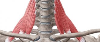
Cauda equina syndrome (CAUDA EQUINA SYNDROME) refers to damage to the bundles of nerve fibers emerging from the terminal part of the spine - the part that is a continuation of the spinal cord, starting from the upper lumbar vertebrae. This part consists of many bundles of nerve fibers gathered together and looks like a horse's tail (hence the name of the syndrome).
These bundles of nerve fibers are responsible for the motor activity and sensation of many organs, especially in the lower part of the body, as well as their proper functioning, and therefore their damage can lead to numerous consequences, such as:
- Severe and prolonged pain (neuropathic pain) in the lower extremities
- Weakness in the lower extremities
- Impaired knee and ankle reflexes
- Backache
- Pain in the buttocks, genitals and rectum
- Numbness in the groin area (loss of sensation in the inner thighs, “rider’s saddle” type)
- Incontinence
- Sexual dysfunction
Each of these symptoms, and especially the appearance of several symptoms at the same time, requires urgent treatment to avoid exacerbation and deterioration. When nerves are damaged, recovery is difficult (sometimes impossible to recover at all) and it is therefore important to get treatment as soon as possible.
Prognosis and prevention
The prognosis of the disease directly depends on the root cause and the level of spread of damage. Complex treatment allows you to reverse the neurological deficiency. Lack of treatment causes irreversible destructive changes. Preventive measures are aimed at preventing spinal injuries and accurately performing spinal surgeries.
A single registration center for MRI/CT provides a convenient service for choosing a clinic for diagnostics of the body. On the mrt-v-msk website you can find out the prices for the examination and sign up for a diagnostic service online at a convenient time. Telephone operators will answer all your questions and provide up to 1,000 rubles for diagnostics.