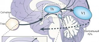
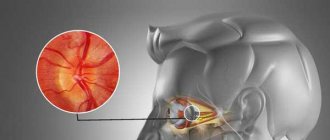
Like each of the sense organs, the eyes have a main receptor - the retina, which consists of photoreceptors: rods and cones, capable of transforming a beam of light into electrical impulses. These impulses are then transmitted to a number of intermediate nerve cells and reach the primary visual center, through which reflex reactions are provided in response to light stimulation. The ultimate goal of nerve impulses is the central office in the cerebral cortex, which is responsible for the final recognition of their characteristics, which is ensured by the complex work of the nervous system. The result of such a long journey is a real image of the surrounding world.
In other words, the visual pathway is the path of nerve impulses from the photoreceptors: rods and cones of the retina to the nerve centers localized in the human cerebral cortex.
Structure of the visual pathway
The origin of the visual pathway is in the retina. Nerve cells here are photoreceptors - rods and cones, capable of translating light signals into the format of nerve impulses through complex chemical reactions. These impulses then travel to the bipolar and ganglion cells of the retina - the second and third links of the visual pathway.
Ganglion cells have long processes - axons that collect information from the entire surface of the retina. Next, the million available axons join together to form the optic nerve.
Groups of axons of the optic nerve are strictly ordered. A special role here belongs to the so-called papillo-macular bundle, which carries signals from the macular zone of the retina. Initially, this bundle lies on the outside of the optic nerve, gradually shifting to its central part.
The optic nerve enters the skull through the optic canal, lying above the sella turcica; here the crossing of the nerve fibers of the two optic nerves occurs, forming the so-called chiasm. The chiasm is characterized by a partial decussation of nerve fibers that come from the inner halves of the retina, including part of the papillo-macular fascicle. When they exit to the opposite half, they merge with the fibers carrying information from the outer halves of the retina of the other eye, forming the visual tracts. Externally, the chiasm is limited to the internal carotid arteries. The peculiarity of the location of the chiasm and the crosshairs of the nerve fibers is the cause of the characteristic loss of halves of the visual field (external and internal), with lesions of the sella turcica or internal carotid arteries, which are commonly called bitemporal or binasal hemianopsia.
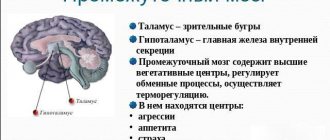
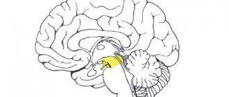
When following further, the optic tracts bypass the cerebral peduncles and end in the posterior part of the visual thalamus - the external geniculate body and the anterior quadrigeminal. The tasks of the primary visual center, in the external geniculate body, are performed by nerve cells. The primary, not yet conscious sensation of light that arises here is necessary for reflex reactions, for example, turning the head towards a sudden flash of light.
Specific groups of cells of the lateral geniculate body form visual radiance, which then carries information to the cells of the cerebral cortex. The area of the cerebral cortex responsible for vision is localized in the avian (calcarine) groove of the occipital lobe. It is here that the visual center is localized, which is involved in the final decoding of nerve impulses originating in the retina.
Brain structure
The brain represents one of the most complex and interesting mysteries science faces. There is not even a clear idea of how much it has been studied and how many discoveries remain to be made. Probably, for several decades, or even centuries, scientists will study the brain and find something new in it, hold discussions, debunk old myths and create new ones.
Our brain performs a huge number of functions. Here are just a few of them:
- Processing sensory information.
- Attention.
- Memory.
- Planning.
- Coordination.
- Formation of positive and negative emotions.
- Thinking.
Yes, the highest property of the brain is the ability to think. However, even thinking comes in several types. If you want to not only learn about this, but also learn how to think differently, take our cognitive science course.
The brain is conventionally divided according to two principles:
- The division of brain functions into hemispheres is the so-called lateralization. When they say that the brain is divided into two hemispheres, it is true, physically it is true. Both hemispheres are similar in appearance and actively interact, but are responsible for different functions and processes. It is now fashionable to argue that the theory that people with developed right hemispheres are more creative is inaccurate. However, in fact, it has clear justifications, we will see this later.
- Distribution of functions across different areas of the brain. Although it may seem that the brain works as a single whole, upon closer examination it turns out that there are so-called lobes (zones, departments), each with its own functions. The division of roles in this case can be seen very clearly.
First, let's look at our brain from the perspective of lateralization.
Right hemisphere
With the help of the right hemisphere, you can metaphorically get from point A to any other point.
It performs the following functions:
- Spatial orientation . Thanks to the right hemisphere, you navigate the area and create a map in your imagination. You can also estimate the distance to an object and become aware of your position in space.
- Processing nonverbal information . We process images and symbols and can manipulate them for our own purposes.
- Metaphors . When we hear a metaphor, the right hemisphere understands it not literally, but allegorically. However, some people have great difficulty understanding metaphors, and the reason for this is that the right hemisphere is not functioning correctly.
- Musicality . One of the abilities for which both hemispheres are responsible. However, for the most part, it is the right that performs many functions, because music is closely related to poetic imagination and the ability to compose metaphors.
- Emotions . The story here is the same as with musicality: both hemispheres are involved, but it is the right one that is more “emotional.”
- Imagination . Any fantasy essentially begins with the words “What if...”. After them, the right hemisphere begins to work to the limit, because all the skills that it possesses are required. Artists, musicians, poets and writers, while logical and consistent, must have great imagination to create a work of art.
- Dreams . When we dream about something, it is the right hemisphere that allows us to do it. Which, however, does not exclude the work of the left - the sequence of events, even in the imagination, is very important.
- Control of movements of the left half of the body . When we move our left arm or leg, it means that the right hemisphere is working. This is why ambidexterity is so popular for those involved in self-development - exercises that involve both hands. For example, writing different letters or numbers at the same time.
Left hemisphere
This is where logical, critical and scientific thinking comes into play.
The left hemisphere is responsible for our language abilities: speech, writing and reading. Also able to remember dates, names and facts. Using the left hemisphere, we can get from point A to point B. If we get to point B, most likely it will be an error in reasoning.
- Mathematical abilities . Logic, analytics, statistics, the ability to solve mathematical problems - all this is possible thanks to the left hemisphere.
- Analytical thinking . We analyze all possible facts, apply logic and draw correct or incorrect conclusions.
- Literal understanding of words . The opposite of understanding metaphors.
- Control of movements of the right half of the body . We raise our right hand, which means the left hemisphere is working.
- Sequential information processing . We process information sequentially and in stages. A missed step leads to an incorrect conclusion and an incorrect decision.
A harmonious personality has well-developed hemispheres. She knows how to dream and create incredible ideas, but at the same time she thinks logically and critically.
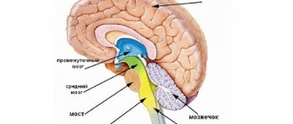
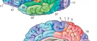
The second principle of dividing the brain is into lobes. There are four of them: occipital, parietal, temporal and frontal.
Occipital lobe
This lobe is responsible for processing visual information. In fact, we see not with our eyes, but with this part of the brain: an electrical impulse is transmitted there and forms a picture.
It should be said that when we see poorly, these are the consequences of eye disease (visual acuity), but the perception of color, shape and movement is the lot of the occipital lobe. Accordingly, if you are a brilliant artist, even if you partially lose your visual acuity, you will still remain one. The main thing is that the occipital lobe is in good condition.
Parietal lobe
The parietal lobe has a dominant and non-dominant side. The dominant one is responsible for the ability to put parts together into a whole. For example, to read you need to put letters into words, words into phrases, phrases into sentences. It's the same with numbers. It is also responsible for the sequence of related movements.
The non-dominant side provides a three-dimensional perception of the surrounding world. If a person has damage to this part of the brain, he may forget how to recognize the faces of even close people and objects.
Both sides are involved in the perception of cold, heat and pain.
Temporal lobe
Given its proximity to the hearing aid, it is not difficult to guess that the temporal lobe is responsible for auditory sensations. It is also responsible for speech, so if the temporal lobe is damaged, it can lead to speech problems. Thanks to it, you can also detect a hint of anger in a person’s voice and facial expression.
This is where the hippocampus is located, which controls long-term memory. Our memories are stored here.
Frontal lobe
Here are the centers responsible for:
- Initiative;
- Independence;
- Critical thinking.
If a person's frontal lobes atrophy, he becomes apathetic and loses interest in everything that happens. This state should not be confused with laziness.
The frontal lobe is also responsible for controlling and directing behavior. When you are in society, it works in full force and prevents the commission of antisocial acts.
Finally, the frontal lobe is responsible for planning and mastering skills. When we practice time management, we develop this part of the brain. Also, when an action is repeated repeatedly, it becomes automatic and a habit or skill is created.
Study the brain, this is important for those interested in self-development. Read the following books to start your exciting journey:
- Dick Swaab "We are the brain";
- Chris Frith "Brain and Soul";
- Theo Comnernolle "The Brain Unchained";
- David Rock "Brain" Instructions for use."
We wish you good luck!
We also recommend reading:
- Storytelling
- How metaphors change the way we think about our own experiences.
- Tony Buzan's Key Ideas
- How to get more done in 24 hours
- Neurolinguistics: Brief Description and Key Ideas
- Creativity and logic: the myth of functional asymmetry of the cerebral hemispheres
- Mental arithmetic
- “Are you out of your mind?” or 32 exercises for brain development
- Brain training: useful tips and online games
- Logic vs intuition
- Brain development: useful tips and exercises
Key words:1Cognitive science
Symptoms of visual pathway diseases
- Preservation of vision in one eye while the other is blind is observed with extensive damage to the optic nerve on the corresponding side.
- Binasal hemianopsia is damage in the outer areas of the chiasm.
- Bitemporal hemianopsia - damage to the central part of the chiasm.
- Right- or left-sided hemianopsia is damage to the optic tracts or optic radiation, respectively on the left or right.
- Loss of certain quadrants of the visual fields - damage on a certain side of half of the visual radiance.
There may be even more complex variations in visual field loss, taking into account the strict ordering of the nerve fibers along the visual pathway.
The peculiarity of damage to the visual pathway is its absolute painlessness, due to the absence of nerve endings.
