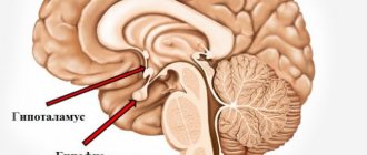
A brain lipoma is a tumor consisting of adipose tissue and is localized in the brain under the frontal cortex. The common name for lipoma is wen. The cavity of the formation is filled with a fatty substance located in the subcutaneous capsule. The size of the wen reaches several millimeters at an early stage of development and five centimeters at a late stage.
This pathology is characterized by painlessness, mobility, slow growth and a latent form of development, due to which the lipoma cannot be detected independently at the initial stage of the disease. Gender, age and body weight are not determining factors in the occurrence of intracranial wen. The ICD-10 code D17 defines a wen as a benign neoplasm. Despite this, the disease can transform into a malignant pathology.
Types of intracranial lipoma
There are a number of types of brain lipoma with characteristic features and different locations in the head.
- Interhemispheric lipoma forms in the callosal tissue of the brain. The most common form of wen is multiple. The tumor interferes with the normal functioning of the cerebellum and provokes disturbances in human behavior and cognitive functions. Wen provokes the occurrence of massive mental disorders, affecting both hemispheres of the brain. Against the background of the disorder, the appearance of hallucinations, memory disorders, and even progressive dementia is observed.
Brain lipoma
- A lipoma of the interhemispheric fissure of the brain is discovered by chance during a full profile examination of the human body, since it is secretive at the initial stage of development. Most people can live with a brain tumor for many years and be unaware of the pathology. Increasing in size, the lipoma interferes with normal blood circulation in the intracranial area, which is fraught with frequent loss of consciousness, dizziness, nausea and headaches. The mutated tissue of the wen adversely affects the central region of the brain, causing visual and auditory hallucinations, behavioral disturbances and motor dysfunction. The fatty tissue puts pressure on the central nervous system, disrupting the circulation of cerebrospinal fluid. The human brain is considered the most dangerous place to localize a wen, as it causes mental disorders and, spreading to the pituitary gland, can lead to paralysis.
- The pericallosal tumor does not affect the corpus callosum and is localized nearby. The pathology is characterized by large size and rapid growth. This type of pathology is recognized as congenital.
- A spinal cord tumor is a neoplasm that is localized in the spine and has the ability to grow deep into the spinal canal. The wen is soft and elastic to the touch. The disease is easily diagnosed by palpation. Most often the disease occurs in newborns. Spinal cord fatty tissues have three types: intradural, filum terminale and lipomyelomeningocele. The pathology of the filum terminale consists of a fatty consistency and is located in the upper region of the intergluteal fold. The main symptom of this disease is the appearance of a skin growth over the buttocks. Skin pathology does not have hair. Wen of the filum terminale is considered a rare phenomenon and occurs in only 5% of the population, mainly in children. Spinal cord adipose tissue occurs as a result of traumatic brain injury, hereditary predisposition, pathological changes in surrounding tissues and diseases of the internal secretion of glands and diabetes mellitus. The pathology can be congenital or acquired. In a child, the first symptoms appear as they grow up in the form of headaches, pelvic dysfunction, pain and compression in the area where the pathology occurs.
Spinal cord lipoma
- Lipoma of the falx cerebri, or Falx's wen, is a capsule filled with fatty fluid, which is located in the area of the frontal lobe of the skull. This type of wen causes frequent headaches, motor dysfunction and involuntary twitching of the limbs. The size of Falx lipoma can reach five centimeters in diameter. The first symptom of the presence of a falx lipoma is behavioral disturbances in a child, which is caused by the localization of the tumor.
- Lipomas of the corpus callosum of the brain spread to the subcortical formations and adjacent parts of the cerebral cortex, which complicates the timely diagnosis of primary symptoms. Location: frontal, temporal and occipital regions of the skull. Located in the frontal lobe, the tumor provokes the occurrence of apathy, lethargy, dysarthria, early pelvic disorders and bilateral motor disorders. Lipoma of the occipital lobe does not have a negative effect on the human mind, but sensory and psychosensory disorders are noticed. The fatty tissue located in the temporal lobe provokes memory impairment and impairs the quality of speech. Any type of lipoma of the corpus callosum is accompanied by the occurrence of mental disorders.
- Intracranial quadrigeminal lipoma is a congenital pathology of low density, located in the occipital region. This phenomenon occurs in adults and children. The pathology is characterized by slow growth. The first symptoms manifest themselves in impaired functioning of the respiratory organs, decreased mobility of the limbs and the occurrence of regular headaches. The localization of this pathology provokes visual impairment.
- A calcified lipoma has a capsule consisting not only of adipose tissue, but also of calcium. Pathology of this type can form necrosis of the corpus callosum.
- Interhemispheric tumor occurs as a result of underdevelopment of the corpus callosum and occurs mainly in children. Lipoma of this type is considered congenital.
Brain lipoma in section
- Fibrolipoma is a congenital pathology of the spinal cord. The disease is characterized by fatty transformation, thickening, shortening and loss of elasticity of the dorsal filament. Most often occurs in children during progressive growth and development - from 4 to 18 years. The greatest growth of the tumor occurs in the first year of a child’s life - the size can reach 24 centimeters in diameter. During a general examination of the skin, 30% of hidden skin abnormalities are detected. Symptoms of fibrolipoma include pain in the lumbar region and thoracic spine, functional disorders of the pelvic organs, the occurrence of meningitis, hair growth in the area where the pathology appears and pain in the lower extremities during physical activity.
Symptoms and causes of scalp lipoma
The origin of formations from fat cells in the brain has not been definitively established. The cause of lipomas is considered to be disorders of embryonic development. During this period, embryonic fat cells or remnants of the primitive membrane are positioned incorrectly for as yet unknown reasons. Heredity plays a certain role in this process. Unfavorable factors can presumably provoke the growth of lipoma, namely:
- exposure to toxins (from the environment, bad habits, etc.);
- inflammatory diseases of the brain;
- infections;
- metabolic disorders;
- diseases of the endocrine system;
- poor nutrition;
- head injuries;
- mental disorders;
- chronic stress;
- ionizing radiation.
For small tumors there are no symptoms. Signs appear when the tumor grows and begins to press on surrounding areas of the brain.
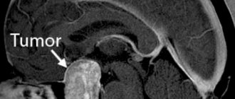
Lipoma of the corpus callosum on MRI (indicated by arrows)
Manifestations depend on the type of lipoma. Common nonspecific signs of the development of an intracranial formation from adipose tissue, which require MRI of the brain, are:
- dizziness;
- cephalgia (sudden, regular, appearing or increasing early in the morning);
- fainting, convulsive attacks;
- visual and auditory hallucinations;
- memory impairment;
- loss of ability to concentrate;
- speech disorders, etc.
A growing tumor causes increased pressure inside the skull, which manifests itself:
- movement disorders (obsessive movements, poor coordination, unsteadiness of walking, paresis and paralysis);
- convulsive attacks;
- sensitivity disorders;
- autonomic dysfunction (pressure surges, limb tremors, instability of appetite, sweating, etc.);
- psychoneurotic disorders (apathy, depression, sleep disorders, irritability, lethargy, etc.).
Symptoms of the development of a tumor from fat cells in the brain are very varied. The manifestations are influenced not only by the location and size of the formation, but also by the growth rate or the presence of other central nervous system pathologies in the patient.
Agenesis of the corpus callosum
This phenomenon often accompanies various brain defects, including lipomas. Agenesis of the corpus callosum develops in the fetus and is predominantly found in children. The disease manifests itself in the form of delayed physical and mental development of the child, convulsive seizures and paralysis. Isolated agenesis is asymptomatic and is discovered accidentally during a profile examination.
Pathology occurs at the 12th week of fetal development. In medical practice, total agenesis occurs, when the posterior part of the corpus callosum is absent, and partial agenesis, in which the anterior part of the corpus callosum is absent.
Brain pathology negatively affects the functioning of the nervous system and significantly slows down mental development in children: cases of mental retardation, idiocy, and dysfunction of the speech apparatus have been recorded.
Types and associated symptoms
The symptoms of lipoma development do not differ in specific signs and have a similar clinical picture with various pathologies of the central nervous system. For this reason, the disease is difficult to diagnose.
In the first stages, the tumor develops without any special manifestations. Sometimes people live with a lipoma in the brain for many years without complaining at all about feeling unwell.
As a rule, the patient learns about the wen by chance, as a result of a brain examination for other reasons. As the tumor grows, the condition worsens, which should not be ignored.
The neoplasm is painless, mobile, soft in consistency. Depending on the location, a distinction is made between interhemispheric and interhemispheric fissure lipoma. The tumor forms only in 2 sections of the intracranial box, rarely in the quadrigeminal cistern.
Interhemispheric
The interhemispheric wen forms in the corpus callosum in children or adults. During diagnostic procedures, a cluster of several tumors is often detected.
Patients are concerned about visual and auditory hallucinations, uncontrolled movements of the limbs, lethargy, and an increase in the production of pituitary hormones.
Interhemispheric fissure
Lipoma of the interhemispheric fissure of the brain affects certain brain centers. This pressure leads to a sharp drop in vision and hearing, sleep disturbances, constant surges in intracranial pressure, weakness, memory impairment, and severe headaches. The formation of an interhemispheric fissure can reach the size of a chicken egg.
Some experts believe that it is possible for a wen to appear in the falx cerebri. Falx lipoma provokes severe headaches localized in the frontal lobe. The person is bothered by twitching of the left limbs and a feeling of a lump in the throat.
Lipoma of the left and right frontal lobe of the brain necessarily provokes a change in mental state and a malfunction of vital organs. Due to cerebrovascular accident, there is a high risk of stroke.
Of particular concern is the addition of persistent vomiting, double vision, increased drowsiness, and disturbances of consciousness. With the above signs, it is important to exclude the presence of malignant tumors and other serious abnormalities.
General symptoms of the disease
Common symptoms of the disease by which wen can be recognized include:
- aching pain in the frontal region of the head;
- involuntary movement of limbs;
- nausea and vomiting;
- dizziness;
- compression in the temporal region of the head;
- deterioration of vision and concentration;
- decreased mental abilities;
- a sharp increase and decrease in blood pressure;
- insomnia, lethargy, apathy;
- lack of appetite and constipation.
News
The task of the radiologist who conducts and interprets the results of an MRI or CT examination is to describe everything that is visible in the images. The task of a neurologist or neurosurgeon is to explain to the patient what the study showed and, if necessary, determine treatment tactics.
However, the human brain is a notoriously mysterious organ and sometimes certain anomalies are found in it. Most often, they do not pose a threat to the health and life of the patient, but even experienced neurologists cannot always explain the nature of such findings.
There are several conditionally pathological formations in the brain, which, as a rule, do not affect the patient’s well-being and do not require special treatment.
An arachnoid cyst is a liquor cyst, the walls of which are formed by the cells of the arachnoid membrane. This is actually a bubble of fluid that has accumulated between the adhered layers of the meninges. Such cysts remain after inflammation of the membranes of the brain, hemorrhages or injuries. They usually turn out to be random finds. A study with intravenous contrast helps to distinguish a cyst from a tumor: a tumor accumulates contrast, but a cyst does not. However, if the fluid pressure in the cyst is higher than the general intracranial pressure, it can compress the cerebral cortex and cause unpleasant symptoms. Most of the bones found do not require treatment. If the examination shows that the size of the cyst is changing, and there are symptoms associated with the presence of the cyst, then treatment is prescribed for the disease that causes the formation of the cyst.
Rathke's pouch cyst.
Rathke's pouch is an anatomical structure that eventually forms the anterior and posterior pituitary glands, as well as the pituitary infundibulum. This pouch usually disappears early in fetal development, but some cells in the pouch often remain as a small cavity within the pituitary gland. Sometimes this condition can lead to the formation of a large cyst called a Rathke's pouch cyst. This is a very rare type of formation and occurs in less than 1 percent of cases. However, such a bone must still be distinguished from other visually similar formations (meningeal cyst, cystic pituitary adenoma or craniopharyngioma). This can be done by injecting a contrast agent (the cyst does not accumulate it). A Rathke's pouch cyst usually causes no symptoms and does not require treatment. However, in some cases it can lead to blurred vision, symptoms of pituitary dysfunction and headaches.
Septum pellucida cyst.
This is a type of arachnoid cyst located between the lateral ventricles of the brain. The septum pellucidum is a medulla consisting of two very thin plates. They are separated by a slit-like cavity, which is located between the corpus callosum and the anterior part of the brain. Actually, a cyst of the transparent septum of the brain is an accumulation of fluid between the plates that we mentioned. Modern medicine does not classify a cyst of the transparent septum as a pathological abnormality and considers it an anomaly and does not pose any threat to the patient’s life. There is a variant of the anatomical structure of the brain. When a cyst is the result of a previous injury or disease rather than a congenital condition, it can cause pain, noise in the head, a feeling of tightness and hearing loss. In this case, a dynamic MRI study and, if necessary, neurosurgical treatment are necessary, because the uncontrolled growth of such a cyst can provoke a disruption in the functioning of the brain.
Pituitary microadenoma.
This is a benign brain tumor measuring less than 1 cm and is seen quite commonly by radiologists. Small hormonally inactive adenomas are usually asymptomatic and therefore are not diagnosed or are diagnosed by chance. The main cause of pituitary adenoma has not yet been clarified. But there are many proven factors that cause the disease. For example, a pituitary adenoma can develop against the background of uncompensated hypothyroidism. A serious danger arises if the pituitary adenoma enlarges, because it begins to compress the tissues adjacent to the pituitary gland, which can lead to severe headaches, impaired coordination of movements, and various visual disturbances. If the pituitary adenoma has reached an alarming size, caused compression of the optic nerves and causes serious clinical manifestations, surgical treatment is indicated.
Calcification of the falx is the formation of salt deposits in the area of the interhemispheric fissure, because salts are deposited not only in the kidneys or joints, but also in the brain. The process is benign, intracranial calcifications are physiological, unaccompanied by any signs of disease and without obvious pathological causes. However, basal ganglia calcification in individuals younger than 30 years of age may be associated with underlying metabolic disorders such as hyper- or hypoparathyroidism, congenital disorders, and infections. Therefore, the presence of basal ganglia calcification in such patients should prompt a thorough clinical evaluation.
A pineal gland cyst is a benign formation, the inside is lined with collagen fibers, glial cells and normal gland cells. The cyst forms in the area of the pineal gland, that is, between the two main hemispheres of the brain. This gland is directly connected to the endocrine system and is responsible for the production of the hormone melatonin, the main task of which is to ensure normal quality sleep. Visually, a pineal gland cyst resembles a sac. In most cases, such cysts do not cause any particular concern and do not require treatment. But if there are prerequisites for the development of hydrocephalus or other forms of disease progression, it is possible to use a surgical method to prevent the patient’s health from deteriorating.
Lipoma of the interhemispheric fissure is a benign formation from fat cells in the area of the interhemispheric fissure (clinical manifestations are possible if the size of the lipoma increases). Small sizes, only dynamic observation is recommended.
Lateroventriculoasymmetry is asymmetry of the lateral ventricles. Mild asymmetry (difference in the sizes of the right and left lateral ventricles at the level of the foramen of Monroe) within the age norm is observed quite often and is not a sign of pathology. But pronounced asymmetry may indicate the presence of various diseases: increased intracranial pressure, a brain tumor, anything that can deform the ventricle of the brain from the outside or inside and increase its size.
Similar articles:
News → Lie or truth? Functional MRI or lie detector (polygraph)?
News → Blue color stimulates brain function.
Glossary → Acromegaly (Marie-Léry syndrome)
News → The human brain on LSD was studied for the first time using MRI
Articles → Brain abscess
Discuss on the forum
Causes
The factors influencing the occurrence of wen have not yet been fully elucidated, but probable causes include:
- heredity;
- bad habits, especially abuse of alcoholic beverages and tobacco products, as they have a direct effect on the brain;
- infectious diseases of the central nervous system increase the risk of tumor development;
- foci of inflammatory processes located in the brain;
- polluted environment;
- unhealthy diet, especially abuse of fatty, sweet and spicy foods;
- physical and chemical effects on the brain;
- presence of mental illness, especially schizophrenia;
- stress, lack of sleep, nervous tension.
Causes of lipomas
Experts identify a set of prerequisites that can provoke the formation of lipomas. Among the most common:
- genetic factors, hereditary predisposition;
- disruption of metabolic processes in the body and fatty tissues;
- insufficient level of personal hygiene;
- malfunctions of the thyroid and pancreas;
- chronic pathologies that significantly affect the body’s immune forces (diabetes, hepatitis, HIV, etc.);
- dependence on alcohol, tobacco, drugs;
- poor nutrition;
- obesity, excess fatty tissue;
- injuries, damage;
- sedentary lifestyle, lack of physical activity.
Despite the fact that lipoma is a benign formation, there is always a risk of developing its malignant form. This probability is especially high for people who are included in the relevant risk groups: they have precancerous diseases (polyps, dysplasia), have already suffered from oncological pathologies, have a hereditary predisposition to the formation of tumors, and are under the constant influence of carcinogens, radiation and other harmful environmental factors.
Diagnostics
Wen in the brain at an early stage of development is difficult to detect due to the lack of symptoms. Cases have been recorded where people lived with a tumor for decades and were not aware of the existence of the phenomenon. The pathology is discovered accidentally during a profile examination of the patient. You should not wait for the first symptoms of a tumor, because modern methods for diagnosing such pathologies are known.
CT scan
CT is an informative and safe examination of the intracranial area. The method allows you to make an accurate diagnosis and replaces ultrasound or radiography, or saves time and makes it possible to find out the results immediately. Using computed tomography, the tumor is visualized in detail, down to the vessels and nerve endings.
Neurosonography
Ultrasound examination of the brain, or neurosonography, is actively used in pediatrics and helps to obtain information about the location, size and degree of tumor growth within the cranial space. Already in the first month of life, a newborn should be examined for the presence of congenital tumors. With the help of neurosonography, even a small tumor at an early stage of development can be detected. It is advisable to undergo examination for children born with low body weight, birth injuries, developmental pathologies and intrauterine infection.
MRI
Magnetic resonance imaging, or MRI, provides accurate information about the extent of tumor development. Diagnosis using this method is much more effective than CT or ultrasound, since MRI makes it possible to obtain a three-dimensional image of a lipoma, which is a more detailed image. The wen has a sharply defined shape in the resulting images. The main sign of lipoma detection is the high signal intensity of adipose tissue when pathology is detected.
Methods for diagnosing lipoma
The clinical picture of lipomas is quite typical. As a rule, this is a palpable formation in the subcutaneous tissue that raises the skin. The tumor has a soft consistency, lobular structure, is easily displaced and is not connected to the skin. Pain and increased local temperature are always absent.
Diagnostic methods include inspection and palpation.
For an experienced surgeon, making a diagnosis is not difficult.
Additional diagnostic methods
Ultrasound examination of soft tissues and MRI of soft tissues of the organ where the tumor is localized have additional diagnostic capabilities.
Sign up for diagnostics To accurately diagnose the disease, make an appointment with specialists from the Family Doctor network.
Treatment
Most often, patients seek medical help when the lipoma needs to be removed. This is due to the absence of symptoms at an early stage or medical wait-and-see tactics. Doctors do not recommend treating at the initial stage of development, so as not to provoke changes or growth of pathology, and observe the patient for a long time until the first symptoms appear. Increasing in size, the wen brings discomfort and pain, and then the question arises of removing the tumor.
Surgical intervention
The fatty tissue poses a threat to the life and mental health of a person, increasing to a substantial size, so surgeons carry out radical removal along with the capsule to avoid further relapses. During the operation, craniotomy is performed, that is, part of the skull is removed.
Brain surgery
Minimally invasive intervention
This method includes endoscopic tumor neutralization, which is carried out using an endoscope and a video camera. The doctor removes the fat along with the capsule using this method. Minimally invasive intervention also includes the puncture-aspiration method of removal.
During the procedure, a hole is made in the patient's skull through which the operation takes place. A tube is inserted into the hole and the pathological fatty tissue is removed using an electric suction. The only drawback of the operation is that the wen capsule remains in the human brain and this causes the recurrence of lipomatosis.
Drug therapy
Proponents of the medicinal method are confident that the brain wen can be removed without surgery. To do this, the doctor prescribes medications that help reduce the size of the lipoma. Most often, hormonal treatment is used, since hormone imbalance is one of the reasons for the appearance of wen, according to theoretical assumptions. Therapy gives results at the initial stage of development, but at the same time it turns out to be ineffective when the wen grows rapidly. To eliminate the wen, non-traditional methods are actively used: people are sure that herbal decoctions and compresses cause complete resorption of the tumor, but often folk remedies are ineffective. Treatment should be carried out comprehensively and only as prescribed by a doctor.
Radio wave method for removing wen
A bloodless and painless method to remove subcutaneous lipoma using high frequency radio waves. It has advantages over laser lipoma removal in terms of less trauma and postoperative complications. The operation is performed under local anesthesia and is therefore completely painless. After the operation, the patient can go home immediately.
A relative disadvantage can be considered the fact that each wen is removed through a separate incision, as with the classical method. But unlike conventional excision, there is less traumatic effect on the tissue, which eliminates infection, reduces bleeding and the risks of postoperative hematomas, and most importantly, reduces the likelihood of the formation of rough scars and scars. The cosmetic effect is better than with classical excision. Postoperative scars after complete healing are almost invisible .
But in each specific case, predicting the reaction of the skin to any mechanical impact - an incision, be it with a scalpel, or a laser or a radio wave, is quite difficult. Even with the best and most modern suture material and threads that can dissolve on their own, the skin can react unpredictably with the formation of a hypertrophic scar up to a keloid.
But medicine does not stand still. What can modern surgery offer?
A technique that is much more effective than laser and radio waves.