How to recognize SMA
Muscle atrophy in children diagnosed with SMA (spinal muscular atrophy) can begin at different times.
The most severe form of muscle atrophy in newborns appears in the first six months of life. The baby is lethargic and sucks poorly. At 3-4 months the child does not roll over on his own and does not attempt to crawl. With spinal muscular atrophy, the baby's posture resembles a “frog”. There is a type of disease that appears after 7-18 months. Muscle atrophy leads to regression of acquired skills in the child. The baby, who was crawling and starting to get up, suddenly becomes inactive. Over time, he stops sitting up straight. With spinal atrophy, reflexes from the upper and lower extremities disappear.
Symptoms of muscle atrophy with spinal muscular atrophy may appear closer to two years of age. Patients have already mastered the skills of standing and walking. At the same time, muscle atrophy confines them to a wheelchair. Intelligence, urination and defecation functions are preserved. SMA after 2 years is the mildest type of the disease.
Muscle atrophy (spinal muscular atrophy) has the following symptoms:
- Appears in infancy or childhood;
- Accompanied by disturbances in walking, running, and standing;
- Tremors and fasciculations (twitching) are detected;
- The “rollback” of motor skills is determined;
- With spinal atrophy, no impairment of intelligence or autonomic functions is detected.
If a child has these signs, he should be consulted with specialists. The diagnosis of SMA is made based on DNA testing.
Characteristics of the pathology
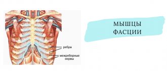
Spinal muscular atrophy, or SMA, involves damage to the neurons in the spinal cord that control muscle movement. The muscles of the legs and neck usually suffer the most, but the muscles of the upper extremities are less often affected. Patients have problems with movement, swallowing, and holding the head, but sensitivity is preserved and there are no delays in mental development. But if with other forms of SMA patients have a chance to live to old age, albeit with disability, then with Werdnig-Hoffmann amyotrophy the maximum life expectancy does not exceed 30 years.
Damage to neurons in the spinal cord leads to severe dysfunction of skeletal muscles
The pathology is quite rare - in one case out of 80-100 thousand. But there are much more carriers of the gene responsible for the development of the anomaly. The disease is inherited in a recessive autosomal manner, and for a child to exhibit Werdnig-Hoffmann amyotrophy, both parents must be carriers of the gene. Although in this case the probability of illness in the baby is only 25%. The cause of the disease is solely a genetic predisposition; no connection between SMA and injuries, infections and other factors has been found.
Causes of development of Werdnig-Hoffmann amyotrophy
WERDNIG-HOFFMANN SPINAL AMIOTROPHY.
Inherited in an autosomal recessive manner. Underdevelopment of the cells of the anterior horns of the spinal cord, demyelination of the anterior roots, and similar changes in the motor nuclei and roots of the Y, YI, YII, IX, X, XI, XII cranial nerves are detected. In skeletal muscles, neurogenic changes are characterized by “bundle atrophy,” an alternation of atrophied and preserved bundles of muscle fibers.
CLINIC
There are three forms of the disease:
- congenital;
- early childhood;
- late childhood
.
In congenital form
children are born with flaccid paresis. From the first days of life, generalized muscle hypotonia and decreased or absent tendon reflexes are evident. Bulbar disorders are detected early, manifested by sluggish sucking, weak cry, tongue fibrillations, and decreased pharyngeal reflex. The disease is combined with osteoarticular deformities: scoliosis, funnel chest, joint contractures. The development of static and locomotor functions is sharply slowed down. Reduced intelligence. Congenital malformations are often observed: congenital hydrocephalus, cryptorchidism, hemangioma, hip dysplasia, clubfoot, etc.
The course is rapidly progressive and malignant. Death occurs before the age of 9 years. One of the main causes of death are severe somatic disorders (heart and respiratory failure), caused by weakness of the chest muscles and a decrease in its participation in the physiology of breathing.
With early childhood form
The first signs of the disease appear in the second half of life. The disease develops subacutely, often after infection or food intoxication. Flaccid paresis is initially localized in the legs, quickly spreading to the muscles of the trunk and arms. Diffuse muscle atrophy is combined with fasciculations, fibrillations of the tongue, fine tremor of the fingers, and tendon contractures. Muscle tone and tendon reflexes decrease. In the later stages, generalized muscle hypotonia and symptoms of bulbar palsy occur.
The course is malignant, death occurs by 14–15 years of age.
In late form
signs of the disease appear at 1.5 - 2.5 years. The disease begins unnoticed. Movements become awkward and uncertain. Children often trip and fall. The gait changes - they walk with their legs bent at the knees (the gait of a “wind-up doll”). Flaccid paresis is initially localized in the proximal muscle groups of the legs, then relatively slowly moves to the proximal muscle groups of the arms and trunk muscles; muscle atrophy is usually subtle due to the well-developed subcutaneous fat layer. Fasciculations, fibrillations of the tongue, fine tremor of the fingers, bulbar symptoms - fibrillations and atrophy of the tongue, decreased pharyngeal and palatal reflexes are typical. Tendon reflexes fade in the early stages of the disease. Osteoarticular deformities develop parallel to the underlying disease. The most pronounced deformation of the chest.
The course is malignant, but milder. Patients live up to 20 - 30 years.
Diagnostics.
Autosomal recessive type of inheritance, early onset, the presence of diffuse atrophies with predominant localization in the proximal muscle groups, generalized muscle hypotonia, fasciculations, fibrillations of the tongue, absence of pseudohypertrophies, progressive, malignant course, electromyography data and morphology of skeletal muscles, revealing the denervation nature of the changes.
Spinal muscular atrophy (amyotrophy) Werdnig-Hoffmann is a hereditary malignant disease, the onset of development of which occurs from birth to 1-1.5 years. This is one of the most severe forms of muscle atrophy. There is a diffuse increase in muscle atrophy throughout the body. The child loses the ability to sit and move independently, and paresis progresses.
The disease was first described by scientists Werdnig and Goffman. They proved the morphological essence of spinal amyotrophy. But they assumed the existence of only one form of the disease. Later, other scientists Welander and Kueckelberg described another form of spinal muscle atrophy. All variants of the disease have the same genetic nature. Today there are no methods that can completely cure this pathology. Therapeutic measures are aimed at improving the trophism of muscles and nervous tissue.
Symptoms of pathology
The congenital type of the disease (SMA I) begins to appear before the baby is six months old. Before this, such a child may move sluggishly. It is not so rare that spinal amyotrophy in a baby can be noticed at the very beginning of the postembryonic period of his life - his deep reflexes fade away:
- The baby's cry is not loud enough;
- it is difficult for him to suck;
- he can't hold his head up.
This pathology can be detected in a baby from the first days of life.
If this becomes noticeable later, which rarely happens, the baby can learn to hold his head, or even sit, but pathology quickly reduces such skills to zero. The following is also typical:
- early speech problems;
- worsening of the pharyngeal reflex;
- fascicular twitching of the tongue.
This type of pathology can be associated with oligophrenia, as well as pathologies of skeletal development:
- deformities of the chest (funnel-shaped/keeled shape);
- curvature of the spine (scoliosis);
- joint contractures.
Other congenital diseases are also common. For example:
- hemangiomas;
- hydrocephalus;
- clubfoot;
- hip dysplasia;
- cryptorchidism.
SMA I is the most “harmful”: it is accompanied by developing paralysis, as well as paresis of the muscles responsible for breathing. Because of the latter, respiratory failure develops, due to which the patient may even die. And impaired swallowing can cause food to enter the respiratory tract and develop aspiration pneumonia. It can also lead to death.
SMA II begins to appear after the baby is six months old. At this age, the baby is already satisfactorily developed, he can stand, hold his head, sit down, and roll over. However, almost never a sick baby does not have time to master walking skills. In most cases, this disease manifests itself after the baby suffers some acute infectious pathology. For example, food poisoning.
When SMA II just begins to manifest itself, the baby develops peripheral paresis in the legs, which then quickly appears in the arms and torso. Diffuse muscle hypotonia appears, deep reflexes begin to disappear. You can also notice the following:
- tendon contractures;
- tremor of fingers;
- fasciculations (involuntary twitching) of the tongue.
Important! Later, bulbar syndrome begins to appear and respiratory failure develops. Due to the slow development of this type of SMA, compared with the congenital form, a sick person usually lives up to fifteen years.
SMA III , also known as Kugelberg-Welander amyotrophy, is the least harmful type of spinal amyotrophy. It begins to appear when the child turns two years old. Sometimes it can be asymptomatic even up to 30 years. However, in this case the patient is no longer a child, which, however, does not make him healthier. With SMA III, the development of the psyche is not delayed; the patient can move for a long time without outside help. He even has a chance to be able to take care of himself in his old age.
SMA IV , also known as the adult form of spinal amyotrophy, is a slowly developing pathology. It usually begins after a person turns 35 years old. This form, if it shortens life, does so only slightly. On the other hand, the patient exhibits the following picture:
- weak proximal muscles;
- fasciculations;
- deterioration of tendon reflexes;
- loss of walking ability.
The third type of pathology can be detected much later - it happens that the disease does not manifest itself for decades
The electromyogram shows the “picket fence rhythm”—spontaneous rhythmic activity. This way you can identify pathology of the anterior spinal horns. If you conduct a morphological study of muscle biopsies, you can notice atro- and hypertrophied fibers of the first and second types. Small round fibers also accumulate, which alternate with hypertrophied fibers - this is “bundle” atrophy. If a pathomorphological study is carried out, swelling/shrinking/atrophy of the motor neurons of the spinal anterior horns becomes noticeable, and quite often also the nuclei of the nerves that exit the brain.
Treatment
The main goal of research aimed at treating spinal muscular amyotrophy is related to increasing the level of SMN protein. Currently, medications are being tested, and official Russian medicine does not use them.
Treatment today includes medications that improve the passage of nerve impulses. Nootropic drugs are prescribed, the main task of which is to improve brain function. Dietary supplements are prescribed to improve metabolism. Vitamin therapy is indicated, in particular, taking B vitamins.
Drugs affecting neuromuscular conduction:
- Alpha lipoic acid
- Acetyl L-carnitine
- Alpha glycerophosphocholine
Vitamins and vitamin complexes:
- Thiamine (B-1)
- Pyridoxine (B-6)
- B-complex
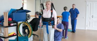
Important treatment methods are massage, physiotherapy, and neuromuscular stimulation. Exercise therapy is prescribed. Physical exercise helps maintain strength, on the other hand, doing it in society, going to the pool helps to socialize and communicate with other people.
Patients with SMA are advised to follow a diet. Food is a source of substances needed by muscles. Thus, the necessary amino acids are found in grains, meat, fish, mushrooms, nuts, and dairy products. Recommended dishes made from oats, wheat and brown rice.
We advise you to study - Lumboischialgia: what is it?
Spinach, broccoli, herring, onions, grapefruit, and watermelon will help to naturally maintain and grow muscles. To increase testosterone, men are recommended to take dill, parsnips, ginseng, and parsley.
Treatment methods
Treatment of spinal amyotrophy is symptomatic and aimed at stabilizing the patient's condition.
Medicines prescribed :
- improving metabolism - cerebrolysin, lipocerebin, aminalon;
- affecting the trophism of muscle tissue - potassium orotate, glutamic acid, methionine, tocopherol acetate;
- promoting neuromuscular conduction - prozerin, galantamine, dibazol;
- stimulating blood circulation in capillaries - complamin, nicotinic acid;
- supporting the viability of motor neurons - valproic acid, riluzole, L-carnitine.
Article on the topic: Breast fibroids
Patients are prescribed orthopedic procedures in combination with warm baths, therapeutic exercises, gentle massage, oxygen therapy, and sulfide baths are indicated.
Treatment
A practical patient's guide to the legal basis for providing medical care to patients with SMA. This directory contains articles and materials on the legal basis for providing medical care and drug provision to patients, requirements for the preparation of medical documentation and its forms, answers to the most frequently asked questions. Foundation "Families of SMA"
Intellect in SMA disease remains completely intact; it develops in the same way as in healthy people. Thus, despite physical limitations, children can live a full life: communicate, play, do physical therapy, and go for walks.
At the moment the disease is incurable. Specialists from different countries are working on drugs to treat SMA, but so far there are no publicly available drugs that can completely cure the disease.
At the end of 2021, the first drug for the treatment of SMA, Spinraza (Nusinersen), appeared in the world. This medicine can slow the progression of the disease, and in some cases improve the condition, but still does not cure the disease completely. At the moment, Spinraza has not yet been registered in Russia - this is a matter for the near future.
In addition, several innovative drugs for the treatment of SMA are currently in development and are at the stage of clinical trials. Detailed and up-to-date information about this can be found on the website of the SMA Families Foundation in the “Research” section.
Nevertheless, we can already do a lot, and with the help of symptomatic therapy and a variety of supportive techniques, slow down the progression of the disease and, in some cases, prevent the development of complications.
Helping organizations
The SMA Families Charitable Foundation is the only organization in Russia that specializes in helping families dealing with spinal muscular atrophy. It provides charitable, informational and psychological support to families, and advises specialists on the disease and methods of working with patients with SMA.
Hospice Assistance Fund "Vera". Charitable and advisory assistance to families with terminally ill children and terminally ill adults.
CSCH No. 1 Department of palliative care for children, Yekaterinburg, Sverdlovsk region. Medical, informational, social and psychological assistance is provided to families raising a disabled child with a palliative condition.
“Research Clinical Institute of Pediatrics named after Academician Yu.E. Veltishchev" FSBEI AT RNRMU named after N.I. Pirogov. The institute is located in Moscow. Residents throughout Russia can seek medical help for children with SMA and other neuromuscular diseases.
Clinic "Chaika". Consultations with pulmonologist Vasily Andreevich Shtabnitsky for children and adults with SMA.
Children's hospice “House with a Lighthouse” (Moscow, near Moscow region). Medical, psychological, legal, social, and charitable assistance to families with terminally ill children and young adults (up to 25 years of age).
Marfo-Mariinsky Medical Center (Moscow). Medical, psychological, legal assistance, nanny assistance, events, spiritual support, charitable assistance to families with terminally ill children.
You will find more detailed information on the website of the SMA Families charity foundation and their special project about life with spinal muscular atrophy
The special project is intended for those who have been diagnosed with SMA and would like to know all the most important things about this disease: which specialists and where to go, how to care, what to watch for, what to remember, what therapy exists today.
To study Back massager on a chair
Diet for spinal amyotrophy
At this time, no diet has been proven to be beneficial for SMA.
According to a large number of parents, a diet that includes a lot of protein or special food additives can increase the strength of the child's muscles. But, despite the obvious need for good nutrition for a sick child, it has not yet been proven that he needs a specific diet. Moreover, some products can even harm his body.
For example, the amino acid menu is sometimes fraught with even greater problems for those children who have too little muscle tissue in their bodies. According to some experts, if there is a lack of muscle tissue, it cannot properly process amino acids and then their level in the blood rises too much.
Doctors have not proven that any diet will improve the condition of a patient with SMA, but proper nutrition can make his life easier.
Some children find it healthier to eat small amounts, more often than three or four times a day. You just need to divide for the patient the entire amount of food taken by a healthy peer of the patient per day into several parts.
Principles of treatment of spinal amyotrophy
Unfortunately, this is an incurable hereditary disease. At the present stage, research is being conducted that may help regulate the synthesis of the SMN protein, but there are no results yet.
The following help alleviate the condition of patients with spinal amyotrophy:
- periodic course intake of drugs that improve the metabolism of nervous tissue and muscles (Cerebrolysin, Cytoflavin, Glutamic acid, ATP, Carnitine chloride, Methionine, Potassium orotate, Tocopherol acetate, etc.);
- B vitamins (Milgamma, Neurovitan, Combilipen);
- anabolic steroids (Retabolil, Nerobol);
- agents that improve neuromuscular conduction (Proserin, Neuromidin, Galantamine, Dibazol);
- massage and physical therapy courses;
- physiotherapy (electrical muscle stimulation, carbon sulfide baths);
- methods of orthopedic correction (with the development of joint contractures and spinal deformities).
Werdnig-Hoffmann spinal amyotrophy, like other forms of this disease, is a pathology that is inherited. The appearance of the disease in a child is explained by the presence of a mutant gene in both the mother and father. The disease is characterized mainly by muscle weakness, which causes immobility and respiratory problems. The disease is currently incurable.
Muscle atrophy treatment and prevention
With reduced tone of the swallowing muscles, nutrition is administered through a vein.
Treatment of Werdnig-Hoffmann spinal amyotrophy is symptomatic. It is impossible to avoid the development of the disease or significantly slow down the growing disorders, but it is possible to relieve the severity of symptoms. Therapy is carried out in three main ways - drug treatment, physical therapy and devices that make the life of sick children and their relatives easier.
Medications used for spinal muscular atrophy type 1 include nootropic drugs and neurometabolites - they improve the transmission of impulses at neuromuscular synapses. If the swallowing reflex is impaired, the only possible route of drug administration is injection.
Therapeutic exercise and massage help increase muscle tone, improve blood circulation and slow down the process of atrophy. Rapidly progressing spinal amyotrophy in Werdnig-Hoffman disease does not allow children to perform active movements. Gymnastics is based only on passive actions - they are carried out by parents.
In the absence of spontaneous breathing, ventilators are used
In the later stages of the disease, patients' lives are greatly facilitated by portable ventilators. This allows children to be at home in a familiar environment. In patients with milder forms, which appear after six months or during adolescence, automated wheelchairs are used.
Since the cause of spinal amyotrophy is a genetic defect, the disease can be prevented in only one way - to prevent the birth of children who have been diagnosed with the pathology. Genetic counseling plays an important role. Patients with the mildest form of genetically determined neuronal death (type 3, Kugelberg-Walander disease) survive to reproductive age and have a chance of having children with more severe forms than themselves.
A common problem with all diseases that can be diagnosed before birth is that not all parents accept termination of pregnancy, even for medical reasons. Additionally, testing for spinal muscular atrophy type 1 in children is not mandatory. Most of the diagnoses of this disease are made after the birth of the child, and parents are faced with the sad fact that the child suffers from an incurable disease.
Diagnostics
Manifestations of proximal spinal amyotrophy often resemble the course of other neurological and congenital diseases, as well as traumatic injuries to the structures of the spinal cord and brain. Diagnosis of this disease is especially difficult in newborns and young children.
The key points in diagnosing spinal amyotrophy are the following studies:
- Careful history taking. The presence of cases of spinal amyotrophy in relatives allows us to suspect this hereditary disease.
- Electroneuromyography is a special study of the neuromuscular system. In this case, primary muscle damage is excluded and signs indicating pathology of the motor neurons of the spinal cord are identified.
- Computed and magnetic resonance imaging. These methods sometimes make it possible to detect atrophic changes in the anterior horns of the spinal cord. However, more often they are used to exclude other pathologies from the structures of the spinal column and brain.
- Muscle biopsy followed by histological examination of the biopsy sample. Specific muscle changes are revealed, consisting in the alternation of bundled atrophic and unchanged muscle fibers. In addition, compensatory hypertrophied muscle areas can be detected, as well as replacement of muscle tissue with connective tissue.
- Genetic analysis. Allows you to identify the exact cause of the disease: DNA testing reveals a gene mutation on the fifth chromosome.
If there have been cases of the birth of children with spinal amyotrophy in the family, when planning a subsequent pregnancy, the married couple is sent for consultation with a geneticist. Prenatal fetal DNA testing is also mandatory. Detection of Werdnig-Hoffman syndrome at the stage of prenatal diagnosis serves as an indication for termination of pregnancy.
How is the diagnosis made?
To detect the disease in utero, amniotic fluid is collected.
Werdnig-Hoffmann spinal muscular atrophy can be detected before or after the birth of the child. Prenatal diagnosis is based on identifying a genetic defect. This requires obtaining the baby's genetic material from umbilical cord blood, amniotic fluid or chorionic villi. Invasive diagnostic methods are dangerous for the development of pregnancy, so parents often refuse them. If pathology is detected, this is an indication for abortion.
The birth of children with Werdnig-Hoffmann spinal muscular atrophy (SMA) is possible for several reasons. Not all hospitals have the opportunity for prenatal diagnosis of the disease and genetic counseling for parents before conceiving a baby. Parents carry out the analysis mainly at their own request and may not know until the moment of birth that the baby will be seriously ill. Sometimes the diagnosis is made at a late stage, when it is too late to terminate the pregnancy. The fundamental opposition to abortion among some parents also plays a role - even knowing about the future pathology, they decide to give birth.
After birth, the diagnosis is made by a neonatologist or pediatric neurologist. The neurological status of a small patient is important - impaired motor activity and extinction of reflexes while maintaining sensitivity. To make a final diagnosis, a DNA test is prescribed. MRI and CT scans of the spine are important to distinguish SMA from other pathologies. Disturbances in the motor nuclei of the spinal cord are not visualized.
Spinal amyotrophy of Werdnig-Hoffmann - is this disease curable?
Spinal amyotrophy Werdnig-Hoffmann is a hereditary pathology of the nerves that affects the part of them that controls the skeletal muscles. It is characterized by weakness of all muscles in the body at once. More than half of the nerve cells that control muscles are located in the spinal cord.
That is why the disease is called “spinal”. It is also called “muscular” because of the detrimental effect on the muscles: they do not receive any signals from these same nerves. "Atrophy" is a medical term that refers to the wasting or shrinking of something that is not used. Here it concerns inactive muscles.
A person with such a disease has no ability to sit, move, or even take care of himself. There is no cure for this. By conducting prenatal diagnostics, you can prevent the birth of a baby with this disease.
Here we will talk about how this pathology is inherited, what its manifestations are, and also about how you can help a sick person.
We advise you to study - Idiopathic scoliosis at a young age
Spinal amyotrophy Werdnig-Hoffmann is named after the two scientists who first described it. In the second half of the 19th century, they proved the morphological essence of the disease. At first, both scientists believed that this pathology had only one form.
But already in the 20th century, scientists Kueckelberg and Welander discovered another clinical form of it, with a genetic cause similar to that discovered by Werdnig and Hoffman. Several clinical forms of spinal amyotrophy are now known.
They are united by a common hereditary defect.
This pathology is hereditary. It is based on a mutation on chromosome 5. The gene that produces the SMN protein is mutated. This protein is necessary for motor neurons to develop exactly as needed.
If the fifth chromosome mutates, it will negatively affect motor neurons, interfering with their development, or even completely destroying them. As a result, the muscle cannot receive control signals from the nerves, and therefore cannot function.
It turns out that not a single movement associated with it is performed.
Advice
The mutated gene has an autosomal recessive mode of inheritance. The phrase is deciphered as follows: for the development of spinal amyotrophy, it is necessary that both parents have a mutant gene.
To put it simply, the disease will not develop if at least one of the parents was not a carrier of the mutated gene. At the same time, they themselves do not get sick: people have paired genes, and the healthy gene dominates in the father and mother of the child.
In this case, a sick baby is born in about a quarter of cases. Scientists estimate that about 2% of living people are carriers of a gene with such a mutation.
Classification
Three types of this pathology are known.
- The most severe, manifesting itself earlier than others.
- Medium-heavy.
- The mildest, manifesting itself at a very late age.
According to some doctors, there is another type: moderate/mild SMA, which manifests itself in an adult.
It is worth noting that in addition to Werdnig-Hoffmann spinal amyotrophy, there are other types of SMA, which differ in symptoms and types of inheritance. They are listed in the table below.
| SMAX1 | X-linked recessive | It is observed mainly in the elderly, affects the bulbar nerves of the skull, causing descending paralysis. |
| SMAХ2 | X - clutch. recessive | Congenital aggressive form, leading to death before 3 months. Causes weakness, areflexia, contractures and fractures. |
| SMAX3 | X - clutch. recessive | It mainly affects boys. Atrophy of all distal muscles. Slow increase in symptoms. |
| Distal DCMA1 | Autosomal - recessive | Congenital, mainly affects the hands, and severe respiratory disorders are possible. |
| Distal forms DCMA2 - DCMA5 | Autosomal - recessive | All four forms are characterized by slow progression; DCMA5 is diagnosed in young people. |
| Juvenile SMA (HMN1 type) | Autosomal dominant | Occurs in youth |
| Congenital spinal amyotrophy | Autosomal dominant |
What is this?
Photo 1. Werdnig-Hoffmann amyotrophy in a 5-year-old boy
Spinal amyotrophy is a genetic disease in which mutations are detected on chromosome 5 in the SMN1 and SMN2 genes . These genes are responsible for the preservation and normal functioning of motor neurons - nerves that send impulses to skeletal muscles. As a result of lack of muscle stimulation, they atrophy. Patients experience degenerative processes in the spinal cord: motor neurons are destroyed, the functioning of the anterior horns of the spinal cord is disrupted. In the brain, changes occur in the motor nuclei.
In Werdnig muscular amyotrophy, the SMN1 gene is mutated , which prevents the death of motor neurons. The SMN2 gene performs this function only partially and its capabilities are quickly depleted. The course and severity of the pathology depends on the location and extent of the lesion in the gene apparatus.
Photo 2
Spinal amyotrophy, regardless of its type and form, is a genetic congenital disease . Often, the first signs of pathology are detected in infancy or during pregnancy of the child’s mother.
Equally common in both men and women. The main condition for its development is the presence of a defective gene in both parents, who can be healthy and are only carriers of the defective chromosome.
As a result of disruption of the conduction of nerve impulses from motor neurons to muscles, atrophy of the muscles of the lower and upper extremities, diaphragm, gastrointestinal tract, heart and others occurs. Against this background, bones and joints are deformed. The most dangerous manifestation is disturbances in respiratory function and heart function .
Depending on the severity of the course, localization of defects and clinical manifestations, 4 forms of amyotrophy are distinguished . Patients with any form of amyotrophy are disabled. They are not able to care for themselves and require constant care and medical supervision.
What is SMA?
SMA (spinal muscular atrophy) is a genetic neuromuscular disease that affects the motor neurons of the spinal cord and leads to increasing muscle weakness. The disease is progressive, weakness begins in the muscles of the legs and the whole body and, with the development of the disease, reaches the muscles responsible for swallowing and breathing. At the same time, the intelligence of patients with SMA is absolutely preserved.
Depending on the severity of symptoms, there are 3 main types of proximal SMA: SMA 1, SMA 2, SMA 3. The earlier the first signs of the disease appear, the more pronounced the symptoms, the more severe they are and the faster the disease progresses.
SMA I (WERDNIG-HOFFMAN DISEASE)
The most severe form. Age of onset of disease: up to 6 months
Description
- Severe muscle hypotonia; "floppy child" syndrome; can't hold his head up; does not achieve the ability to sit and roll over; saggy body when held suspended on the stomach;
- Weakened cough, sucking and swallowing reflexes; choking; respiratory disorders;
- There may be a history of decreased intrauterine activity of the fetus. Deformation of joints and limbs may occur due to intrauterine hypotension.
Flow
- Severe delay in motor development;
- Rapid development of contractures and deformities of the chest;
- Progression of bulbar and respiratory disorders, problems with swallowing food and saliva, sputum discharge;
- High risk of developing aspiration pneumonia;
- Rapid increase in respiratory failure, especially when an infection occurs.
Forecast
- The most severe form: in the absence of respiratory support, most children do not survive beyond 2 years of age;
- Death occurs, as a rule, due to increasing respiratory failure and the development of pneumonia;
- Timely respiratory support can increase a child's life expectancy;
- Such children require palliative care.
SMA II (DUBOVITZ DISEASE)
Age of manifestation of the disease: 6-18 months.
Description
- Delayed motor development;
- The ability to sit without support, sometimes to crawl or stand, but these abilities are reduced as they grow older;
- Finger tremor may occur;
- Muscular and skeletal deformities;
- Breathing disorders.
Flow
- Delayed motor development, its stop and regression;
- Weakness of the intercostal muscles, shallow diaphragmatic breathing, weakening of cough function, and over time the development of respiratory failure;
- Increased risk of complications after a respiratory infection;
- Chest deformities, contractures, scoliosis.
Forecast
Timely care and respiratory support increase life expectancy.
Yulia Samoilova. The most famous person with SMA in Russia. Photo: https://www.instagram.com/jsvok/
SMA III (KUGELBERG-WELANDER DISEASE)
Age of onset of disease: after 18 months
Description
- Ability to walk independently (loses over time);
- Difficulty with complex motor skills (eg, climbing stairs, running);
- As the disease progresses, you may experience difficulty chewing and swallowing, as well as breathing and coughing problems.
Flow
- Progresses slowly;
- By adolescence, most patients use a wheelchair, but some may retain the ability to walk independently into adulthood;
- Over time, pronounced contractures and scoliosis appear;
- Risk of complications after respiratory infection.
Forecast
With proper care they have a normal lifespan.
Signs and symptoms
The symptoms and progression of SMA type 1 or Werdnig-Hoffmann disease vary from person to person. Affected infants are weak until 6 months of age. Early signs include general muscle weakness, decreased muscle tone (hypotonia) leading to “flaccidity,” abnormal joint flexibility (hypermobility), absent tendon reflexes, tongue twitching (fasciculation), and a frog-like posture with hips apart and knees bent. , as well as a wary (anxious) appearance. The facial muscles are not initially affected. Mental development is usually normal. The child usually has no head control, cannot roll over, and cannot sit or stand. In addition, children with SMA may have difficulty sucking, swallowing, and breathing; increased susceptibility to respiratory infections, or other complications that can lead to potentially life-threatening abnormalities in the first months or years of life.
Infants who appear to be developing normally in the months before muscle weakness begins may have a more slowly progressive course. The muscles of the lower extremities are disproportionately affected. As the disease progresses, decreased muscle tone and weakness can gradually spread and affect almost all voluntary muscles, with the exception of certain muscles that control eye movements.
The rate of progression of Werdnig-Hoffmann disease varies. Difficulty breathing (shortness of breath) and constipation may develop within a few months. The baby may be unable to swallow. Respiratory failure may occur, or food entering the lungs (aspiration) may cause suffocation. Most affected children die before age 2 years, but survival may depend on the degree of respiratory function.
Diagnostics
In the early development of the disease, it can be difficult to make an accurate diagnosis, since the symptoms are similar to other diseases. First of all, the child should undergo a consultation with a neurologist. If the baby has a disease at birth, then an approximate diagnosis can be made in the maternity hospital. The specialist conducts an examination and checks for violations of the motor system.
The following studies are being carried out:
Magnetic resonance imaging is ordered to check the spine. It is necessarily used for spinal amyotrophy, because it allows you to understand the state of the area of interest. The procedure is considered safe because it does not worsen a person’s health. Moreover, it is recommended even for children who have Werdnig's amyotrophy. If specialists and parents understand that the minor will not be able to lie still for about an hour, then the question of using anesthesia may arise. In any case, you should not refuse an MRI; it will allow you to learn a lot of useful information about your health status and the development of Werdnig’s amyotrophy.
We advise you to study - Tachycardia in osteochondrosisElectroneuromyogaffy helps to study the state of nerve and muscle endings. This is also required in order to understand how severe the disease is.
If a person has spinal amyotrophy, then it is important to collect as much information as possible about the state of the body. In particular, you will have to undergo this examination.
Genetic diagnostics makes it possible to identify gene mutations. This is relevant for cases where Werdnig's amyotrophy is present. Of course, the examination is not the simplest, and not all clinics can carry it out. Moreover, it is mandatory for those people who are faced with spinal amyotrophy.
Congenital pathology can be detected before the baby is born. Diagnosis is carried out if a girl experiences weak fetal movement. Then the pregnant woman should go to the hospital for a full examination.
Fazio-Londe disease
This is a special variant of the manifestation of atrophy. Pathology begins to develop, as a rule, by the age of three, and in some cases in adolescence. The disease is characterized by weakness of the facial muscles, including the masticatory muscles. There is difficulty swallowing and voice changes. The pathology is accompanied by atrophy of the tongue, and in some cases ophthalmoplegia may appear. The disease progresses very quickly. After 6-12 months, death occurs. Paralysis and paresis in the limbs may be added to bulbar disorders. In some cases, these symptoms do not even have time to develop. However, an autopsy always reveals a lesion in the cells of the anterior spinal horns along its entire length.
Causes and factors of the disease
Why does spinal amyotrophy appear? What risk factors can be identified? What is the main cause of the disease? The disease is genetic in nature and is transmitted in an autosomal recessive manner. In this case, both parents must have the defective gene. In this case, spinal atrophy occurs in the child in 25% of cases.
How do genes influence the development of the disease? Spinal amyotrophy occurs when there is a deficiency or complete absence of the SMN protein. It ensures the survival of motor neurons. Its deficiency is the main cause of muscle atrophy in SMA. Brain cells die and there is no signal from them to the muscles.
When the long arm of chromosome 5, on which the SMN 1 gene is located, is deleted, the protein is not produced. Spinal amyotrophy develops.
Risk factors:
- Family history is significant for stillbirths;
- The disease was detected in close relatives;
- Familial infant mortality cases;
- Spinal amyotrophy in an older child.
The likelihood of muscle atrophy occurring in younger children is 1:4.
Causes
All forms of spinal muscular atrophy are caused by mutations of the SMN1 gene (from the English Survival of motor neuron 1, which means survival motor neuron 1) in the chromosomal locus 5q11-q13. The second gene, known as the SMN2 (survival motor neuron 2) gene, plays a role in the development of SMA. The SMN2 gene is located next to the SMN1 gene on chromosome 5. Although SMA is caused by mutations in the SMN1 gene, there has been evidence that SMN2 influences the severity of the disease; people with more copies of the SMN2 gene tend to have a milder form of spinal muscular atrophy.
Chromosomes present in the nucleus of human cells carry its genetic information. Cells in the human body usually have 46 chromosomes. The pairs of human chromosomes are numbered 1 to 22, and the sex chromosomes are designated X and Y. Males have one X and one Y chromosome, and females have two X chromosomes. Each chromosome has a short arm, designated "p", and a long arm, designated "q". Chromosomes are further divided into many numbered bands. For example, "chromosomal locus 5q11-q13" refers to bands 11-13 on the long arm of chromosome 5. The numbered bands indicate the location of the thousands of genes present on each chromosome.
Genetic diseases are determined by a combination of genes for a certain trait, which are located on chromosomes received from the father and mother. All SMA is inherited in an autosomal recessive manner. Recessive genetic disorders occur when a person inherits the same abnormal gene for the same trait from each parent. If a person receives one normal gene and one disease gene, they will be a carrier of the disease, but usually asymptomatic. The risk for two carrier parents of passing on the defective gene and therefore having an affected child is 25% in each pregnancy. The risk of having a child who will be a carrier of the disease, like the parents, is 50% with each pregnancy. The probability that a child will receive normal genes from both parents and be genetically normal for a given trait is 25%.
The parents of several people with Werdnig-Hoffmann disease were close relatives. All people carry 4-5 abnormal genes. Consanguineous parents are more likely than unrelated parents to have the same abnormal gene, increasing the risk of having children with the recessive genetic disorder.
The specific underlying cause of Werdnig-Hoffmann disease is unknown. In SMA, the SMN1 and SMN2 genes produce (encode) a protein that is necessary for the proper functioning of motor neurons. The SMN1 mutation causes the gene to produce a defective protein that cannot perform its function. The SMN2 gene is thought to produce a partially effective protein that motor neurons need to function. This is why people with more copies of SMN2 have a milder form of SMA.
Additional genes may influence the development of SMA. Deletion of the NAIP (neuronal apoptosis inhibitory protein) gene, which is close to the SMN gene, may also be associated with SMA. A larger number of patients with Werdnig-Hoffman disease (SMA type 1) have NAIP deletions. Some researchers have suggested that loss of the NAIP gene and/or various mutations in the SMN gene may play a role in influencing disease severity. Some researchers also indicate that other genetic factors may contribute to the variable clinical manifestation of the disorder.
Types of spinal amyotrophies
Spinal muscular atrophy is one of the most dangerous genetically determined diseases that is found in infants, adolescents, and adults.
It's scary to find out that the baby will never sit, stand, or run. It’s even more scary to see how a normally growing and developing child suddenly begins to slowly fade away, constantly fall, after a few months cannot climb the stairs, and one day loses the ability to simply stand up.
Conventionally, proximal and distal forms of SMA are distinguished. 80% of all types of spinal amyotrophy are of the proximal form.
These include, in addition to Werdnig-Hoffmann disease:
- SMA 3 or Kuldberg-Welander disease - occurs between the ages of 2 and 20, and the pelvic muscles are the first to suffer. There is tremor of the hands and lordosis.
- Lethal X-linked form - described in 1994 by Baumbach, is inherited in a recessive manner, predominantly affecting the muscles of the pelvis and shoulder girdle.
- Infantile degeneration - reflexes of sucking, swallowing, breathing are impaired. Death may occur before the age of 5 months.
- SPA Ryukyu - the linkage gene has not been identified, there is a lack of reflexes, muscle weakness of the limbs after birth.
Distal spinal amyotrophies include progressive Fazio-Londe paralysis, Brown-Vialetta-van Laere disease, SMA with diaphragmatic paralysis, epilepsy and oculomotor disorders.
Symptoms
There are four types of spinal amyotrophy, they differ in symptoms and life expectancy of the patient. All forms of pathology are manifested by a common feature: a violation of mental functions and sensitivity.
There are no changes in the area of the pelvic organs. All signs appear with a violation of the motor system.
Spinal amyotraphy type 1
There is a violation of swallowing and sucking functions. It becomes difficult for the child to move the tongue and it is noticeable that a wave-like contraction appears on it. The baby's cries are faintly audible. If swallowing functions decrease, then there will be problems with nutrition, as food will enter the respiratory system. As a rule, this leads to the development of aspiration pneumonia and the baby may die because of this.
If the intercostal muscles are damaged, there will be disturbances in the respiratory system. This may not be noticeable at first, but over time the child's condition will worsen. As a rule, the facial muscles that respond to eye movements are not impaired. The child does not sit, cannot hold and turn his head and reach for toys. If there were any movements developed before spinal amyotrophy, then they will disappear.
In addition to these disorders, deformation of the thoracic region also occurs. If the disease becomes noticeable immediately after the child is born, then he lives no more than six months. If the pathology developed after three months, then the baby will live for about two years. Death can occur much earlier, even due to an infectious lesion of the respiratory system. Spinal amyotrophy of Werdnig can occur together with other congenital pathologies.
Spinal amyotraphy type 2
The disease develops in a child from six months to two years. Until this moment, no changes are noticeable. The child independently holds his head, sits, rolls over and even walks.
As with the first type of spinal amyotrophy, damage to the eye and facial muscles does not occur. There may be trembling in the hands, twitching of the tongue, arms and legs. Further muscle weakness develops in the neck and this leads to the fact that the head hangs down. There may also be thoracic deformity and scoliosis. The form of the disease is benign and most often there may be a violation of the respiratory system in adolescence.
To study Hernia of the thoracic spine: symptoms and treatment
Spinal amyotrophy type 3
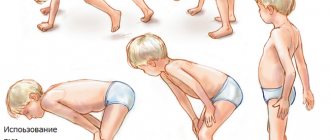
The disease often occurs in patients between the ages of two and fifteen years. Symptoms include abnormal gait and muscle weakness in the limbs.
Later, damage to the upper extremities may also occur. Then a violation of the facial muscles occurs, and the patient moves his eyes without problems. The reflex of those muscles that are already affected disappears. Deformation of the skeleton and joints may occur. With this disease, if the necessary treatment is given, the patient will live to about forty years.
Spinal amyotrophy type 4
This type of spinal amyotrophy occurs in adulthood after 35 years. Pathology manifests itself in the form of muscle weakness in the limbs and decreased reflexes.
Clinical picture
The symptoms of SMA 1 and SMA 2 are very different. Manifestations of SMA 1 are often detected during pregnancy, since the fetus in the womb is inactive and moves very rarely. After birth, the child experiences respiratory failure, often he cannot breathe on his own, and other innate reflexes are absent. The main manifestations of the disease can be considered:
- Decreased muscle tone.
- Retarded physical development.
- Inability to hold the head upright.
- Cannot independently roll over onto its side, stomach or back.
- Mild paralysis of the limbs.
- Lack of swallowing reflexes.
- Lack of sucking reflex.
The child takes a characteristic position in which he is almost constantly. In most cases, paralysis of the diaphragm may also occur. All this over time leads to disruption of skeletal development, scoliosis appears, the presence of a hump is often noted, and the shape of the chest changes. Only 12% of such children live to be 5 years old.
Type I amyotrophy
The most malignant and common type of pathology is Werdnig-Hoffmann amyotrophy. Diagnosed in young children or in utero. After birth, children experience a decrease and extinction of all reflexes. They find it difficult to breastfeed, which often makes it necessary to feed the baby through a tube.
Muscular hypotonia leads to weakness of the neck muscles - children cannot learn to hold their heads, roll over independently, sit, stand and walk. Most children have difficulty swallowing. In most cases, amyotrophy is diagnosed within 6 months after birth. Parents pay attention to the child’s lethargy and inactivity, and a progressive decrease in muscle tone. Babies also have trouble gaining weight.
It is characterized by damage to the diaphragm, which is involved in the respiratory act. Many patients cannot breathe on their own. Due to severe respiratory failure, many children do not live to see one year of age. It is possible to prolong their life with the help of mechanical ventilation and feeding through a feeding tube. 95% of children die before age 4.
The pathology is accompanied by progressive deformations of the musculoskeletal system.
In addition to damage to muscles and bones, mental retardation is often detected.
Reference. The diagnosis can be suspected even during the mother's pregnancy, when there is weak fetal movement, cardiac dysfunction, and developmental delays.