What is it and what does it consist of
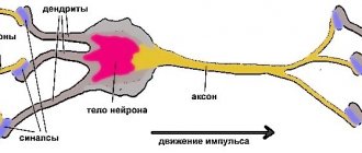
The human brain consists of two types of nervous tissue - gray matter and white matter. The gray matter of the nervous system is a collection of nerve cells responsible for most of the functions of higher nervous activity in humans. The function of white cells is to transmit electrical impulses to different parts of the brain. The thickness of gray brain tissue reaches about half a centimeter in the population. Topographically, the gray matter is the membrane of the brain, under it there is a cluster of long processes (axons), that is, the matter is white.
The gray matter is formed by an accumulation of soma neurons, small capillaries, glial tissue and short processes - dendrites. In addition, the gray matter includes long unmyelinated processes - axons. Unlike gray matter, which does not have myelin fibers, white matter is called white because its color is given by the sheaths of axons consisting of myelin.
Gray matter nuclei are histological structures, concentric clusters of nerve cell bodies that perform a specific function in the nervous system. Anatomically, two subtypes of nuclei are distinguished: nuclei in the central nervous system and those in the structure of the peripheral nervous system. Each nucleus is a regulator of a specific body function, be it the act of urination or the center of the heartbeat.
There is a partially erroneous belief that gray matter consists of long processes of neurons. Specialized processes, equipped with a fast conductor myelin, are found in the structure of the white matter of the brain and spinal cord, while in the gray substance there are only dendrites and long unmyelinated fibers. The bottom line is that myelinated long axons are not needed in the cortex, because the gray matter of the brain consists of clusters of adjacent neuron bodies, and information from cells to cells is transmitted by short processes (dendro-dendritic synapses), because the main task of long processes is the transmission of electrical impulses from one center to another. There, the function of transmitting and receiving information is served by axo-axonal or axo-dendritic synapses. Gray matter is the same in all parts of the brain. It is the same in different departments. Therefore, the gray matter of the telencephalon includes that set of elements that are also inherent in other brain structures.
How does COVID-19 affect the brain?
The fact that coronavirus has a devastating effect on the brain has been known relatively recently: scientific research has shown that SARS-CoV-2 damages the brain, causing headaches, strokes and seizures. Thus, according to a study published in the New England Journal of Medicine, patients with confirmed COVID-19 experienced neurological symptoms (neurological disorders, confusion). This trend was identified back in Wuhan, at the very beginning of the pandemic.
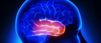
With the emergence of new coronavirus variants, the situation has become more complicated. A new study based on data collected by the UK Biobank suggests that COVID-19 survivors may suffer loss of gray matter in the brain over time. Gray matter is part of the central nervous system and essentially controls all brain functions.
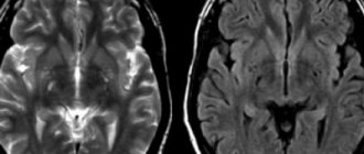
MRI scan of the brain of a patient with COVID-19. Data from a 2020 study published in the journal Neurology
The authors of the scientific work note that this is the first study to compare brain images both before and after the subject was diagnosed with COVID-19. Researchers found loss of gray matter in three areas of the brain.
“The findings are consistently associated with gray matter loss in areas of the limbic cortex, areas of the brain associated with the perception of tastes and smells.”
The long-term experiment, which involved 782 volunteers, compared brain scans of people before the pandemic. To provide an analogy between pre-pandemic and post-pandemic brain scans, the researchers then invited 394 COVID-19 survivors to return for follow-up scans, as well as 388 healthy volunteers.
You may be interested in: Is it true that coronavirus damages the brain stem?
Among those participants who recovered from COVID-19, the researchers saw a significant impact of the virus on gray matter, or more specifically gray matter loss in three brain regions over time. Gray matter allows people to control movement, memory and emotions, so the identified abnormality could have an impact on communication skills and brain cells.
The new coronavirus, according to another assumption of scientists, can cause a brain tumor and accelerate the development of an existing one.
It should also be noted that the study has not yet been subjected to rigorous peer review and published on a preprint server. The work, however, suggests that loss of gray matter in areas of the brain associated with memory " may increase the risk of dementia in these patients in the long term."
This finding follows a study published by the Lancet Psychiatry journal last year suggesting that severe COVID-19 infections can damage the brain, leading to long-term complications such as stroke or dementia-like symptoms. However, the authors noted that more data is needed to adequately assess the impact of COVID-19 on brain health.
This is interesting: Scientists have discovered why some people believe in conspiracy theories about coronavirus
I will also note that most of those who took part in the study reported mild or moderate symptoms or no symptoms at all. Scientists also cannot confirm whether the loss of gray matter is a result of the virus spreading to the brain or some other effect of the disease, Reuters reports.
Where is it located in the brain
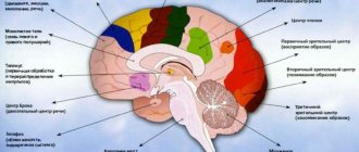
The question of where the gray matter of the brain is located is answered by several basic theoretical medical sciences - normal and topographic anatomy and histology. Other brain sciences study its function rather than its location and structure. Gray matter is the cerebral cortex. On average, the layer of dark fabric is about 3-4mm (from 1.5 to 5mm). It has the most pronounced thickness in the region of the anterior central gyrus. Due to the arrangement of many convolutions and fissures, the area of gray matter increases significantly. In addition to the brain, a layer of gray matter is located inside the spinal cord.
In the cerebellum, the bulk of the gray matter is located by analogy with the brain: the gray matter is the cerebellar cortex and is located on the surface of the structure itself, being its shell, when the white matter is located inside the cerebellum. In addition, the cortex, the coordinating center of the human body, consists of three layers - the molecular ball, piriform neurons and the granular layer.
The cerebral bulb, like other parts of the brain, also has gray substance. The medulla oblongata is one of the first evolutionarily formed brain structures. This part is located at the level of the foramen magnum and passes into the spinal cord. The gray matter of the medulla oblongata forms some nuclei and nerve centers, among which are the nuclei of the cranial nerves and the reticular formation. The nuclei formed by the dark tissue include the hypoglossal, accessory, vagus and glossopharyngeal nerves. It should be noted that all these centers are neither lower nor higher centers of regulation - they occupy an intermediate position in the hierarchy of regulatory systems of the brain.
The structure located above the oblong is called the pons. At its junction with the adjacent structure, several nerves emerge, including the vestibulocochlear nerve. The gray matter of the pons forms its own centers of a mixed nature: the nucleus of the trigeminal nerve, the facial and abducens nerves. Such nerves are responsible for the innervation of the facial (facial) muscles, the scalp (its scalp), some eye muscles and certain parts of the tongue. In addition to such functions, the task of the pons is to maintain the correct posture and partially preserve the location of the body in space. The gray matter of the midbrain is represented by red nuclei and substantia nigra. These structures are collectors of conscious and unconscious movements: the nuclei have rich connections with the cerebellum. In general, these structures are part of the striopadlidal system of the brain.
The cortex, consisting of gray matter, covers many brain structures, including:
- brain;
- cerebellum;
- thalamus;
- hypothalamus;
- subthalamus;
- pale globe;
- basal ganglia;
- shell;
- brain stem structures (red nucleus and substantia nigra);
- cranial nerves.
The conclusion suggests itself that any structure that has a specific regulatory function is covered with an accumulation of gray substance.
Location in other parts of the brain
The medulla oblongata regulates all automatic functions, protective and pro-survival reflexes (for example, heartbeat and sneezing). There is also a center that distributes tension between certain muscles responsible for holding the body in any position. The gray matter here forms the nuclei of the olive, several pairs of the lower head and vagus nerves, as well as the reticular formation, the sphenoid and thin bodies.
The hindbrain consists of the pons and the cerebellum. The cores of the bridge are dispersed between fibers directed in different directions. The cerebellar cortex has three sections: external (molecular), ganglionic and granular (granular). The outer layer contains several scattered cell nuclei. The ganglion contains a number of Purkinje cells, their processes extend deep into the cerebellum. The white matter contains the subcortical nuclei: spherical, mushroom, dentate, tent nucleus.
The midbrain has a cavity in its center filled with central gray matter. The so-called aqueduct is located here; at its bottom there are several nuclei, the processes of which mainly innervate the organs of vision, as well as the nuclei of the III and IX cranial nerves. In front of it are the cerebral peduncles, divided by the substantia nigra into a base and a tegmentum, where the red nucleus stands out. Together with the substantia nigra, they belong to the extra pyramidal system and control unconscious movements and muscle tone with the help of the hormone dopamine. The visual and auditory centers are located in the colliculi of the midbrain roof.
Useful to know: Midbrain: structure, functions, development
The nervous tissue of the diencephalon forms its four parts. The first of them is the Thalamus, where the nuclei of the thalamus lie. The epithalamus - the second part - includes the leash, its commissure and triangle, as well as the epiphysis, otherwise called the pineal gland. It is interesting for its mystical reputation - esotericists believe that it is this gland that causes clairvoyance and other supernatural abilities. In fact, the pineal gland produces the hormone melatonin, which is responsible for regulating sleep and wake cycles. The third part, the Metathalamus, contains the geniculate bodies. The latter, the Hypothalamus, has two sections. In the anterior one there is the so-called gray tubercle, and in the posterior one there are the mastoid bodies, containing two nuclei.
to contents ^
What role does gray matter play?
Millions of years of evolution, natural selection and the origin of species have given the human being a unique structure - a relatively thick cerebral cortex. It is known that the structure of gray matter is properly developed only in representatives of the human species. Unlike lower and even higher mammals, the gray substance has endowed a person with the opportunity to have a unique property of matter, the object of study of all neurosciences and philosophy - consciousness and self-awareness, the resulting abstract thinking, developed memory, inner speech and many other specific attributes of higher nervous activity a reasonable person.
It must be remembered that gray matter is a collection of nerve cells, namely neurons. When talking about the function of gray matter, we are talking about the function of all clusters of neurons with short processes. So, the functions of gray matter are diverse:
- Physiological tasks: generation, transmission, reception and processing of electrical signals.
- Neurophysiological: perception, speech, thinking, memory, vision, emotions, attention.
- Psychological: personality formation, worldview, motivation, will.
For a long time, scientists have wondered what the gray matter of the brain is responsible for. Back in the 18th century, Franz Gall drew attention to the dark brain substance. For the first time, the scientist managed to localize some mental functions on the cortex. Subsequent studies were carried out by removing a section of the cortex and observing which brain function was lost. A serious impetus for further research was the study of the work of the cortex by Academician Pavlov, who studied basic reflexes and the principles of strengthening the conditioned reflex. In parallel, his French colleagues found a speech center in the cortex - the lower part of the frontal gyrus. Modern science, although it knows many properties of the cerebral cortex, claims that the percentage of knowledge on it is no more than one thousandth.
One blind spot in the empirical knowledge of the brain and its formation is the question of what heterotopia of the gray matter of the brain is. In particular, this question is often raised in the field of clinical medicine, where treatment is only symptomatic, that is, the symptoms are removed alone. As is known, heterotopia is a defective accumulation of neurons that have stopped in a certain place and have not reached their histological place. So, if the cause of the pathology is found, there will be an etiological treatment. A variant of the manifestation of heterotopia is childhood epilepsy.
Composition and functions of gray matter
The gray matter of the brain consists of neurons, glial cells, neuropil (a substance that looks like felt, mainly consisting of dendrites), and thin capillaries.
Contrary to popular belief, it is not gray, but brownish in color, surrounded by white matter formed from myelin fibers. It differs significantly in composition: the gray matter contains 84% water, 16% dry matter, half of which, that is, 8%, consists of proteins, 5% lipids and 1% mineral compounds. White contains 70% water, 30% dry matter, only one third of which (9%) consists of proteins, as much as 17% are lipids and 2% are minerals.
Gray matter determines muscle movements, sensory perception (visual, auditory, tactile), cognitive functions (memory, attention, thinking), verbal communications and emotional-sensory response.
to contents ^
Difference from white matter
This section is intended to calibrate concepts and answer the question of what gray and white matter of the brain are.
Gray matter
- Created by the nuclei of nerve cells and its relatives.
- It is located mainly in the central parts of the nervous system.
- Makes up no more than 40% of the total mass of the brain.
- Consumes about 3-5 ml of oxygen per minute.
- A structure that has a regulatory function.
White matter
- Formed by long myelinated axons.
- It is located primarily in the peripheral nervous system.
- Makes up more than 60% of the weight of the human brain.
- Consumes less than 1 ml of oxygen per minute.
- Responsible for conducting nerve impulses through the nervous system
It should be remembered that unlike the structure of the cerebral cortex, where the gray matter is a shell and covers the white substance, in the spinal cord the gray matter is surrounded by the white matter of the brain.