content .. 101 102 103 104 105 106 ..
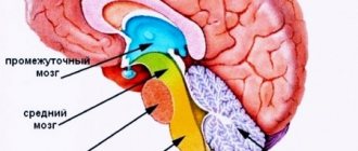
Diencephalon (human anatomy)
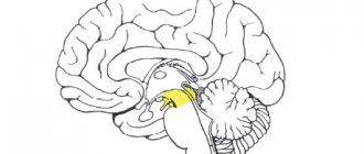
The diencephalon is located under the corpus callosum and fornix, fused on the sides with the cerebral hemispheres. It includes the thalamus (visual thalamus), epithalamus (supra-tubercular region), metathalamus (sub-tubercular region) and hypothalamus (sub-tubercular region). The cavity of the diencephalon is the third ventricle.
The thalamus is a paired, ovoid collection of gray matter covered by a layer of white matter. The anterior sections are adjacent to the interventricular foramina, the posterior sections are expanded to the quadrigeminal. The lateral surfaces of the thalamus grow together with the hemispheres and border the caudate nucleus and the internal capsule. The medial surfaces form the walls of the third ventricle, the lower ones continue into the hypothalamus. In the thalamus, there are three main groups of nuclei: anterior, lateral and medial, and there are 40 nuclei in total. In the epithalamus lies the upper appendage of the brain - the pineal gland, or pineal body, suspended on two leashes in the recess between the upper colliculi of the roof plate. The metathalamus is represented by the medial and lateral geniculate bodies, connected by bundles of fibers (handles of the colliculi) with the superior (lateral) and inferior (medial) colliculi of the roof plate. They contain nuclei that are reflex centers of vision and hearing.
The hypothalamus is located ventral to the thalamus and includes the subcutaneous region itself and a number of formations located at the base of the brain. These include: the terminal plate, the optic chiasm, the gray tubercle, the infundibulum with the lower appendage of the brain extending from it - the pituitary gland and the mastoid bodies. In the hypothalamic region there are nuclei (supravisceral, periventricular, etc.) containing large nerve cells capable of secreting a secretion (neurosecretion) that flows along their axons into the posterior lobe of the pituitary gland and then into the blood. In the posterior part of the hypothalamus lie nuclei formed by small nerve cells, which are connected to the anterior lobe of the pituitary gland by a special system of blood vessels.
The third (III) ventricle is located in the midline and is a narrow vertical slit. Its lateral walls are formed by the medial surfaces of the thalamus and the subtubercular region, the anterior - by the columns of the fornix and the anterior commissure, the lower - by the formations of the hypothalamus, and the posterior - by the cerebral peduncles and the supracuberous region. The upper wall - the lid of the third ventricle - is the thinnest and consists of the soft membrane of the brain, lined on the side of the ventricular cavity with an epithelial plate (ependyma). The soft shell has a large number of blood vessels here, forming the choroid plexus. In front, the third ventricle communicates with the lateral ventricles (I - II) through the interventricular foramina, and behind it passes into the cerebral aqueduct.
Physiology of the diencephalon (human anatomy)
The thalamus is a sensitive subcortical nucleus. It is called the “collector of sensitivity”, since afferent pathways from all receptors converge to it, excluding olfactory ones. In the lateral nuclei of the thalamus there is a third neuron of the afferent pathways, the processes of which end in the sensitive zones of the cerebral cortex.
The main functions of the thalamus are integration (unification) of all types of sensitivity, comparison of information received through various communication channels, and assessment of its biological significance. The nuclei of the thalamus are divided according to their function into specific (the ascending afferent pathways end on the neurons of these nuclei), nonspecific (nuclei of the reticular formation) and associative. Through the associative nuclei, the thalamus is connected with all the motor subcortical nuclei: the striatum, the globus pallidus, the hypothalamus - and with the nuclei of the midbrain and medulla oblongata.
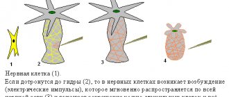
The study of the functions of the thalamus is carried out by cutting, irritation and destruction. A cat in which the incision is made above the diencephalon is very different from a cat in which the highest part of the central nervous system is the midbrain. She not only gets up and walks, that is, performs complexly coordinated movements, but also shows all the signs of emotional reactions. A light touch causes an angry reaction: the cat whips its tail, bares its teeth, growls, bites, and extends its claws. In humans, the thalamus plays a significant role in emotional behavior, characterized by peculiar facial expressions, gestures and shifts in the functions of internal organs. During emotional reactions, blood pressure rises, pulse and breathing quicken, and pupils dilate. The facial reaction of a person is innate. If you tickle the nose of a fetus for 5-6 months, you can see a typical grimace of displeasure (P.K. Anokhin). In animals, when the thalamus is irritated, motor and pain reactions occur: squealing, grumbling. The effect can be explained by the fact that impulses from the visual thalamus easily transfer to the associated motor subcortical nuclei.
In the clinic, symptoms of damage to the thalamus are severe headache, sleep disturbances, disturbances in sensitivity (increased or decreased), movements, their accuracy, proportionality, and the occurrence of violent involuntary movements.
The hypothalamus is the highest subcortical center of the autonomic nervous system. In this area there are centers that regulate all vegetative functions, ensuring the constancy of the internal environment of the body, as well as regulating fat, protein, carbohydrate and water-salt metabolism. In the activity of the autonomic nervous system, the hypothalamus plays the same important role as the red nuclei of the midbrain play in the regulation of skeletal-motor functions of the somatic nervous system.
The earliest studies of the function of the hypothalamus belong to Claude Bernard. He discovered that an injection into the diencephalon of a rabbit caused an increase in body temperature of almost 3°C. This classic experiment, which made it possible to discover the thermoregulation center in the hypothalamus, was called heat injection. After the destruction of the hypothalamus, the animal becomes poikilothermic, that is, it loses the ability to maintain a constant body temperature.
Later it was found that almost all organs innervated by the autonomic nervous system can be activated by irritation of the subtubercular region. In other words, all the effects that can be obtained by irritating the sympathetic and parasympathetic nerves are observed when irritating the hypothalamus.
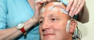
Currently, the method of implanting electrodes is widely used to stimulate various brain structures. Using a special, so-called stereotaxic technique, electrodes are inserted into any given area of the brain through a burr hole in the skull. The electrodes are insulated throughout, only their tip is free. By connecting electrodes in a circuit, you can locally irritate certain areas.
When the anterior parts of the hypothalamus are irritated, parasympathetic effects occur: increased intestinal movements, separation of digestive juices, slowing down heart contractions, etc.; when the posterior sections are irritated, sympathetic effects are observed: increased heart rate, constriction of blood vessels, increased body temperature, etc. Consequently, parasympathetic centers are located in the anterior sections of the hypothalamus, and sympathetic centers in the posterior sections.
Since stimulation with the help of implanted electrodes is carried out on the animal without anesthesia, it is possible to judge the behavior of the animal. In Andersen's experiments on a goat with implanted electrodes, a center was discovered, the irritation of which causes unquenchable thirst - the thirst center. When irritated, the goat could drink up to 10 liters of water. By stimulating other areas, it was possible to force a well-fed animal to eat (hunger center).
The experiments of the Spanish scientist Delgado on a bull became widely known. An electrode was implanted into the bull's fear center. When an angry bull rushed at a bullfighter in the arena, the irritation was turned on and the bull retreated with clearly expressed signs of fear.
American researcher D. Olds proposed modifying the method: allowing the animal itself to make contact (self-irritation method). He believed that the animal would avoid unpleasant stimuli and, on the contrary, would strive to repeat pleasant ones. Experiments have shown that there are structures whose irritation causes an uncontrollable desire to repeat. The rats worked themselves to the point of exhaustion by pressing the lever up to 14,000 times. In addition, structures were discovered whose irritation apparently causes an unpleasant sensation, since the rat avoids pressing the lever a second time and runs away from it. The first center is obviously the center of pleasure, the second is the center of displeasure.
Extremely important for understanding the functions of the hypothalamus was the discovery in this part of the brain of receptors that detect changes in blood temperature (thermoreceptors), osmotic pressure (osmoreceptors) and blood composition (glucoreceptors).
Reflexes arise from receptors “turned into the blood” aimed at maintaining the constancy of the internal environment of the body - homeostasis. “Hungry” blood, irritating glucoreceptors, excites the food center: food reactions arise, aimed at searching and eating food.
One of the common manifestations of hypothalamic disease is a violation of water-salt metabolism, manifested in the release of large amounts of low-density urine. The disease is called diabetes insipidus.
The subcutaneous region is closely related to the activity of the pituitary gland. The hormones vasopressin and oxytocin are produced in large neurons of the supravisual and paraventricular nuclei of the hypothalamus. Hormones travel along axons to the posterior lobe of the pituitary gland, where they accumulate and then enter the blood.
A different relationship between the hypothalamus and the anterior pituitary gland. The vessels surrounding the nuclei of the hypothalamus unite into a system of veins, which reach the anterior lobe of the pituitary gland and here again break up into capillaries. With the blood, releasing factors, or releasing factors, enter the pituitary gland, stimulating the formation of hormones in its anterior lobe.
Reticular formation. In the brain stem and diencephalon, between its specific nuclei, there are clusters of neurons with numerous highly branching processes, forming a dense network. This system of neurons is called a network formation, or reticular formation. Special studies have shown that all the so-called specific pathways that carry certain types of sensitivity from receptors to sensitive areas of the cerebral cortex give off branches in the brain stem that end on the cells of the reticular formation. Streams of impulses from the periphery from extero-, intero- and proprioceptors support constant tonic excitation of the structures of the reticular formation.
Nonspecific pathways begin from the neurons of the reticular formation. They go up to the cerebral cortex and subcortical nuclei and down to the neurons of the spinal cord.
By irritating individual structures of the reticular formation, it was possible to reveal its function as a regulator of the functional state of the spinal cord and brain, as well as the most important regulator of muscle tone. The role of the reticular formation in the activity of the central nervous system is compared to the role of a regulator on a TV: without giving an image, it can change the volume of sound and illumination.
Irritation of the reticular formation does not cause a motor effect, but affects existing activity, inhibiting it or enhancing it. If in a cat a protective reflex is induced by short, rhythmic stimulation of the sensory nerve - flexion of the hind leg, and then against this background the reticular formation is irritated, then depending on the zone of irritation, the effect will be different: the spinal reflexes will either sharply intensify, or become weaker and disappear, i.e. e. will slow down. Inhibition occurs when the posterior parts of the brain stem are irritated, and reflexes are strengthened when the anterior parts are irritated. The corresponding zones of the reticular formation are called inhibitory and activating zones.
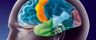
The reticular formation has an activating effect on the cerebral cortex, maintaining a state of wakefulness and concentrating attention. If you turn on stimulation of the reticular formation in a sleeping cat with electrodes implanted into the diencephalon, the cat will wake up and open its eyes. On the electroencephalogram, slow waves characteristic of sleep will disappear, and fast waves characteristic of the waking state will appear. The reticular formation has an ascending, generalized (encompassing the entire cortex) activating effect on the cerebral cortex. According to I.P. Pavlov, “the subcortex charges the cortex” (Fig. 115). In turn, the cerebral cortex regulates the activity of the network formation.
Rice. 115. Cat's brain (diagram). Facilitating (5) and inhibitory (4) zones of the reticular formation of the brain stem, as well as connections going to it from the cortex (1), subcortical nuclei (2) and cerebellum (3) (according to Magoon)
content .. 101 102 103 104 105 106 ..
Functions of the thalamus
The main task of the thalamus is to receive signals from receptors (sensory organs) both external - exteroceptors, and located inside the body - interoceptors. After entering the thalamus, the signals undergo primary processing, are identified and sent to the corresponding part of the cerebral cortex: visual, auditory, tactile, etc. Here they are further processed, converted into sensory images, comprehended and transmitted to the hippocampus for storage in long-term memory.
But regulating the flow of sensory information is not the only function of the thalamus. This part of the brain also has completely nonspecific tasks that are not related to processing signals from receptors:
- Providing the necessary level of excitation of the areas of the cerebral cortex responsible for processing sensory signals.
- Controlling involuntary movements and maintaining muscle tone.
- Some of the nuclei of the thalamus are connected to the limbic system and the hippocampus, so this department is involved in the formation of an emotional assessment of sensations and the processes of storing sensory images in memory.
- We must thank the thalamus for pain sensations, since it is it that regulates their intensity and area of distribution.
- By maintaining the activity of the cerebral cortex, this department is involved in regulating excitation in the central nervous system as a whole.
- The thalamus influences both attention processes and the change in sleep-wake cycles.
Research in recent years has shown that despite the ancient origin of the thalamus (all vertebrates have it), in the human brain this section is closely connected with higher mental functions. Thus, the interaction of a number of thalamic nuclei influences the processes of speech activity. In particular, this concerns the regulation of the motor sphere of articulate speech and the provision of speech movements.
Along with speech motor skills, the thalamus is involved in the control of motor activity associated with the sensory sphere, for example, eye movement when looking at an object. However, this area of thalamic functions is still very poorly studied, and there are more assumptions than knowledge.