Description: What is the conducting function of the spinal cord? Although almost every schoolchild knows the answer to the question, an ordinary person is unlikely to be able to answer right away. Its conducting function is simple - it is the transmission of a nerve signal. It is because of this feature of the NS that a person is a single system.
And to ensure control over organ functions, the ability to move, timely transmission or receipt of reflex, sympathetic impulses, pathways are needed. Failures in the transmission of impulses entail serious disruptions in the functioning of the body.
What is the conducting function?
Indeed, what is the conductive function of the spinal cord? The very concept of “pathways” implies a common thread of nerves that conduct signals to different areas of the gray matter. The descending and ascending pathways of the spinal cord are subordinated to one function - the transmission of impulses. As a rule, there are 3 types of fibers:
- projection;
- commissural connections;
- associative pathways of nerves.
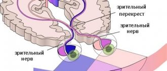
However, in addition to this classification, there is another one. It distinguishes between motor and sensory pathways. The former provide a reflex reaction and organize the supply of impulses from the brain to the spinal cord, as well as to muscle tissue. They are also responsible for coordinating movements. In addition, these threads stretch to the optic nerve and to the plate of the roof of the midbrain, whose task is to ensure the function of vision and hearing. Thanks to the nerve fibers that make up the sensory pathways, a person is endowed with the ability to recognize the following 4 types of impulses: pain, temperature, tactile feeling, joint-muscular feeling (movement, body position).
What does the spinal cord look like?
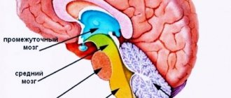
Not everyone knows what the spinal cord looks like. Moreover, not all people have an idea of what its role is in the life of every person. It is therefore worthwhile to fill this knowledge gap. In addition, many people mistakenly believe that the brain and spinal cord are separate parts.
To find out what the reflex function of the spinal cord is for, let's try to determine what it looks like. It is impossible to clearly understand where the spinal cord begins and ends. It starts from the first vertebra just below the skull, smoothly connecting to the brain in this area. The division into the spinal cord and the brain is formal, but in reality the spinal cord smoothly passes into the brain. Thus, we can conclude that these two parts are a single whole.
What are the pathways formed by?
The main pathways of the spinal cord are formed by bundles of cells - neurons. It is this structure that guarantees the required speed of impulse transmission. The division of the functions of the paths is associated with the peculiarities of their purpose. Ascending pathways perceive and transmit signals from the skin, mucous membranes, and human organs. In addition, they are responsible for the functioning of the musculoskeletal system.
The descending pathways of the spinal cord transmit signals to human organs - tissues, glands. They connect to the cortical gray matter. The transmission of signals to organs is carried out due to the spinal neural connection.
The spinal cord is characterized by a double direction of such pathways, due to which rapid impulse transmission of information from systems under control occurs. The conductive spinal function becomes possible only due to the transmission of signals through nerve tissue. The following designations are often used for these pathways in medicine.
- Corticospinal tract. This is a collection of nerve threads that is responsible for movement functions. Based on its purpose and direction, it is divided into several parts: lateral, cortical-nuclear, anterior cortical-cerebral system.
- Tectospinal tract. It is represented by the descending projection NS, which originates in the midbrain cortex, passes through the trunk, cord of the hemispheres, and ends on the anterior horns of the ridge. Otherwise it is called the tectospinal tract.
- vestibulospinal tract. Manages the work of the vestibular apparatus. It begins from the lateral nucleus of the prespinal nerve.
- Reticular-spinal tract. Provides muscle tone, and the task of its fibers can be called the transmission of nerve impulses to the gray matter of the cerebral hemispheres.
- Pyramid path. Its components include the lateral and direct bundle of nerve fibers.
Conducting function of the spinal cord.
It is carried out according to the Bell-Magendie law : afferent information enters the spinal cord through the dorsal roots, efferent impulses are transmitted through the anterior roots.
· The ascending (sensitive) tracts of the spinal cord are located in the posterior columns of the white matter and carry information from the external world and the internal environment of the body:
− from skin receptors (pain, temperature, touch, pressure, vibration);
- from proprioceptors (muscle spindles, Golgi tendon receptors, periosteum, joint membranes);
− from receptors of internal organs - visceroreceptors (mechano- and chemoreceptors).
· Descending (motor) pathways are located in the anterior columns and transmit impulses to the skeletal muscles about voluntary (conscious) movements, tonic influences on the muscles, impulses that ensure the maintenance of posture and balance. Autonomic influences (on internal organs) are also transmitted through descending pathways.
Conductive functions are similar in other stem structures (medulla oblongata, midbrain and pons): afferent pathways pass through the posterior group of white fibers, and efferent pathways pass through the anterior group.
Functions of the medulla oblongata.
The medulla oblongata contains the nuclei of the VIII-XII cranial nerves, therefore the medulla oblongata carries out protective reflexes (coughing, sneezing, vomiting, lacrimation, blinking, constriction of the pupil).
The medulla oblongata performs sensory functions : reception of skin sensitivity of the face, primary analysis of taste. The medulla oblongata receives signals from chemoreceptors and baroreceptors of blood vessels, interoreceptors of internal organs and vestibuloreceptors. The influence of these structures determines the functioning of the respiratory and cardiovascular centers .
The structures of the reticular formation also perform the functions of regulating the tone of skeletal muscles .
Complex brain stem reflexes.
With the participation of the brain stem, complex somatic reflexes are carried out, each of which involves the nuclei of several cranial nerves.
· Oculomotor reflexes. As a result of these reflexes, friendly eye movements occur in different directions. They are carried out by the work of the III-VI and VIII cranial nerves .
· The reflex act of chewing is provided by muscles that cause movements of the lower jaw and hold food between the dentition. Afferent impulses arise from various receptors of the oral mucosa and proprioceptors of the masticatory apparatus and spread to the chewing center , which causes rhythmic excitation of motor neurons of the muscles that raise and lower the lower jaw. The work of the chewing center can be launched not only from the receptors, but also from the chewing area of the frontal cortex, which provides voluntary control of chewing. It is carried out by the work of the VII and XII cranial nerves .
· The reflex act of swallowing ensures the passage of food from the mouth to the stomach. When a bolus of food moves from the oral cavity to the esophagus, sequential stimulation of the receptors of the root of the tongue, soft palate, pharynx and esophagus occurs. The impulse enters the swallowing center, resulting in a strictly coordinated sequence of contractions of the corresponding muscles: the muscles of the soft palate, pharynx, larynx and epiglottis, and esophagus. The swallowing center is functionally connected with the breathing center, which stops during each swallowing act. It is carried out by the work of the V, IX and X cranial nerves .
· The gag reflex is a protective reaction that occurs when receptors in the root of the tongue, pharynx, stomach, intestines, peritoneum, and vestibular apparatus are irritated. Vomiting can also be caused by direct irritation of the vomiting center by a local pathological process or by certain chemicals, for example, apomorphine. Efferent impulses from the vomiting center enter the esophagus, stomach, intestines and through the spinal motor centers to the diaphragm and the muscles of the abdominal wall, the contraction of which leads to the movement of the contents of the stomach. Carried out by the work of the VIII-X cranial nerves .
· The cough reflex (“watchdog” of the lungs) is a protective reflex that occurs when the receptors of the larynx, trachea and bronchi are irritated. The impulse excites the cough center of the medulla oblongata, which triggers a hard-coded sequence of reactions:
− deep breath;
− contraction of the expiratory muscles against the background of a closed glottis and narrowing of the bronchi, which leads to a sharp increase in pressure in the lungs (up to 140 mm Hg);
− active exhalation against the backdrop of instant opening of the glottis, creating a powerful air flow directed by stretching the soft palate through the mouth.
It is carried out by the work of the X cranial nerve .
· The sneezing reflex occurs when receptors in the mucous membrane of the nasal cavity are irritated. The sneezing center is similar to the cough center, but the flow of air during forced exhalation against the background of rapid opening of the glottis and lowering of the soft palate is directed primarily through the nose. It is carried out by the work of the V cranial nerve .
Functions of the hindbrain.
Bridge functions.
· Conductor function.
· Ensures maintaining posture and maintaining body balance in space when changing speed.
· Provides tone to the neck muscles.
· Regulates chewing and swallowing.
· Contains vegetative centers for the regulation of respiration (pneumotoxic center), heart rate, and gastrointestinal tract activity.
· Plays an important role in the activation of the cerebral cortex (including in a state of anxiety).
· Limits sensory inflows of nerve impulses to the cerebral hemispheres during sleep.
The locus coeruleus, or locus coeruleus , is a nucleus located in the brain stem at the level of the pons and is part of the reticular formation. Its ascending (afferent) fibers project to the structures of the cortex, diencephalon and cerebellum, descending (efferent) projections go to the spinal cord to the sympathetic centers and motor neurons. Responsible for the physiological response to stress and anxiety, and is involved in sleep mechanisms. Many of its neurons are noradrenergic.
Functions of the cerebellum.
· Maintaining body posture and balance.
· Coordination of targeted movements.
· Construction of fast ballistic targeted movements.
· Regulation of muscle tone.
· Regulation of autonomic functions (heartbeat, vascular tone, intestinal motility, etc.).
· Conductor function.
The functions of the cerebellum are mainly associated with the organization of motor acts . From the motor cortex and basal ganglia, the cerebellum receives information about the planned movement, as well as afferentation from the somatosensory system. The cerebellum ensures mutual coordination of purposeful movements , as well as correction of the movement being performed (necessary, because when performing a motor act, moving parts of the body are influenced by inertial forces, which disrupts the smoothness and accuracy of the movement performed).
The cerebellum is especially important for constructing fast, ballistic, purposeful movements (for example, throwing a ball at a target, jumping over an obstacle, playing the piano). In such cases, correction during the movement is impossible due to the short time parameters, and the ballistic movement will be performed only according to a pre-prepared program. It is formed and fixed in the cerebellum.
The connection of the cerebellum with higher autonomic centers and with some endocrine glands ensures its participation in the regulation of autonomic functions : digestion, respiration, cardiac activity and vascular tone, thermoregulation, metabolism.
Localization of pathways
The entire set of nerve tissue is located in the white, gray matter, uniting the spinal horns and the cerebral cortex. Pathways are often understood as a set of threads and tissues of nerves located in certain areas of the gray, white matter of the brain. Impulses are transmitted through neural communication.
The morphofunctional characteristics of the descending pathways make it likely that impulses are transmitted in strictly one direction. Irritation of synapses occurs from the presynaptic to the postsynaptic membrane. Such possibilities and the location of ascending and descending pathways can be compared with the conductive function of both brains.
- Associative paths. They are called “bridges”, which unite the zones between the cortex and the nuclei of gray matter. They include long and short fibers. Thus, short fibers are located within one half or lobe of the hemisphere, but long fibers can transmit impulses through 2-3 segments of gray matter. Spinal neurons form intersegmental bundles.
- Commissural fibers. They make up the corpus callosum and unite the newly formed sections of the two brains. As a rule, they diverge in rays and are located in the white matter of the brain tissue.
- Projection fibers. They, located in the spinal cord, allow signals to reach the brain at maximum speed. By their nature and specific functions, fibers are divided into ascending (called afferent pathways) and descending. In turn, they are divided into interoceptive (provide communication with organs), proprioceptive (responsible for movements), exteroceptive (vision, hearing). Such receptors are located between the ridge and the hypothalamus.
The descending pathways of the brain of the back, as a rule, include the pyramidal and central motor neurons, as well as the spino-cerebellar filaments of nerves. The pyramidal neuron originates in the cerebral cortex and descends through the entire trunk. Each of its bundles ends on the horn of the spinal substance. The central motor neuron connects the cerebral cortex with the anterior horns of the brain through axons - nerve roots.
If we talk about the spinocerebellar filaments of nerves, then they cover a thin and wedge-shaped path that connects the legs and the spinal cord of a person. The specifics of such paths are very difficult for a person who does not have a medical education. However, you need to realize: neural signaling is what turns the human body into one whole.
Introduction
The pathways of the nervous system and the complex reflex arcs consisting of them are the most important and complex section of neurology. It is important because it affirms the cellular nature of the nervous system (neural doctrine) and shows the ordered nature of the arrangement and connections of neurons (in the form of reflex arcs), which underlies its regulatory function.
There is a significant difference from the method of descriptive anatomy. The latter makes it possible to demonstrate the shape, size and localization of a particular formation of the nervous system, as well as its belonging to gray or white matter, but does not reveal at all the structural organization of the nervous system and the mechanisms of its functioning.
This “separation” of structure from function, which is dangerous for the worldview, eliminates the systematic approach to the nervous system in the form of the study of reflex arcs. Here the emphasis is placed precisely on the presence of connections between neurons, on their interaction, leading to the functioning of both the nervous system itself and the entire organism. However, at the same time, the number of mental operations among students increases (synthesis is added to analysis), which increases the complexity of mastering the material and its subjective complexity. Nevertheless, only the study of the nervous system as a set of reflex arcs allows us to understand its organization and functional significance. Finally, only knowledge of neural connections and interactions allows for topical diagnosis of damage to the nervous system, i.e. take a meaningful approach to the diagnosis and treatment of nervous and many other diseases and injuries.
How do the concepts of “conducting path” and “reflex arc” relate to each other? Here it should be clearly understood that any conductive path is part of one or another reflex arc. Since there are two main links in the reflex arc: afferent and efferent, the pathways are classified into afferent and efferent. Taking into account the hierarchical principle of construction of the central nervous system (the presence of higher and lower nerve centers subordinate to them) and the possibility of closing reflex arcs at the level of higher nerve centers, it is clear that both afferent and efferent pathways should be localized in both the peripheral and central parts nervous system. Since the closure of somatic reflex arcs (the connection of afferent and efferent links through interneurons) always occurs in the central nervous system, the latter also contain an associative link and corresponding associative pathways, localized only within the central nervous system.
Afferent nerve pathways conduct impulses from the receptor to the nerve center and are sensitive. Afferent nerve pathways ending in the projection centers of the cerebral cortex are classified as pathways of conscious sensitivity. The same afferent pathways that end in the subcortical sensory nerve centers are classified as pathways of unconscious sensitivity.
Efferent nerve pathways conduct impulses from nerve centers to the working organ. Since we are talking here only about the somatic nervous system, the working organ is the skeletal muscle, therefore the efferent nerve pathways are called motor. Depending on which nerve centers the efferent pathways are connected to, the latter are responsible for performing both conscious and unconscious movements.
Any conducting pathway (afferent, associative or efferent), depending on the level of closure and complexity of the reflex arc, can be single-neuron or multi-neuron (several neurons connected in series in a chain). If we consider a multineuron pathway as a chain, then within its boundaries we can distinguish links represented by the corresponding neurons. Compactly located neuron bodies form nerve centers (nodal, nuclear or screen type), and axons collected in bundles form nerve tracts. Thus, a multineural pathway consists of nerve centers and tracts. In this case, the nerve centers and tracts of the same pathway are localized in certain but different parts of the nervous system. Each tract within the central nervous system conducts nerve impulses usually in one direction and in most cases - of the same functional content. It should be clearly understood how the tracts within the central nervous system differ from the bundles of fibers that form the cranial or spinal nerves. Nerves contain both afferent and efferent fibers, and different afferent fibers can carry different sensory impulses.
In the future, material will be presented that concerns primarily the somatic part of the nervous system.
What happens when tracks become damaged?
To understand the neurophysiology of motor and sensory pathways, one must understand the anatomy of the spine. In its structure, the brain of the back resembles a cylinder surrounded by muscles. Paths pass through its gray matter, thanks to which movements and control of the functioning of organs are carried out. Associative pathways make tactile sensations and pain possible, and movements provide reflex functions of the body.
Due to injury, illness or pathology of brain development, impulse conductivity may decrease or even disappear. This phenomenon occurs due to the death of nerve branches. As a rule, conduction disturbances are accompanied by paralysis and lack of sensation in the limbs. In addition, there are disturbances in the functions of organs that were controlled by damaged nerve fibers. Thus, with damage to the lower back brain, urinary incontinence and spontaneous defecation are possible.
The reflex, conductive function of the brain undergoes changes immediately after the appearance of degenerative changes. There is a death of nerves, which are very difficult to recover in the future. The disease progresses rapidly, causing conduction disturbances to become noticeable. That is why treatment in this case should be started without delay.
Location of the spinal cord and its membrane
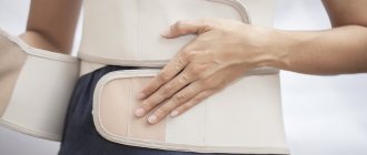
The brain is protected by the cranium, and the spinal cord is hidden in the spine and surrounded by three membranes. The first of them is the most delicate, thin and soft. It contains blood vessels that deliver nutrients to the brain. In other words, the spinal cord is a kind of “courier” for delivering food.
Continuing to talk about how the reflex function of the spinal cord works, we cannot ignore the analysis of the structure of the second arachnoid membrane. There is a special space here called the subarachnoid space. Along the entire length of the spine it is filled with cerebrospinal fluid (CSF). It is this that is taken during puncturing for analysis in order to determine the health status of the spinal cord.
The last shell is located on the outside and has a harder surface, which allows it to provide protective functions against various types of external damage.
Is it possible to restore conductivity?
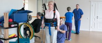
Therapy for non-conduction is aimed at stopping the death of nerves and eliminating the causes that provoked the pathology.
Medication therapy
This type of treatment involves prescribing medications that counteract the death of brain cells and also provide blood flow to the damaged area of the brain in the back. In the process of such therapy, the specificity of the conductive function of the brain is taken into account, which is associated with the patient’s age, as well as the severity of the disease or injury. In order to stimulate nerve cells, therapy is prescribed using electrical impulses to help maintain muscle tone.
Surgery
The operation performed to restore conductivity has 2 goals:
- eliminate factors that provoke paralysis of neural connections;
- This is brain stimulation to restore lost functions.
As a rule, before performing an intervention, doctors conduct an examination of the body to identify the location of the degeneration process. Since the list of paths is very large, the neurosurgeon tries to narrow the search area using diagnostics. In case of severe injuries, it is very important to quickly remove the causes of spinal compression.
ethnoscience
The means of such medicine for pathology of impulse conduction should be used with caution so as not to provoke a deterioration in the patient’s condition. Often when this problem occurs:
- apitherapy;
- herbal medicine;
- hirudotherapy.
Apitherapy is a treatment with bee stings, which helps restore efferent pathways, in particular when the pathology is caused by a growing hernia, radiculitis or other similar ailments. Bee venom has another useful feature - it provides blood flow to the problem area. In the case of herbal medicine, medicinal plant herbs that improve metabolism and help normalize blood flow are suitable. Hirudotherapy, which involves the use of leeches, helps eliminate congestion, which is inevitable with problems in the structure of the spine.
Complete restoration of neural connections after serious injury is not an easy task. A lot depends on immediate medical attention and timely assistance from a qualified neurosurgeon. But it is important not to forget: the more time has passed since the onset of degenerative changes, the less chance there is of restoring the functional capabilities of the spinal cord.
Gray matter
Gray matter or substantia grisea is represented by several columns connected to each other by two plates (anterior and inferior), called commissures. On a cross-section of one of these pillars, you can see that the gray matter in its shape resembles a butterfly with spread wings or the Latin letter H.
In addition, you can also notice that there are projections extending from the substance, which are otherwise called horns. They can be either front, located on the front wall, or rear, running along the back wall. Both the first and second pairs, and have a narrow and wide shape. But in addition to the rear and anterior ones, there are also lateral horns, which contain the centers of the autonomic nervous system.
What is the reflex function of the spinal cord? The fact is that in the anterior horns there is a special type of motor neurons, the processes of which form nerve roots.
In the middle of the gray matter there is a central canal, which is also filled with cerebrospinal fluid. In the upper part, the canal is connected to the ventricles of the brain. In this case, all sections: the ventricles, the central canal and the subarachnoid space take an active part in the circulation of cerebrospinal fluid.
How our brain works
Ascending and descending pathways are responsible for the fast and correct functioning of our body. The latter flows are formed using the red nuclear and lateral pathways. It is thanks to these pathways that the reflex and conduction functions of the spinal cord are carried out. Thanks to the red nuclear spinal tract, involuntary motor impulses are produced. While the lateral corticospinal tracts are responsible for voluntary impulses.
All roots are supplied by personal veins and arteries, which as a result form neurovascular bundles. Each such beam is responsible only for its own segment and works autonomously, analyzing incoming information and transmitting the necessary impulses.
Damage to these bundles leads to serious pathological and sometimes irreversible changes in the human body. And so that specialists can determine which particular bundle is damaged and localize the pain, it is necessary to conduct a whole range of studies.
White matter
White matter - substantia alba, envelops the gray matter, is formed by a collection of nerve fibers, which also come in three types:
- front;
- rear;
- lateral.
Moreover, all the roots have a different direction, and some of them are connected directly to the brain and central nervous system (hereinafter simply the CNS). And if the reflex function of the spinal cord is to transmit signals from the motor neurons of the gray matter, then the task of the white matter neurons is the prompt delivery of impulses from the muscles and joints to the medulla oblongata. Thus, the transmission of all commands along the entire spinal cord is realized.
Here are the paths through which all information regarding sensitivity and pain is transmitted. Only before entering the cerebral cortex, the information first reaches the diencephalon, and only then rushes further to its destination.
Internal structure of the spinal cord
The central sections of the spinal cord are made of gray matter. On a preparation of a section of an organ, this substance resembles a butterfly in outline. This component of the brain consists of nerve cell bodies (intercalary and motor type). This part of the nervous system is divided into functional zones: anterior and posterior horns. The former contain motor-type neurons, the latter have intercalary nerve cells. Along the segment of the spinal cord from the 7th cervical segment to the 2nd lumbar segment there are additional lateral horns. It contains centers responsible for the functioning of the autonomic nervous system (nervous system).
Internal structure of the spinal cord
The posterior horns are characterized by the heterogeneity of their structure. These zones of the spinal cord contain special nuclei made of interneurons.
The outer part of the spinal cord is formed by white matter, made of axons of butterfly neurons. The spinal grooves conventionally divide the white matter into 3 pairs of cords, known as: lateral, posterior and anterior. Axons unite into several conducting tracts:
- associative fibers (short) - provide connection between different spinal segments;
- ascending fibers, or sensory fibers, transmit nerve signals to the head section of the central nervous system;
- descending fibers, or motor fibers, transmit impulse signals from the cerebral cortex to the anterior horns that control the executive organs.
From the editor: How neuralgia manifests itself and is treated in children
The posterior cords contain only ascending conductors, and the remaining two pairs are characterized by the presence of descending and ascending pathways. The number of conducting tracts in the cords varies. The table below shows the location of the conduction tracts in the dorsal part of the central nervous system.
Lateral cord of conductors:
- spinocerebellar tract (posterior) – transmits proprioceptive impulse signals to the cerebellum;
- spino-cerebellar tract (anterior) - responsible for communication with the cerebellar cortex, where it transmits impulse signals;
- spinothalamic tract (external lateral) - responsible for transmitting impulse signals to the brain from receptors that respond to pain and temperature changes;
- pyramidal tract (outer lateral) – conducts motor impulse signals from the cortex of the large hemispheres to the spinal cord;
- red nuclear spinal tract - controls the maintenance of skeletal muscle tone and regulates the performance of subconscious (automatic) motor functions.
Front cord of conductors:
- pyramidal tract (anterior) – transmits the motor signal from the cortex of the upper parts of the central nervous system to the lower ones;
- spinothalamic tract (anterior) – transmits impulse signals from tactile receptors;
- vestibulospinal - carries out coordination of conscious movements and balance, and is also characterized by the presence of a connection with the medulla oblongata.
Posterior cord of conductors:
- thin bundle of Gaulle fibers - responsible for transmitting impulse signals from proprioceptors, interoreceptors and skin receptors of the lower torso and legs to the brain;
- wedge-shaped bundle of Burdach fibers - is responsible for transmitting the same receptors to the brain from the arms and upper torso.