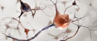
The dendrite of a neuron (dendrum - branch) is a process of the neuron body through which it receives a signal from other cells. The dendrite receives a signal from the axon of another neuron or a receptor protein that responds to the environment.
Answering the question of what dendrites are, we can say that traditionally dendrites are considered as antennas of a neuron. Information exchange occurs in one direction: from axon to dendrite. The more dendrites a neuron has, the more information channels, the more complex decisions the neuron makes.
Synaptic cleft
You may be interested in: Semantic barriers and ways to eliminate them
The signal from other cells reaches the body of the neuron along one of its dendrites. The dendrite in the human nervous system usually receives a chemical signal (neurotransmitter) from the axon. The junction of a dendrite and an axon is called a synapse.
Synapses allow precise messages to be passed from neuron to neuron. Thanks to synapses, there is neuroplasticity and the ability to fine-tune the functions and behavior of the body.
The dendrite contains receptors that receive the neurotransmitter. Receptors are specialized proteins that capture a neurotransmitter molecule and, depending on their type, trigger further reactions in the cell.
Functions
The axon and dendrite, despite their different morphological structures, have similar functions - they serve as connecting elements, thanks to which interaction between all cells of the body is maintained, and all physiological processes are integrated. The main function of the dendrite is to perceive signals from other nerve cells and receptors of internal organs. Dendritic branches also perceive external stimuli.
As a result, synaptic connections of several types are formed - axonodendritic (dendrite-axon contact), dendro-dendritic (dendrite-dendrite contact), axospinous (axon-dendritic spine contact). The received impulses travel to the body of the neuron. The terminal segments of dendritic processes serve as a site of synaptic contact, from where inhibitory and excitatory stimuli arrive at the soma. Thanks to synaptic contacts, one neuron is connected to numerous (over 20 thousand) nerve cells.
Recent studies show that dendritic processes are capable of independently generating signals. Previously, it was believed that only the cell body generates impulses; the role of the processes is reduced to signal transmission. Scientists discovered that dendritic spines were much more active than somas when they studied the nature and strength of signals transmitted within nerve tissue. The signal passing along the trunk of the process may change.
Dendritic spines
Small growths called spines form on the dendrites. The latter can take many forms, but the most persistent is that of a fungus.
The number of dendritic spines ranges from 20 to 50 per 10 µm of dendrite length. The spines are very variable in shape and volume.
There are 86 billion neurons in the brain. Axons, dendrites and cell bodies of neurons form huge neural networks.
Dendrites are responsible for learning and memory, and also control the balance of the system. When there is a local strengthening of connections between certain neurons, it is in the dendrites that the production of a protein increases, regulating the decrease in the activity of other synapses.
How the structure of a neurocyte is related to its functions
Electron microscopic research methods confirmed the fact of high specialization and complex structure of an open biological system called a nerve cell. It contains a body (soma), one long branch - the axon and many short processes. Each of them is connected to the cytoplasm of the neuron body. This is a dendrite. The structure and appearance of a collection of short shoots resembles the crown of a tree. Through them, bioelectric potentials from other nerve cells enter the neuron body through synapses.
Training and Spikes
Dendritic spines are responsible for the possibility of learning and memory formation. Thanks to the spines and their plasticity, a neuron can easily connect to one or another neighbor and quickly disconnect from them, controlling the possibility of receiving a signal.
It would be logical to assume that if synaptic connections are responsible for memories, then their plasticity is a problem for maintaining memories of the past. In 2009, Nature published a publication in which the authors examined how learning experience affects synaptic connections in mice.
The work showed that a large number of new spines formed from a new experience disappeared over time if the experience was not repeated periodically. But those that were preserved were most likely responsible for the acquired skills.
Moreover, if the training was repeated for a long time, spines were removed; apparently, the removed spines were responsible for incorrect actions. Learning and daily sensory experience leave permanent marks in the form of a small group of spines formed at different stages of learning.
What are dendrites if not a huge library of memories? But the main problem of dendritic spines is that they are very sensitive to any mechanical and chemical influences. Therefore, brain injuries, even if localized in one place, usually affect the entire neural network.
Morphology and types
According to modern histological studies, dendrites are the branching ends of a neurocyte that not only receive, but also transmit information encoded in the form of electrical impulses through a multichannel system of anatomically and functionally interconnected nerve cells. They contain a large number of protein-synthesizing organelles - ribosomes. Some types of short processes, for example in pyramidal neurocytes, are covered with special structures - spines.
According to the classification proposed by the Spanish neurohistologist S. Ramon y Cajal, two dendrites can extend from the body of a nerve cell in opposite directions (bipolar neurocytes). If there are many dendrites, then they diverge radially from the soma. This structure is characteristic of interneurons. In the cerebellar Purkinje cells, processes extend from the body of the neurocyte in the form of a fan. Each dendrite, whose structure is three-dimensional, differs from neighboring branches in the amount of electrical charges accumulated on it.
Sleep and learning
A 2014 study by ZG Yang showed how, after 24 hours of training and sleep, new dendritic spines appeared in mice and some of the existing ones disappeared. The authors note that the rate of formation of new spines in mice trained to learn a new behavior was significantly higher within 6 hours of training compared to untrained mice.
In addition, the authors showed that spines form much more slowly when mice are deprived of sleep. And neither new skill training nor late sleep can correct the situation.
Dendrite structure
Based on the study of microscopic preparations of nerve cells, it was established that most of the processes are cylindrical in shape. Their diameter averages 0.9 microns. The length of dendrites varies widely. For example, stellate neurons of the gray matter of the cerebral cortex have short (no more than 200 μm) branches of the dendritic tree, while the processes of the motor neuron entering the anterior horn of the spinal cord are about 2 mm.
Special formations - spines, formed on the branches of neurocytes, lead to the appearance of a large number of synapses - slit-like places of contact with the axon, dendrite or soma of another neuron. Synapses can be located on the body of the dendrite and are called stem synapses or directly on its spines. As we already know, dendrites are branched processes of neurocytes that can receive excitation. The transfer of biopotentials occurs in them with the help of molecules of chemical compounds - mediators, for example, GABA or acetylcholine. In the membrane covering the dendrite, ion channels were found that selectively transmit calcium, sodium and potassium cations, which are involved in the passage of nerve impulses through the neuron.
Dendrite as an independent unit
What dendrites are is still being figured out. The fact is that it is difficult to study the behavior and functions of dendrites in living objects.
If the size of a neuron is about ten microns, then the length of a dendrite can reach up to a thousand. Dendrites are usually understood as not very active participants in the process.
In 2021, a study was published in the journal Science that reconsiders the classic view of dendrites. It turned out that dendrites generate signals several times more often than the neuron body does, which suggests that information is encoded at the dendritic level as well.
It was previously discovered that if during an experience the cell bodies of neurons were activated and the dendrites were silent, then long-term memory was not formed regarding this experience. It was suggested that the activity of neurons is associated more with real time, with current experiences, and dendrites - with what will remain in memory.
What are dendrites, given the new data? These are amazing structures that make up 90% of the nervous tissue and perhaps take on most of the work of storing and transforming experience.
quiz
1. What are dendrites? A. Projections from neurons that transmit information to postsynaptic neurons. B. Projections of neurons that receive information from presynaptic neurons. C. Projections of neurons that release neurotransmitters. D. Neuronal projections that allow movement.
Answer to question #1
B is correct. Dendrites are tree-shaped projections of neurons that receive chemical signals from presynaptic (ascending) neurons.
2. What are the main functions of dendrites? A. Receive information (chemical signals). B. Process information. C. Transfer of information to the soma (cell body). D. All of the above.
Answer to question No. 2
D is correct. Dendrites receive information (chemical signals) from presynaptic neurons, then process this information and transmit it to the soma in the form of electrical impulses.
What is a dendrite?
Dendrites are an important part of any neuron, since they are responsible for receiving an electrical signal from the axon of a neighboring cell through the synapse connection, and transmitting this signal to the soma of that neuron.
From the ancient Greek language, the word “dendrite” means tree. They are numerous elongated and branched formations that grow from the body of the nerve cell. In the case of spinal cord neurons, they grow and branch from long axons. At the cytoplasmic ends of the branched dendrite there are sites for the formation of synapse connections. The system of short processes also has cytoplasm within itself, which contains mitochondria, microtubes, vesicles and other organelles. In addition, it contains special chemoreceivers responsible for initiating the appropriate chemical reaction in the dendrite when it receives an electrical impulse from the axon of a neighboring neuron.