Spina bifida is a pathology in which a ruptured intervertebral disc protrudes in a person, resulting in compression of surrounding tissues and nerve fibers. Due to pinching of the nerve processes, severe pain occurs, motor activity decreases and sensitivity is lost.
Today, there are several versions of the occurrence of this disease, however, according to medical research, the main role in the development of the pathological process in children belongs to a deficiency of vitamins, primarily folic acid. The main cause of the development of spina bifida in adults is considered to be osteochondrosis.
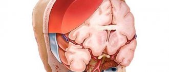
The severity of the disease is determined by the degree of damage to the spine and the volume of spinal tissue that formed the hernial sac.
If you are concerned about spina bifida, you can undergo treatment at one of the best medical clinics in Moscow - the rehabilitation center of the Yusupov Hospital. Thanks to the experience and highly qualified specialists, a multidisciplinary approach to the diagnosis and treatment of diseases, as well as the ultra-modern technical equipment of the Yusupov Hospital, our patients are able to achieve excellent results in a short time.
Causes of spina bifida in adults
Spina bifida can be congenital or acquired.
Acquired spinal hernia is characterized by slow development, which is not accompanied by any particular discomfort. However, at some point the hernial protrusion makes itself felt.
Spina bifida in adults can occur due to the following reasons:
- lack of treatment for osteochondrosis;
- previous spinal injuries;
- carrying unbearable weights;
- uncomfortable working postures;
- strength training;
- making sudden movements;
- excess weight.
The development of a hernial protrusion may be associated with incorrect posture, an imbalance of substances in the tissues adjacent to the pathological focus, and spinal infections.
Symptoms and complications of spina bifida in adults and children
With congenital latent spina bifida, specific symptoms are usually absent, appearing only in adulthood with intense physical activity. Often, the presence of a congenital defect is discovered during a diagnostic examination when an adult patient complains of back pain.
The following clinical manifestations are characteristic of spina bifida:
- in the spine area a round protrusion with a soft consistency is visualized. The formation is covered by thin, shiny skin of a bluish or red color;
- the patient experiences muscle weakness in the arms or legs, paresis and paralysis, and flexion contractures form;
- Sensitivity of all types is impaired (pain, temperature, tactile);
- reflexes of the hands and feet (plantar, grasping, knee) decrease, up to their absolute extinction;
- the muscles of the limbs atrophy;
- the development of trophic disorders is noted (dry skin, cold extremities, non-healing ulcers);
- changes in pelvic functions (incontinence or retention of urine and feces);
- defects of the lower extremities appear;
- the development of hydrocephalus (dropsy of the brain), associated with excessive flow of cerebrospinal fluid into the sinuses of the brain;
- vision is impaired, epileptic seizures occur, and mental retardation develops.
Patients with severe neurological symptoms require constant care at home, regular observation by medical personnel, and supportive treatment. Such patients often develop diseases of internal organs. Impaired urination is often accompanied by the development of pyelonephritis, which forms severe renal failure. Fluid retention in the brain can lead to an increase in intracranial pressure, as a result of which areas of nervous tissue atrophy and inflammatory processes develop: encephalitis, meningitis. Prolonged bed rest threatens the development of congestive pneumonia (pneumonia) and, as a consequence, pulmonary heart failure.
Army service
We cannot help but touch upon the issue of the army, because the male sex often suffers from this disease. Therefore, we want to immediately inform you about whether they will be recruited into the army if the young man’s medical history includes a herniated disc. In most cases, the conscript is given an exemption.
The pathology we are considering does not allow intense physical training, and is also often accompanied by quite serious complications. A person is shown a special way of life: constant medical supervision, immobilization, proportionate gentle loads and strict adherence to a treatment and prophylactic course.
But it is worth considering that the medical commission considers each case individually, based on the complexity of the specific problem. Uncomplicated, asymptomatic and mild forms of the disease are not an absolute contraindication for enlistment in the military.
To obtain an exemption from the army, you must provide a medical commission with medical information. documentation (together with photographs) from the main place of treatment confirming the diagnosis. Based on the severity of the clinical picture, a verdict will be made: service with restrictions; stock; deferment, when the recruit must recover before the next conscription campaign; complete exemption from conscription (unfit).
Diagnosis of spina bifida
Diagnosis of spina bifida at the Yusupov Hospital is a comprehensive examination. First of all, the doctor collects anamnesis - finds out the patient’s complaints, studies medical documentation, the results of previous examinations and records of specialists.
Then a neurological examination of the patient is carried out: a specialist visually examines the protrusion in the spinal column, assesses muscle tone and strength in the limbs, and determines the intensity of reflexes in them.
Additionally, the following diagnostic studies are prescribed:
- transillumination - the hernia is illuminated, revealing the nature of the contents of the hernial sac;
- computed tomography and magnetic resonance imaging – for layer-by-layer study of the spinal cord in the area of the pathological focus in order to identify concomitant damage to brain tissue;
- myelography - a contrast agent is injected into a peripheral vein and accumulates at the site of pathology, which allows one to assess the degree of damage to the nervous tissue in the hernial sac.
Localization feature l4 l5
A designation such as l4 l5 means that between the 4th and 5th lumbar vertebrae the integrity of the cartilage lining is broken. This object affects the L4 nerve, which is part of the structure of one large nerve - the sciatic. If we take into account the corresponding part of the spinal column, such damage to the l4 l5 disc (treatment is urgently necessary!) occurs in 46% of cases. In 48% - between the fifth lumbar body and S1 (first sacral), which is not much more common.
Zone l4 l5, the focus is indicated by arrows.
A flat subligamentous hernia l4-l5, known in medicine as hernial sequestration, is the final stage of the disease. With such unfavorable development, the contents of the disc (nucleus pulposus) begin to flow in parts into the cavity of the spinal canal, which is fraught with the complete disappearance of voluntary movements in the limb. When the subligamentous process has begun, even sneezing and coughing cause incredible pain to a person in the form of a terrible lumbago in the lower back, not to mention performing physical tasks. In 80% of cases, the final stage ends in disability. If you undergo emergency surgical treatment, and after it are well rehabilitated, you can hope for 70-80% of the restoration of supporting and locomotor functions as close as possible to normal.
Clinically, the disease in this place manifests itself in a special way, so it is not difficult to distinguish it from a problem, for example, with the cervical area C4 C5. Firstly, there is local hyperalgesia (shooting, aching, pulling in the lower back), and there is also pain radiating to the right or left leg. In addition, the patient complains that the leg is numb, especially in the area of the lower leg and foot, and also that the knee bends insufficiently or muscle-ligamentous weakness is felt in the ankle area. Additionally, increased sweating, marbling of the skin, and curvature of the spine may occur.
Do not sleep on a hard bed or lie down or sit on a cool surface. In case of intervertebral hernia, as orthopedic specialists warn, this is strictly contraindicated.
Spina bifida: treatment, patient reviews
To treat this disease, only a radical method is used - the pathological formation in the spinal column is surgically removed; treatment of spina bifida without surgery is ineffective. During surgery, the defect in the spinal column is reconstructed and the unclosed hole in the bone tissue is closed. After a thorough examination of the hernial sac, non-viable tissue is removed from it, and healthy spinal cord structures are placed in the spinal canal.
Patients with spina bifida may develop hydrocephalus, which threatens irreversible changes in the brain. In order to prevent the harmful effects of excessive intracranial pressure, a shunt is installed to divert cerebrospinal fluid into the thoracic lymphatic duct.
To prevent the progression of the disease and improve the general condition of the patient, specialists at the Yusupov Hospital prescribe conservative therapy:
- taking medications that normalize the function of nervous tissue (nootropics, neurotrophics);
- taking vitamins A, C, E, B - to improve metabolic processes in the affected area of the spinal cord;
- physiotherapeutic procedures, according to patient reviews, can significantly restore tissue trophism and motor activity (laser, magnet, etc.);
- physical therapy – for patients diagnosed with spina bifida, exercise therapy helps develop neuromuscular connections in the affected area of the body;
- diet – a large number of foods enriched with coarse fiber are introduced into the diet of patients, which helps to normalize intestinal motility.
basic information
Between the vertebral bodies there are layers of cartilage - these are intervertebral discs. They consist of a ring-shaped element (annulus fibrosus), inside of which there is a jelly-like substance (nucleus pulposus), which has good elasticity. Intervertebral discs are the main shock absorbers of the spine. When walking, running, and even while standing, they help soften the vertical pressure (axial load) on the spinal column and dampen vibrations when the body moves.
Under unfavorable factors that worsen metabolism in the osteochondral components of the spinal system, degenerative and dystrophic processes in the structure-forming units of the shock-absorbing layer are activated, due to which the fibrous ring is destroyed and torn. The gelatinous substance, in turn, begins to put pressure on the damaged area, which leads to protrusion of the disc from a certain side. Since the protruding part is usually located in close proximity to the spinal nerve formations, irritation occurs of these nerves with which the pathological protrusion comes into contact. Thus, a person begins to experience painful symptoms in the affected area of the back or neck, often radiating to the leg or arm.
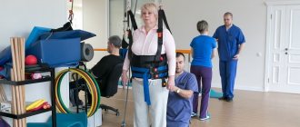
Spina bifida: rehabilitation after surgery
Surgical removal of a hernia is not the only and final stage of treatment. To consolidate the effect of the manipulations performed, the patient needs high-quality postoperative rehabilitation. An individual rehabilitation treatment plan at the rehabilitation center of the Yusupov Hospital is drawn up by highly qualified experienced rehabilitation doctors.
Competent rehabilitation after removal of spina bifida allows you to achieve the following results:
- eliminate pain syndrome as soon as possible;
- prevent possible complications (infections, thrombosis, scars, etc.);
- bring general health indicators back to normal;
- restore muscle strength of the limbs, abdomen, back;
- restore the functionality of the spine;
- master the skills of proper load distribution;
- correct defects in posture and gait;
- return the patient to a full life with a minimum number of restrictions;
- minimize the risk of relapse of the disease.
You can make an appointment with a rehabilitation specialist at the rehabilitation center and find out the cost of medical services by calling the Yusupov Hospital or on the clinic’s website by contacting the coordinating doctor through the feedback form.
Foraminal type and disc protrusion
Foraminal hernia and protrusion are disc bulges facing the aperture where the nerve roots emerge. In other words, the lesion migrates to the narrowest opening in the spine, which in Latin is called “foramen” (intervertebral foramen). This compartment is formed by the posterior arches of two adjacent vertebral bodies. The spinal nerves are freely located in it. When a foreign body appears, especially a larger one, it becomes oppressed, irritated, inflamed, and compressed.
Protrusion.
This is also one of the alarming diagnoses, which differs from others in its rapid progression. It causes the so-called neurocompression syndrome, namely, compression of the roots, which makes itself felt by sharply occurring, pronounced, local attacks of pain, which forces a person to take a forced adaptive posture. When changing position, the pain only intensifies. Analgesics and NSAIDs provide a short-term and insignificant effect.
Surgery relieves persistent excruciating pain that cannot be relieved. Such a fatal stage as sequestration occurs within a fairly short time after the formation of protrusion. Fortunately, this classification of the disease is diagnosed quite rarely; of the total number of all possible variations of protrusion, it accounts for 7%.
Therapeutic drug blockades in the form of hormonal injections, which are given directly to the diseased area, cannot be performed on yourself! This is too complex a manipulation that requires high precision needle insertion and excellent professionalism. Local corticosteroids are indicated in the most extreme situations, if there are debilitating pain symptoms that do not go away after the use of NSAIDs. And remember, hormones do not resolve the hernia, but only relieve local swelling, thereby reducing pressure on the nerve endings.
