Benefits of MRI
Magnetic tomography allows you to obtain a three-dimensional image of the areas under study in three projections. During the procedure, the device takes many slices, the thickness of which can be set individually and is usually 2-4 mm.
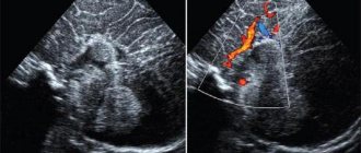
Images taken using a tomograph
Obtaining a large number of sections allows you to examine the entire organ and detect even the slightest abnormalities and pathologies.
What types of tomographs are there?
Modern magnetic tomographs are available in various variations with a wide variety of characteristics.
All tomographic devices are divided into:
- open;
- closed.
Despite the fact that conducting a study in an open tomograph is usually considered more comfortable for the patient, closed devices have greater power and detail. If the patient does not have a strong fear of closed spaces and does not have weight restrictions, it is recommended to conduct the study in a closed-type apparatus.
Tomographs are also divided according to the strength of the magnetic field radiation, the unit of measurement of which is called Tesla. Magnetic tomographs can be:
- low-floor – power up to 1.0 T;
- high-field – radiation intensity above 1.0 T.
Low-field tomographs do not provide a clear and detailed picture. A study using a high-field tomograph will allow you to examine the diagnosed area with the highest accuracy.
Modern high-field tomograph
The DiMagnit clinic has a closed-type tomograph from Philips, whose power is 1.5 Tesla. Using the device, it is possible to obtain images of the highest quality and detail.
Diagnostics in a private clinic
To conduct an MRI in a private clinic, the patient must have a referral from the attending physician, which includes the following information:
- Which organ should be examined?
- Are there any contraindications?
- Does the procedure require a contrast agent?
- Preliminary inspection data.
If there is no referral, the examination is not carried out even in a private medical institution. If the patient has a referral and there are no contraindications to the procedure, then he pays for the medical service and is examined. The patient will not have to wait long in line for the examination; at night MRIs are given a 50% discount.
In a private hospital, the patient will be treated by specialists of the highest category, whose work experience is at least 5 years.
to contents ^
Should you be afraid of the procedure?
Some patients are worried before the test. But their fears are in vain - magnetic resonance imaging is absolutely painless, and the effect of magnetic radiation on the body is safe.
Unlike other types of radiation diagnostics, MRI does not use ionizing radiation. The magnetic field does not have a carcinogenic or mutagenic effect on the cells of the body. Magnetic resonance scanning can be performed as often as needed.
MRI procedure, the essence of the method
A strong magnetic field allows the research to be carried out. The information received by the tomograph is transferred to a computer on which special software is installed. The program converts the received information into a two-dimensional or three-dimensional image.
Thanks to magnetic resonance imaging, organs and tissues of the body are displayed in high quality. Magnetic resonance is the response of hydrogen atoms in the body when a magnetic field is applied to the human body. Hydrogen atoms line up parallel to this field. During the end of the radio waves, the hydrogen protons will return to “their zones” (the usual chaotic position). Then they will begin to emit a radio signal, which is processed in a computer, creating a three-dimensional image of the internal organ.
The diagnostic process will not cause any harm to the human body.
The study is carried out with contrast; the patient should have no contraindications for this substance. The patient lies down on a couch, which is placed in a tomograph, where a magnetic field is applied to the person to obtain information. This is a closed magnetic resonance imaging scan. If a person is afraid of closed spaces, then diagnostics are carried out in open-type devices. The disadvantage of such tomographs is that the results obtained are less accurate.
to contents ^
The difference between MRI and CT and ultrasound
Magnetic resonance diagnostics has a number of advantages compared to ultrasound and computed tomography.
Ultrasound examination allows you to obtain a two-dimensional image of the area under study, but does not allow you to see a three-dimensional image of soft structures.
Computed tomography can be compared to MRI in terms of image clarity, but has a number of serious contraindications. CT is more often used to visualize hollow organs and bone structures, while MRI is much more effective at visualizing soft tissue.
What does an MRI show?
Magnetic resonance imaging is successfully used to diagnose diseases:
- thyroid gland;
- liver;
- gallbladder and ducts;
- pancreas;
- kidney;
- spleen;
- joints;
- spinal cord;
- vessels of the head, neck, abdominal region;
- pelvic organs;
- soft tissues;
- etc.
All of the above anatomical structures are perfectly visualized on MRI images. The diagnostic results make it possible to accurately identify abnormalities in the functioning of the organs being examined.
Is it possible to understand the MRI results yourself?
On Internet forums you can see many posts on the topic “Help me understand the results of an MRI.” Often such “pleas” are accompanied by detailed descriptions of the examination results. Is there any point in this?
A specialist will already understand everything from the attached descriptions, but a patient, in order to understand everything, needs at least a basic level of medical knowledge, and not everyone has it. People who request such services know absolutely nothing about the qualifications of the person who will write them a response. Such hope for a kind uncle can end badly.
The situation is even worse when people, armed with books and the Internet, try to “decipher” something while simultaneously prescribing treatment for themselves. In this case, the result can be completely sad.
To understand what was discovered in your possession, you need to have appropriate education, a sufficient level of knowledge and good experience. Sometimes cancer and harmless pathologies give a similar picture, especially if contrast was not used. MRI specialists are constantly trained and attend special seminars to detect diseases with greater accuracy. What can we say about people who are far from medicine? So, no one other than a doctor can give you a correct diagnosis based on the results of magnetic resonance imaging.
In what cases is MRI prescribed?
The wide capabilities of magnetic resonance diagnostics make its use indispensable in the following cases:
- the need for a primary diagnosis;
- conducting a comprehensive survey;
- preparation for surgery;
- monitoring the effectiveness of the therapy and treatment methods used.
In each individual case, the choice of diagnostic technique is made by the attending physician. Magnetic resonance imaging is more often used than other methods to detect diseases and injuries of soft tissues.
The MRI technique is indispensable for diagnosis:
- Neoplasms.
Magnetic resonance scanning can clearly identify the boundaries and size of the tumor and the extent of its invasion into soft tissue. No other radiation diagnostic technique is capable of providing such a clear and detailed picture of diseases.
MRI also makes it possible to determine the nature of the tumor with a high degree of probability. Malignant neoplasms have unclear boundaries and grow into surrounding tissues. Benign neoplasms, as a rule, are clearly differentiated from healthy tissues.
- Brain diseases.
The greater accuracy of magnetic resonance diagnostics makes it possible to visualize such small anatomical structures as the pituitary gland and the sella turcica. Also, MRI with contrast of the brain shows high efficiency for diagnosing demyelinating diseases (multiple sclerosis, Parkinson's disease, etc.), as it allows one to clearly see the structure of altered nerve tissues.
Brain images obtained with contrast-enhanced MRI of the brain are particularly clear because magnetic waves are difficult to image solid anatomical structures and there are no artifacts from the skull in the brain images.
- Diseases of the intervertebral discs.
MRI images of the spine
Magnetic resonance imaging is the only diagnostic method that allows you to see intervertebral discs. Even modern diagnostic methods, such as computed tomography, allow you to see only the space between the vertebrae, while MRI gives a complete picture of the condition of the discs, the possible presence of hernias and protrusions.
The use of MRI is not limited only to the above diseases, but is used when it is necessary to identify and monitor a wide range of pathologies, congenital developmental anomalies, consequences of injuries and previous surgical interventions.
If an MRI of the brain showed...
There is probably no person in the world who has never had a headache in his life. “Can you do an MRI of the brain?” is one of the most common questions received by our diagnostic centers. But even more questions arise for those who have undergone the examination. Serious diagnoses are not always hidden behind the terms used by MRI diagnostic doctors when describing images. That’s why we always insist: don’t take your fears with you. We are always there to help you figure it out and refer, if necessary, to a doctor of the required specialization; explain what is hidden behind this or that, sometimes frightening name.
In this article, we have collected questions regarding magnetic resonance imaging of the brain and its vessels that patients ask us most often.
Answered by Mikhail Kosygin – radiologist at MRT Expert Lipetsk LLC
— What does a brain MRI diagnose?
MRI of the brain allows us to identify space-occupying formations of various origins, congenital developmental anomalies, ischemic and hemorrhagic strokes, hemorrhages in the area of the meninges, inflammatory and demyelinating processes, and much more.
We talked about the topic of MRI of the brain on the pages of our website earlier. They raised the question “why undergo an MRI of the brain when nothing hurts” and talked about “how MRI will help a patient with a traumatic brain injury and its consequences.”
— Will MRI help identify the causes of high blood pressure?
If the increase in pressure is a consequence of arterial hypertension of idiopathic (i.e., unclear) origin, then this cannot be determined during an MRI study. But there is symptomatic arterial hypertension, which is not an independent disease, but accompanies, as a symptom, another disease. Based on their origin, such diseases are divided into four groups: renal, endocrine, neurogenic and hemodynamic. Most of them can be identified on MRI and it can be assumed that they are the cause of the patient’s increased blood pressure.
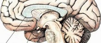
— What is the quadrigeminal plate in the brain?
The quadrigeminal midbrain is a plate-shaped formation located in the roof of the midbrain. The quadrigeminal tubercles are independent structures. In this case, the superior colliculi function as subcortical formations and work as centers of the visual analyzer. The lower tubercles serve as subcortical formations, working as centers of auditory analyzers.
— What to do if an MRI showed the presence of an angioma?
Brain angioma is a vascular formation of benign origin and looks like a tangled tangle of blood vessels. Such formations come in various sizes and degrees of fullness.
If MRI reveals an angioma, we recommend monitoring the formation once a year in order to prevent the occurrence of extremely rare, but still occurring complications in the form of hemorrhages.
Answered by Alexander Samaev, radiologist at MRI Expert Lipetsk LLC
— What is the pathology of the intracranial arteries of the brain?
The arteries supplying blood to the brain have cervical (extracranial) and intracranial (intracranial) sections. Consequently, this is a pathology of the intracranial sections of the arteries supplying blood to the brain.
— Should there be focal changes on MRI in ischemic stroke?
Ischemic stroke can manifest itself in both focal (up to 1.0 cm) and focal (more than 1.0 cm) changes and even larger areas. Therefore, the presence of focal changes on MRI depends on the caliber of the affected vessels (the main large arteries of the brain or small capillaries are affected).
— What is caudal displacement of the cerebellar tonsils?
Normally, the lower part of the cerebellum, which is the tonsils, is located at the level of or above the foramen magnum (i.e., the hole through which the spinal cord exits).
Caudal displacement of the cerebellar tonsils is a displacement below the level of this foramen, into the lumen of the spinal canal. The condition is diagnosed by space-occupying formations in the brain and indicates that there is not enough space in the skull.
— What are the dangers of a pineal gland cyst?
We find this formation quite often during MRI of the brain and classify it as an accidental finding. That is, the patient came for the study because he was bothered by some symptoms associated with this disease. A pineal gland cyst most often does not manifest itself with any symptoms.
However, if it begins to grow rapidly, this can lead to disruption of liquor dynamics and be the cause of internal hydrocephalus.
Therefore, if the diagnosis reveals a pineal gland cyst, then the recommendation to monitor it is justified. There is another reason for constant dynamics in this pathology - in the initial stages, tumors of the pineal gland can look the same as a cyst.
Answered by Vladimir Mikolaichuk – radiologist at MRI Expert Lipetsk LLC
— In what cases is it necessary to study the veins of the brain?
Examination of the veins (or MR venography) is necessary if thrombosis of the sinuses (“large veins”) of the brain is suspected; for the diagnosis of vascular malformations (so-called structural and developmental anomalies); when assessing the degree of germination of a brain tumor into a venous vessel.
In order to assess the nature of the changes, there is a need to compare MR venography data with the picture revealed during an MRI study of the structure of the brain.
For example, the narrowness of the sinus of congenital origin and its thrombosis may look almost identical when performing MR venography. But with an MRI examination of the brain, we will see a pathological signal from a thrombosed vessel.
— Is it possible to detect meningitis using MRI?
Yes, you can. If a patient has meningitis, an MRI of the brain with a contrast agent will identify structural changes in the membranes of the brain, accompanied by intense accumulation of contrast.
But we see a similar picture in other diseases. For example, with sarcoidosis, cancer, intracranial hypotension and a number of other pathological conditions, therefore, an important factor in making a diagnosis will be a comparison of MRI diagnostic data with the general clinical picture, anamnesis and, of course, laboratory test data.
***
Especially for our patients, the #GKExpert Encyclopedia was created on the company’s official public pages on social networks Vkontakte, Facebook, Odnoklassniki, within which a team of doctors from the medical group answers questions from our patients. You don't know what's wrong with you? us.
When to Apply Contrast
Magnetic resonance diagnostics can provide a very high degree of clarity of the resulting images. In most cases, the use of contrast is not required.
But when it comes to diagnosing tumors and small anatomical structures, a contrast agent can still be used.
The staining agents are made from the rare earth metal gadolinium and are injected intravenously into the patient during an MRI.
MRI contrast agents are much better tolerated than CT contrast agents. This makes the use of the dye safe even for patients with kidney pathology and does not require a preliminary test for creatinine, which is necessary for CT diagnostics with contrast.
MRI with contrast is used in the following cases:
- suspicion of a neoplasm;
- the need for differential diagnosis of a malignant tumor;
- study of the pituitary gland;
- the need to diagnose demyelinating diseases.
The use of contrast allows one to obtain a comprehensive picture of the disease, its course and the effectiveness of the therapy used.
Contraindications for MRI
Despite the fact that magnetic resonance diagnostics is a safe technique, the study has a number of absolute contraindications, in the presence of which diagnostics are prohibited:
- presence of a pacemaker, neurostimulator, insulin pump;
- vascular clips on the arteries of the brain;
- the patient’s inability to maintain a stationary position for various reasons;
- early childhood up to 5 years;
- the patient’s weight is more than 130 kg and body girth is more than 150 cm;
- first trimester of pregnancy;
There are also a number of conditions in which MRI examination is carried out with caution:
- severe pain syndrome, in which it is difficult for patients to remain in a motionless position for a long time;
- fear of confined spaces;
- psychical deviations;
- second and third trimester of pregnancy.
The presence of various prostheses and implants in the patient's body may be a contraindication to MRI if they are made of metals sensitive to magnetic radiation. Modern medical devices are most often made of titanium and other materials that are inert to the effects of magnetic fields. Their presence in the body does not interfere with MRI.
Indications and contraindications
MRI reveals:
- Benign and malignant neoplasms.
- Vascular pathologies, diseases of the nervous system.
- Problems with the heart, kidneys, brain.
- Injuries.
- Diseases of the spinal column, joints and other pathological conditions.
The specialist evaluates the general indications for MRI:
- A disease with a similar lesion and the same symptoms is differentiated.
- The severity is assessed once the diagnosis is established. The study will determine the depth of pathological changes, and other disorders may additionally be revealed.
- The effectiveness of the therapy is assessed.
The doctor determines contraindications for the examination. They are absolute and relative. The absolute ones include:
- Presence of a pacemaker.
- Metal implants, magnetized metal structures, bullets with fragments inside the body.
- Early pregnancy.
- Clips on vessels.
- Relative contraindications include:
- Pregnancy in the second, third trimester.
- Fear of closed spaces.
- Body weight exceeding more than 140 kg.
- Convulsive syndrome.
- Hemolytic type of anemia (some contrast agents have a destructive effect on red blood cells).
- The patient is feeling unwell (if the pain is severe, the person will not be able to lie still).
to contents ^
How is an MRI performed?
Before the procedure, the radiologist questions the patient about the presence of contraindications to the study. The patient is asked to remove any metal accessories, including clothing with metal fittings, and lie down on a couch, which is then placed in the CT scanner tube.
During the diagnosis, it is strictly forbidden to move, as this may affect the clarity of the resulting images.
Scanning procedure
High-field tomographs produce a fairly high level of noise, which can cause some discomfort to patients. The medical department will provide you with headphones that will play pleasant music, drowning out the sounds of the operating device.
The tomograph scans the patient’s body in different projections and instantly transmits the images to the computer screen. The interpretation of the results by the research physician begins even before the procedure is completed.
Getting Results
Scan results are available immediately after the end of the study. Many of the images obtained are carefully examined by a radiologist, and a detailed report is drawn up describing both the normal anatomy of the area under study and possible abnormalities and pathologies.
15-30 minutes after the procedure, the patient is given a written report and a computer disk with the resulting images.
Magnetic resonance imaging is a modern, safe type of radiation diagnostics that allows you to obtain accurate and quick results and thoroughly examine the area under study. MRI helps to identify many diseases and abnormalities even at the initial stages of their development.
| MRI of the pancreas in Rostov-on-Don |
| MRI of the abdomen with contrast |
| MRI of the spine, what does it show? |
| MRI of ureters |
| MRI of the abdominal cavity, which organs are checked? |
| What does an MRI of the knee show? |