What is MRI of the brain based on?
The diagnostic effect of brain MRI is based on nuclear magnetic resonance. In response to the powerful radiation created by the generator, the hydrogen atomic nuclei contained in the tissues line up along the electromagnetic field lines and begin to vibrate. Each atom becomes like a spinning mini-spinning top, emitting energy waves.
Different structures emit different amounts of energy - some give it out more intensely, while others less so. The difference is recorded by a device that takes pictures (slices) in different projections.
To do this, the patient is placed inside a tomograph in which generators maintain a high-frequency electromagnetic field. Special radio emitters generate pulses, and coils record the energy sent by vibrating atoms.
The resulting slice images are combined into a three-dimensional matrix using a special computer program, in which dark or light unhealthy areas are visualized against a gray background.
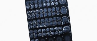
MRI of a child's brain
The data obtained indicate that more than 70% of the examined patients with ASD had either a history of inflammatory processes, which occurred with complications characteristic of neuroinfection (see Fig. 7), or signs of a chronic inflammatory focus (including on moment of examination) in the structures of the facial skull, anatomically associated with various parts of the brain (see Fig. 1,2), which, apparently, could be the cause of intoxication and allergization of various parts of the central nervous system.
This fact is also confirmed by the fact that 40 patients out of 61 examined showed signs of hypertrophy of the adenoid tonsils, which amounted to 65% of the total number of patients (see Fig. 3)
Also noteworthy is the fact that the ratio of changes detected on MRI is almost equal in the group of patients with childhood and atypical autism (see Diagram 2), which once again indicates the possible trigger role of an infectious-allergic factor in the etiology of the development and course of these forms of autism.
In turn, the presence of changes such as midline cysts and dysgenesis of the corpus callosum (in 37% of cases), related to anomalies in the development of the central nervous system (see Fig. 4, 5, 6), is more indicative of a genetically determined cause of autism .
Advantages of magnetic resonance imaging over other methods
An MRI examination gives results much more accurately than x-rays, echoencephalography (EchoEG), ultrasound and other diagnostic options. It allows you to obtain maximum data about existing tumors, diseases, post-traumatic and post-stroke changes. Unlike CT and X-rays, in this case the body is not irradiated.
Only soft tissues are visualized in the finished images. The bones of the skull are not visible, so they do not interfere with analysis and decoding.
The contrast agent used in MRI diagnostics is much less likely to cause allergic reactions compared to X-ray contrast agents used for radiography.
How does the procedure work?
The patient removes all metal jewelry and takes out removable dentures containing metal.
The patient is placed on a movable table and secured with special belts. This measure is necessary because you will have to lie inside the tomograph, remaining motionless, for a long time.
A device equipped with wires that transmit and receive radio signals is placed on the head. The equipment is quite noisy and tires with constant clicks and whistles. Therefore, the patient’s ears are protected with earplugs. After this, the table slides into the device, and the specialist sits down at the computer, in which the transmitted data is analyzed and processed.
The technique takes pictures, the quality of which depends on the characteristics of a particular MRI scanner. The thinner the visual slices the equipment makes, the more accurate the final images will be. The duration of diagnosis is 20-30 minutes, and when contrast is used - up to an hour.
After MRI diagnostics, you can immediately return to your normal life. No side effects subsequently or during the MRI examination occur, with the exception of an extremely rare allergy to gadolinium salts.
Finished photographs are handed out printed or recorded on a magnetic medium - disk or flash card. It can be sent to email with SMS notification.
Types of MRI of the brain
- Standard - done without the introduction of contrasting solutions, but at the same time provides a sufficient amount of information.
- With contrast, before which drugs containing gadolinium salts are injected into the vein - gadopentetic and gadoteric acids, Omniscan, Magnevist, etc. These solutions penetrate into the bloodstream and, once in the rays of the MRI scanner, highlight the resulting “picture”. At the same time, the changed areas become better visible, which simplifies decoding. The technique is most often used to detect vascular abnormalities, multiple sclerosis and tumor formations. The dose of contrast agent is selected individually, taking into account weight.
- MR angiography – is performed to assess the condition of blood vessels in atherosclerosis, aneurysms, blood clots and pre-stroke conditions. It is done with gadolinium contrast to show blood flow problems in detail.
- MR imaging of the pituitary gland, an appendage that is an endocrine gland. The pituitary gland secretes hormones responsible for reproductive function, tissue metabolism and the regulation of human growth. An examination is prescribed if an adenoma is suspected - a benign tumor that causes migraine-like pain, hormonal imbalances, gigantism, infertility, obesity and sexual dysfunction. The same method is used to identify malignant pituitary formations that have similar symptoms and are accompanied by a pronounced deterioration in health.
How to prepare your child for the procedure
No special preparation is required before the MRI procedure. The day before, the baby can eat any food and drink water.
Parents can help their children prepare psychologically for the diagnosis by explaining the purpose and principle of the examination. It must be said that the baby will not experience pain or discomfort. One of the unexpected things that may come as a surprise to children during an MRI is the knocking and buzzing noise that the scanner makes. Explain to your child that there is no need to be afraid of sounds, that you will be nearby all the time, and that it is important for the baby himself to remain motionless.
In some MRI studies for children, specialists use a contrast agent that is injected into the body intravenously. Do not hide from your child that he will be given an injection, but be sure to note that this manipulation will be quick. If you hide the fact of the injection from your baby, he may enter a state of strong emotional stress, which is undesirable before the scan.
If time permits, ask the specialist to give your child a mini tour of the MRI room. This will allow him to calm down and tune in to the diagnosis.
Indications for general anesthesia for MRI
Intravenous or inhalational sedation is only necessary for patients who are unable to hold their body still for long periods of time. Main indications for general anesthesia:
- Claustrophobia is the fear of closed spaces. Such patients, while inside the device, experience panic, which negatively affects their health and makes MRI diagnostics impossible.
- Mental disorders accompanied by unpredictability of behavior and high excitability.
- Uncontrollable involuntary head movements (swaying, shaking, tics).
- Epilepsy and other types of convulsive readiness and seizures - anesthesia is given only intravenously due to the risk of provoking a seizure attack.
- Early childhood. Small children cannot lie still for a long time in an MRI scanner, so light mask anesthesia is indicated for them.
- Severe pain syndrome, in which prolonged stay in one position causes discomfort, cramps, pain and spasms.
Features of MRI for children
It is advisable to describe the procedure algorithm to the child in advance in a language he understands, and also emphasize its importance for making a diagnosis. To avoid frightening your child, you should also tell him about the noise made by the MRI machine. It is better to explain to the child in advance that there is nothing strange or scary in the sounds that he will hear during the study. In addition, for comfort, the child will be offered earplugs or headphones, as well as a rubber bulb or “panic button” (read more about it in this article). We advise you to agree with the clinic staff about the possibility of having a parent or accompanying person in the scanning room during the examination. Young patients may have an easier time undergoing an MRI if they know their parents or relatives are nearby to support them.
It is extremely important to explain to your child the need to remain still during the scan.
It should be emphasized that performing MRI on young children is difficult for several reasons:
- the child will have to be inside the tomograph tunnel, which may seem long and narrow to him, thereby frightening him;
- During diagnosis, patients must lie absolutely still, and this rule is especially difficult for children to follow;
- the operation of the tomograph, as we have already said, is accompanied by noise, which can give the child cause for concern.
In our center, due to such nuances, we accept children aged 8 years and older. But in some cases this limitation varies, because a child can be calm at an earlier age, or hyperactive at an older age. In other words, MRI can be done at any age, the main thing is to remain still during the examination. For the youngest patients, MRI is performed under anesthesia, but we do not perform it in our clinic - it is for this reason that, as a rule, we do not accept children under 8 years of age.
When registering your child for a study, check whether MRI is performed for children at the clinic, at what age children are accepted, and whether it is possible for an adult to be present during the scan.
The medical staff at our center is attentive and careful towards young patients. The doctor can encourage the child and give him confidence. Sometimes among the reviews that our patients leave us, you can find words of gratitude written by a child’s hand. Usually children always clarify that they “were not scared at all.”
Indications for magnetic resonance imaging of the brain
- Neoplasms or their metastases. Diagnosis is prescribed for persistent migraine-like pain, sudden loss of vision and hearing, auditory, olfactory and visual hallucinations, attacks of confusion, sudden reading and writing disorders, often accompanying cancer pathologies.
- Epilepsy and other diseases manifested by fainting, confusion and convulsions.
- Suspicion of single or multiple cystic cavities filled with fluid, bloody or other contents.
- Possible presence of parasites (cysticercus and echinococci) carried through the vascular bed with the bloodstream into the head.
- Inflammations – meningitis, encephalitis, arachnoiditis, myelitis. Lesions caused by infections - measles, herpes, tuberculosis, toxoplasmosis, tick-borne encephalitis.
- Rehabilitation after stroke, traumatic brain injury and surgery. Using magnetic resonance diagnostics, the doctor evaluates the effectiveness of the treatment and predicts long-term results.
- The likelihood of developing multiple sclerosis, Alzheimer's disease and other degenerative processes.
- Children are examined for congenital pathologies and hydrocephalus.
With all these diseases, life and health directly depend on a timely diagnosis. Therefore, at the slightest suspicion of brain disorders in yourself or your child, you need to come to the clinic and be examined.
About the areas of application of MRI studies in pediatrics:
- In cardiology Congenital heart defects, aorta, pulmonary arteries, vascular malformations (arteriovenous malformation);
- In neurology and neurosurgery Birth injuries of the head and nerve plexuses (birth paralysis), cortical dysplasia, congenital sensory hearing loss, benign and malignant tumors of the brain and spinal cord, congenital and acquired hydrocephalus;
- In oncology and hematology Neuroblastoma, lymphoma, leukemia, metabolic bone damage in various diseases, sickle cell anemia;
- In traumatology and orthopedics, pathology and diseases of the joints, household and sports injuries, osteochondropathy.
What the results show
MRI studies, especially those performed with contrast, reveal numerous pathological processes. In the sections, compactions, cystic cavities, and hematomas (collections of blood) are visible in detail. Scars, parasites and their cysts, foci of degeneration, sclerosis and inflammation are identified.
Vascular changes are diagnosed, manifested by impaired patency, narrowing or dilation of blood vessels, the appearance of aneurysms (protrusion of walls) and thrombosis.
The degree of tissue damage in traumatic brain injuries, hemorrhagic and ischemic strokes is determined. The affected areas look lighter and are visible even if they are small in size and have sparse neurological symptoms.
Congenital defects are determined - underdevelopment and hypertrophy of the organ, small and incorrectly located gyri, cysts, holoprosencephaly - lack of division into hemispheres. Hydrocephalus is detected - accumulation of fluid in the ventricles, which with this anomaly are greatly enlarged.
Pathological areas and neoplasms appear as dark or lightish spots of various sizes and shapes, standing out against a grayish background. Oncological seals, especially malignant ones, have blurred, uneven edges and surrounding areas of necrosis.
MR diagnostics is recommended periodically for everyone who has been treated for oncological pathologies of any location. It detects metastases, which usually accompany cancer recurrence.
Contraindications
- Installed pacemakers and other electronic devices that are disrupted by the surrounding electromagnetic field.
- Fixed dentures with metal elements present in the mouth, crowns containing metal, braces and other orthodontic structures. The metal they contain is heated by a magnet and deteriorates, damaging surrounding tissue.
- Tattoos applied to the skin, made with metal-containing ink. Due to the heat caused by the electromagnets, burns may occur in these areas. In the absence of information about the pigment. used when applying a tattoo, it is better not to risk it and do a CT scan, ultrasound or x-ray. Examination is also prohibited for any metal piercing that cannot be removed.
- MRI examinations with contrast are not performed during pregnancy or if contrast agents are intolerant. Such an examination is not prescribed for severe kidney pathologies that make it difficult to eliminate gadolinium.
Magnetic resonance imaging is a safe and highly informative procedure that detects pathologies in the early stages. Therefore, in case of migraine-like phenomena, coordination problems, a sharp decrease in hearing and vision, fainting, or progressive memory deterioration, you must definitely go to the clinic and be examined. The price of MRI diagnostics is low and quite affordable for Muscovites and residents of Moscow Region.
Get an MRI of the brain >>>