Electromyography (EMG) is a mandatory study if damage to the neuromuscular system is suspected. Such disorders accompany various diseases diagnosed at any age. EMG is a painless procedure, does not require preliminary preparation, and its indicators are quite informative for making a diagnosis. This makes muscle electromyography an indispensable study in neurological practice.
At the Yusupov Hospital, EMG is performed using modern electromyographs, and highly qualified personnel interpret the study indicators in the shortest possible time.
Electromyography: what is it?
Electromyography is a method for diagnosing disorders of the neuromuscular system, based on indicators of bioelectrical muscle activity. The study is based on the ability of muscle tissue to create electrical activity with each contraction. Electromyography records these values, resulting in an assessment of the results obtained. Depending on the indicators, taking into account the accompanying clinical picture, the lesion and its location are determined.
EMG is performed using an electromyograph. The device records bioelectrical activity, transmitting it to monitor screens or recording it on paper. Any deviation from normal values indicates a violation of muscle conduction.
Several research methods are used to take electromyography. The choice of the type of EMG is made by the attending physician based on the individual characteristics of the development of the disease. This study of muscle nerve conduction is used in various fields of medicine: neurology, traumatology and orthopedics, cosmetology, dentistry, sports medicine. Electromyography makes it possible to identify the pathological focus in the early stages of the disease. In addition, EMG is used to monitor treatment.
Electromyography allows you to determine:
- Localization of the pathological focus.
- Nature of the pathology. Damage to muscle or nerve fibers is determined.
- The extent of the process.
- Stage of the disease.
- Damage level. Local or systemic disease may be present. Depending on this, the type of research is selected.
- Dynamics of the pathological process. The doctor monitors the prescribed treatment using dynamic electromyography. This allows you to timely adjust or continue previously prescribed therapy.
What is EMG/ENMG?
In what cases is electromyography prescribed?
EMG is usually prescribed for patients who have muscle and/or nerve problems. In cases where you have symptoms such as numbness, chilliness in the limbs, an EMG can show the extent and level of nerve damage. Myography is very informative in identifying the causes: rapid weakness in the zone of innervation of a particular peripheral nerve, motor and sensory disorders of various origins. After the necessary treatment, EMG will also help evaluate its effectiveness. There are still a significant number of indications for EMG. In all cases when you have doubts about the need for myography, consult your doctor.
What happens during the procedure?
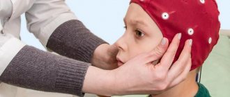
During the procedure, you can lie or sit on a couch, with a doctor nearby with an EMG device and a computer for recording and analyzing data. The testing itself can be roughly divided into two parts.
The first part is called a nerve conduction study or stimulation electromyography (some sources use the term Electroneuromyography to describe this part of the examination). To do this, a weak electric current is used to stimulate the nerve being tested in the accessible area to determine how quickly or slowly the electrical impulse travels. Analysis of the results will indicate the presence or absence of disease in this area. Imagine that the nerves in the body work like electrical wires. The easiest way to identify the problem is to run an electric current through the wire. If there is any damage to the functioning of the nerve, the doctor will register the conduction pathology. To study nerve conduction, the doctor places several electrodes on the surface of the limb being studied, and with another electrode he conducts electrical stimulation of the nerve in different areas of the limb. In itself, conducting an electrical impulse along a nerve is painless, but current stimulation may or may not be painless. You can be sure that this study is absolutely safe for your health. The supplied electrical impulse is so weak that it does not interact with any electrical devices, including artificial pacemakers. The doctor can pause between individual electrical impulses to carry out calculations and additional measurements.
The second part of the procedure is called needle electromyography. It is called that because the study requires the insertion of a very thin needle (needle electrode) directly into the muscle. At this stage, the muscle is examined to determine damage to it, both the result of damage to nerves or neurons (nerve cells), and primary damage to the muscle itself. Typically, the needle is gradually inserted into the relaxed muscle until muscle activity is recorded. When this happens, you may hear an audible signal of muscle activity, amplified by the EMG device. The painful stage of needle myography is caused by the passage of a needle through the skin, where a large number of receptors are located. As soon as the needle is in the muscle, the sensations from the examination will become more uncomfortable than painful. A needle electrode is inserted into the muscle to record the biopotentials of the muscle. No electrical stimulation occurs through it, just as no medications are administered. On average, such a needle examination of one zone takes from 2 to 5 minutes and varies depending on the severity of the pathological problem.
There is also surface electromyography. The technique is similar to needle EMG, but surface myography does not use needle electrodes. This study is absolutely painless, although not as informative as needle electromyography. Often, the use of this type of study is sufficient, but needle EMG provides more information about the state of the neuromuscular system.
How long does electromyography last?
Typically, the first stage (nerve conduction study) requires more time than needle or surface myography because it requires additional calculations and measurements. However, in-depth data analysis and interpretation of needle electromyography can also take a long time. On average, a study of the nerve conduction of one limb takes from 10 to 30 minutes. Needle or surface EMG of one muscle rarely exceeds 5 minutes.
What preparation is required for electromyography?
To successfully conduct an EMG, certain rules must be followed. It is necessary to refrain from using creams and other body products that can leave a thin layer on the skin. This may interfere with the examination. You should not arrive for your procedure on an empty stomach, and at the same time, you should not overeat or take any special foods before the procedure. Alcohol intake is equally excluded. You should be relaxed, not tense. Immediately after the procedure, you can return to your daily activities. It is worth considering that when performing myography, it is necessary to remove clothing from the areas of the body that the specialist is going to examine. Usually these are the limbs: arms - from the fingertips to the armpit; legs - from the tips of the toes to the popliteal fossa. In most cases, if the patient is taking any pharmacological medications, there is no need to stop them before the procedure. However, if you have a bleeding disorder or are taking medications that lead to this condition, you should notify the EMG specialist so that measures can be taken during the needle myography to prevent bleeding. In any case, if you are taking any medications, you must notify the doctor conducting the study. Some of them may affect the readings. The doctor needs to know what medications you are taking so that, if necessary, he can make the necessary adjustments when analyzing the data obtained.
Is it necessary to use painkillers before the study?
In the vast majority of cases, no special anesthetic measures are required before the procedure. You can take the patient’s usual analgesic, but be sure to notify the attending physician. It is recommended that you avoid taking sedatives, even if you have notified your EMG physician. If the drugs are potent, then after a short EMG study the patient will not be able to return to everyday activities, being under the influence of the latter.
How quickly can results from an electromyography study be obtained?
In most cases, an experienced specialist will tell you the main idea during or immediately after the procedure. The doctor issues a more accurate conclusion some time after the procedure (on average up to 30 minutes), although some doctors prefer to analyze the data obtained and give the results to the patient the next day.
Types of EMG
There are several ways to perform electromyography. The choice of method is made by the doctor depending on the existing pathology. The following types of EMG are distinguished:
- Stimulation (surface) electromyography. It is a non-invasive and painless examination. This EMG method allows you to evaluate bioelectrical activity over a large area of muscle. Stimulation myography is performed on the lower and upper extremities to study weakness, fatigue, numbness, and decreased muscle sensitivity. In addition, surface EMG is performed to diagnose nerve damage. This type of study evaluates the condition of the masticatory and facial muscles, which is informative for cosmetologists and dentists.
- Needle (local) electromyography. Used for more accurate research. For this purpose, a needle electrode is inserted into the muscle. In this case, minor pain occurs, which soon disappears. Local electromyography is an invasive research method. In this regard, hematomas or infiltrates may occur after the procedure.
Any of the EMG methods is performed for diagnosis and treatment evaluation.
Type of EMG
Modern devices differ in the types of pass diodes: the range of such parts determines the accuracy of the results obtained. Two types of devices are used for superficial and local examination. Global diagnostics occurs in a non-invasive manner (non-contact) and allows you to see the activity of muscle tissue over a large area of the body. This type of diagnosis is used in cases where the cause of pain or damage within the muscles is unknown. Examination of a wide area allows us to track the dynamics in the treatment of chronic diseases.
Local EMG is carried out using the contact method: the electrode is inserted directly into the part being studied. The area of the body is first numbed and treated with disinfectants. The electrode is a thin needle that makes a minimal puncture. The invasive technique is suitable for examining a small part of muscle tissue.
The choice of technique depends on the expected problem and the doctor’s prescriptions. Indications for EMG are patient complaints, injuries and injuries that affect a person’s walking and mobility. In some cases, to accurately diagnose the problem, 2 types of EMG are prescribed at once: local and global.
Indications for electromyography
The method of recording the bioelectrical activity of muscle tissue has become widespread in various fields. EMG is used in neurology, cosmetology, traumatology, dentistry, and sports medicine. The high accuracy and painlessness of the procedure make it mandatory in the presence of pathology of the neuromuscular system.
General indications for EMG include:
- The appearance of muscle weakness, increased fatigue.
- Presence of convulsive syndrome.
- Impaired sensitivity.
- Reduced muscle volume.
- Muscle pain of varying severity.
The presence of any of the above signs is an indication for electromyography.
Most often, changes in the conductivity of muscle fibers occur in neurological practice. Using EMG, the condition of the nerves innervating the muscles is assessed. Diseases requiring electromyography include:
- Polyneuropathy. Nerve damage causes changes in the EMG waveform. The oscillation amplitude values depend on the degree of damage to the nerve fiber.
- Muscle pathology. This group of diseases includes inflammation, dystrophy, and increased fatigue.
- Degenerative-dystrophic changes in the spine. The presence of a spinal hernia often causes compression of the nerve roots. Such conditions are reflected in electromyography.
- Hyperkinesis. Involuntary muscle movements cause EMG changes characteristic of this syndrome. Using diagnostics, it is possible to establish the localization of the pathological focus.
- Parkinsonism. The tremor that appears in Parkinson's disease significantly changes muscle conductivity. Such a condition must be examined using electromyography.
- Radiculopathy. Nerve root lesions are diagnosed using EMG.
In dental practice, electromyography is prescribed to study the masticatory muscle. Diagnosis is carried out both for diagnostic purposes and to monitor previous treatment. Indications for EMG are:
- Bruxism (teeth grinding).
- Jaw injury.
- Inflammatory dental diseases.
- Damage to the facial nerve.
- Prosthetics.
Sports medicine is another area where electromyography is actively used. Assessing the degree of muscle damage plays an important role in the recovery of athletes after injury. EMG makes it possible to identify a lesion at the initial stage, preventing the development of serious complications.
When selecting a prosthesis, traumatologists and orthopedists always prescribe electromyography to assess the lost functions of the limb. Diagnostics of muscle conductivity is actively used in cosmetology for the administration of Botox.
results
During the rest phase, the muscle is normally inactive. If the patient slowly contracts a muscle, a response occurs from first one and then more motor units. A motor unit is a concept that includes a neuron in the anterior horn of the spinal cord with a motor axon and the varying number of muscle fibers that it innervates. First, the response is reflected in the form of a progressive increase in the number and then an increase in the amplitude of the action potential of motor units with a recognizable structure of potential oscillations.
When the muscle is fully involved in the process, “white noise” appears. At this point, the overall activity of a large number of motor units is so pronounced that both the peaks on the monitor and the noise in the speakers overlap, creating an “interference pattern.” The muscles of the upper limb in a relaxed state in diseases of the nervous system may not have a resting potential. This is manifested by activity during the insertion of a needle electrode. In this case, changes occur that are characteristic of active denervation. They are called fibrillation potentials and positive sharp waves on the oscillogram, which are spontaneously produced by denervated muscle fibers. This is a sign of complete or partial nerve damage. Such changes occur 7-12 days after axonal rupture.
When a spinal nerve root is pinched in a denervated muscle of the upper limb, the number of active motor units decreases in proportion to the number of damaged axons. When performing electromyography, instead of “white noise”, which indicates the full involvement of all motor units, a picture of a decrease in muscle potentials is recorded. In diseases of the muscles of the upper limb, all of the above changes can be recorded, but the graph of action potentials is different. A complete interference pattern occurs with a lower force of active muscle contraction. In chronic neuropathy, with re-sprouting of remaining viable nerve fibers, elongated reinnervated motor units are formed, which are characterized by a multiphasic profile or a profile with increased potential amplitude.
Contraindications
Electromyography is a procedure performed on patients of any age. There are no specific contraindications for its implementation, but there are common ones for all diagnostic procedures. Conditions in which EMG is not prescribed include:
- Acute pathology. Infectious or non-infectious diseases in the acute stage are an absolute contraindication for electromyography.
- Presence of a pacemaker.
- Skin lesions. Inflammatory processes on the skin, accompanied by pustular rashes, exclude the possibility of performing an EMG.
- Epilepsy.
- Cardiovascular pathology. This group of restrictions includes hypertensive crisis, myocardial infarction, and unstable angina.
- Mental disorders. Contraindications are conditions in which the patient cannot control his actions.
Needle electromyography is contraindicated in cases of blood clotting disorders, as well as increased pain sensitivity.
Electromyography: preparation for the study
The advantage of electromyography over other diagnostic methods is the lack of special preparation before the study. However, there are several recommendations, adherence to which will ensure the most accurate recording of bioelectrical muscle activity. These include:
- Refusal to take certain medications. Such medications include tranquilizers, muscle relaxants and other drugs that affect the nervous system. It is recommended to cancel them the day before the study.
- Observe dietary restrictions. For a more accurate diagnosis, it is necessary to stop eating foods that increase nervous excitability several hours before electromyography. These include tea, coffee, caffeinated drinks, chocolate, and carbonated drinks.
In cases where discontinuation of medications is not possible, the attending physician must be notified in advance.
How is electromyography performed?
Electromyography takes from 30 to 60 minutes. The time depends on the number of areas examined, as well as the severity of the lesion. Electromyography is performed using an electromyograph. With its help, the bioelectrical activity of muscle fibers is registered and recorded.
The EMG procedure can be performed in an inpatient or outpatient setting. For this, the patient is asked to take a comfortable position (sitting, lying, half-sitting). The area to be examined is treated with an antiseptic. After this, I do not apply the electromyograph electrodes. In cases where needle EMG is indicated, a needle electrode is inserted into the muscle being studied. This is the only type of electromyography in which minor pain is felt. All other methods are painless.
At the very beginning of the procedure, muscle conductivity at rest is assessed. After this, she is asked to strain, after which the bioelectrical activity is recorded again. The results obtained are an electromyogram, which reflects all the changes occurring in the neuromuscular system. Based on the data obtained, a diagnosis is made or treatment is assessed.
Diagnostic technique
ENMG of the upper extremities and other parts of the body is performed using special equipment. It records the speed of passage of a nerve impulse to tissues. During stimulation, the patient may experience discomfort, but not pain. Discomfort is caused by irritation of the nerve and further contraction of the muscle.
The procedure usually takes 30-60 minutes. The patient is in a special chair sitting, half-sitting or lying down.
All results obtained are recorded in a computer. If desired, they can be burned to disk or printed on paper.
The results of the examination are issued immediately. The doctor does the decoding.
EMG interpretation
The electromyography technique is based on recording muscle activity. The results obtained form an interference curve, reflecting any changes in conductivity. There are several types of EMG curve:
- First. In this case, the curve registers rapid fluctuations in potential. The frequency is about 50-100 Hz. Such an EMG is considered a normal variant. Indicators may vary depending on age, weight and physical development.
- Second. Fluctuations on electromyography occur with a frequency of less than 50 Hz. This indicates decreased muscle conductivity. Such changes are characteristic of neuropathy.
- Third. The EMG curve records a significant decrease in oscillation frequency. On average it reaches 4-10 Hz. These results indicate pathology of the extrapyramidal system.
- Fourth. Electromyography does not cause any hesitation. Bioelectrical activity is completely absent. A similar condition occurs with muscle paralysis. Another name for type 4 EMG is “bioelectric silence.”
The following main diseases are identified in which changes in the amplitude of oscillations are recorded on electromyography:
- Polyneuropathy. The results of the curve depend on the degree of damage to the nerve fiber. With a mild degree, the oscillation frequency is significantly reduced. In cases where the pathology of the nerve has caused its complete death, “bioelectric silence” is recorded.
- Myositis. Inflammation of muscle fibers causes a decrease in their conductivity. Indicators of bioelectrical activity depend on the degree and stage of myositis.
- Amyotrophy. The pathology is characterized by loss of muscle mass. In this case, an increase in the amplitude of oscillations is recorded on the EMG. The curve has the shape of a “picket fence”.
- Myasthenia. Muscle fatigue syndrome significantly affects the electrical conductivity of fibers. On the EMG there is a decrease in the amplitude of oscillations. The values depend on the stage of the disease.
- Tremor. The symptom is characteristic of various neurological pathologies. In this case, a sharp increase in the amplitude of oscillations is recorded. Their frequency depends on the location of the lesion.
- Myotonia. Syndromes associated with delayed muscle relaxation are an indication for electromyography. It identifies curves with low amplitude and high frequency.
Highly qualified doctors interpret EMG results. Based on the data obtained, the specialist is able to establish the localization of the pathological focus, its degree and stage.
Decoding the results
The results of ENMG of the extremities are reflected on the device screen in the form of graphs and lines, which can only be deciphered by a specialist who has extensive experience in the methodology of conducting such research.
When interpreting electroneuromyography, the following indicators are taken into account:
- The ability of nerve fibers to conduct electrical impulses. If impulse non-conduction is observed, it means that the nerve being studied has a pathology.
- An important indicator of ENMG is the determination of the speed of impulses passing through nerve fibers. If the speed is reduced from normal, the likelihood of multiple sclerosis can be assumed.
- The amplitude of muscle activity, their strength and height. Decreased amplitude may indicate nerve damage. With an increase in amplitude, spinal cord pathology can be diagnosed.
- The presence of fasciculations may indicate lesions in the spinal cord.
The results of the ENMG examination are reported immediately after the procedure. An electroneuromyography study can be prescribed by an orthopedist during an examination, a rheumatologist, an endocrinologist to clarify the diagnosis, as well as a neurosurgeon.
Electromyography – price in Moscow
Electromyography is a mandatory study in the presence of damage to the neuromuscular system. The Moscow Yusupov Hospital has the latest equipment that allows diagnostic procedures to be carried out with high precision. The many years of experience of the institution’s specialists allows us to decipher the results of the study and make a correct diagnosis in the shortest possible time. Identifying pathology of muscle fiber conduction at an early stage allows you to begin timely treatment and avoid serious complications. You can sign up for electromyography and learn more about the cost of the procedure by phone.
Indications
Neurologists at the Yusupov Hospital prescribe ENMG of the nerves of the upper extremities if there are symptoms of a muscle or nerve disorder:
- Muscle pain or cramps;
- Tingling;
- Numbness;
- Palpable weakness in the arms;
- Involuntary muscle twitching (tics).
The results of electroneuromyography of the hand help diagnose pathology of the muscles, ulnar, median and radial nerves, disorders that affect the connection between nerves and muscles.
