The scalene muscles are 3 large muscles that are located on both sides of the neck. With their upper edge they are attached to the lateral protrusions of the vertebrae (C2, C3, C4) of the cervical spine. With their lower edge they are attached to the 1st and 2nd ribs of the chest. Between the anterior and middle scalene muscles pass large blood vessels (subclavian artery and vein) and a bundle of nerve fibers (brachial nerve plexus).
Rice. 1. Anatomy of the neck - localization of the scalene muscles
Normally, muscles have elasticity, extensibility, are covered with “slippery” fascia and do not compress nerves and blood vessels. With pathological changes in the bone and muscle-tendon system, compression of these important organs of innervation and blood supply occurs, which causes the appearance of scalene muscle syndrome.
Depending on which muscle undergoes changes, a distinction is made between anterior scalene syndrome and posterior scalene syndrome. When nerves and blood vessels are compressed, neurovascular disorders are observed, accompanied by characteristic symptoms.
Symptoms of scalene muscle syndrome
Scalenus syndrome (SMS) may develop first as a myofascial pain syndrome. Due to overexertion or injury, the muscles spasm, structural changes occur in them, inflammation of the neck muscles develops, and trigger points (increased pain sensitivity) are formed. In this case, the main symptom of the pathology is pain. It radiates to the upper limb from the affected side, spreading from the shoulder joint to the tip of the phalanx of the ring or little finger. Often the soft tissues of the occipital part of the head and chest are involved in the pain process. The pain usually gets worse at night or when moving the head, upper limb, or taking a deep breath. The neck muscles hurt in different ways - from a mild nagging pain to a shooting, constant, burning pain.
Rice. 2. Map of the distribution of pain in scalene muscle syndrome
When a nerve bundle is compressed by a spasmodic muscle, the following manifestations are observed:
- a feeling of numbness, tingling, loss of sensitivity in the fourth and fifth fingers, as well as along the inner surface of the hand and forearm;
- swelling above the metacarpophalangeal joints of the II–V fingers and on the dorsum of the hand;
- stiffness of movements in the fingers, worsening in the morning.
When the great vessels are compressed, vascular disorders are observed:
- coldness of the extremity;
- cyanosis;
- numbness;
- swelling;
- disappearance of the pulse in the radial artery when raising the arm up and tilting the head to the affected side (Adson's test).
If lymphatic drainage is impaired, patients develop a swelling in the neck (Kovtunovich's pseudotumor) in the area of the supraclavicular fossa.
With prolonged disruption of innervation and blood supply, an imbalance of trophic processes occurs, which is accompanied by the following symptoms:
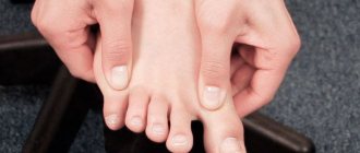
- damage to the nail plate – brittleness, unevenness;
- hair loss on the affected limb.
Long-term compression of nerve endings can lead to:
- hypertonicity of the neck muscles;
- heaviness, weakness in the hand;
- incomplete paralysis of a limb;
- atrophy of the muscles of the distal part of the arm.
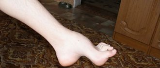
As a rule, SLM is observed only on one side, much less often on both. The most dangerous complications occur when there is pain in the left neck.
Scalenus syndrome as a cause of numbness and cold hands
The scalene muscles are three powerful muscles of the neck (anterior, middle, posterior). When at least one of them is shortened, compression of the nerves occurs, which, in turn, causes pain or numbness in the arm, problems with the shoulder joint, and glenohumeral periarthrosis. About the reasons for the shortening of these muscles and effective methods of treatment.
About the scalene muscles of the neck
The scalene muscles begin posteriorly from the transverse processes of the cervical vertebrae, stretch vertically along the neck and are attached to the two uppermost ribs, the first and second, deep behind the collarbone. This group of muscles is shaped like steps, hence the name.
Why is shortening of the scalene muscles dangerous?
The main feature is that
between them pass the subclavian artery and the nerves of the cervical plexus, which are directly connected to the arms
.
If the muscles are healthy, then nothing threatens the arteries and nerves - healthy muscles are elastic, soft, covered with a dense slippery membrane - fascia. This ensures they slide smoothly relative to each other and protects them from any pinching when moving the head and neck. However, with pathology - shortening - everything changes: the anterior scalene muscle spasms, this causes pain, and a muscle stretched like a string can pinch the nerves and artery
.
Therefore, the first danger of shortening the scalene muscles is radicular pain, akin to that which occurs when the nerves are compressed (squeezed) by hernia and osteophytes of the cervical spine
. We turned our head unsuccessfully, tension in the shortened muscle arose, pressure on the nerve increased, and pain arose. But that is not all.
Blood vessels in the costoclavicular space (at the exit from the chest) may also be pinched. This may impair blood flow to the arms, hence
numbness in the hand, sensory impairment, vertebral artery syndrome.
Often, glenohumeral periarthrosis begins with shortening of the scalene muscles. Gradually, neck instability develops and the head moves forward.
What follows is a typical scenario. To compensate for balance and center of gravity, the muscles in the back of the neck are forced to tighten and become overloaded. Due to overload, painful trigger points are formed and myofascial syndrome develops. If left untreated, this leads to compensatory curvature of the spine in the thoracic and lumbar regions as it is forced to seek balance. Curvature of the spine, in turn, disrupts the distribution of axial loads on the intervertebral discs, displacement of the vertebrae and, thus, opens the way to the formation of protrusions and disc herniations in the cervicothoracic region.
Timely consultation with a doctor, early detection and proper treatment can avoid all these serious complications of shortening of the scalene muscle. Therefore, it is useful to regularly undergo examination by an experienced kinesiologist, who, using a special functional muscle test, determines the locations of spasmed, weakened and shortened muscles, and then, based on the identified problems, draws up an individual treatment program using the method of applied kinesiology.
How signs should alert you. Characteristic signs of scalene muscle syndrome:
- Pain in the neck and shoulder, especially when bending to the side and turning.
The pain may radiate from the shoulder down the arm and into the little and ring fingers.
Sometimes it spreads to the chest or back of the head. It often intensifies at night, as well as when turning the head, moving the arm to the side and taking a deep breath (the scalene muscles contract when taking a deep breath and pull the upper ribs and chest upward).
- Numbness or tingling in the fingers, hands, entire arm, muscle weakness.
- Shifting the head forward.
— Restriction of movement of the chest, since the first and second ribs, to which the scalene muscle is attached, are fixed.
When the blood vessels are compressed, swelling of the hand, cold hand syndrome, pale or blue skin, and a weak pulse in the hand are typical.
How does shortening of the scalene muscles develop?
Long-term overload leads to muscle shortening, which often occurs against the background of weakening of the antagonist muscles - the long extensors of the neck.
. The main function of the long neck extensors is to support the head and its weight when bending forward.
So, in modern man, the long extensors of the neck are chronically overstrained and weakened.
When the long extensors of the neck weaken, other muscles, mainly the scalenes (as the main antagonist), take on increased load and shorten them, and the same
scalene syndrome is formed.
Next important question
: why do our neck muscles weaken and spasm, and why has this problem become relevant for most people, especially in the last few years?
The number one reason is a constantly bent neck forward.
.
The muscles in the back of the neck tense every time we tilt our head forward - to look at a smartphone, read a magazine, when we cook in the kitchen or sit for a long time at the computer with our head stretched forward. In modern humans, the long extensors that support the head experience chronic overload.
Reason number two is weakness of the lower back muscles, a sedentary lifestyle.
Modern man spends most of his life sitting. How are we sitting? Leaning on the back of a chair or in a banana pose, slouching. The back is rounded, the lower back is relaxed, bends back, the head is stretched forward. In this position, there are simply no physiological bends
, the lumbar and cervical spine are in a constant kyphotic state. And we spend several hours a day in this position.
As a result, the back muscles (back extensors, quadratus, lumbar muscles) weaken, and a person cannot support his lower back in a sitting position. In such conditions, the long neck extensors are also not included in the work, the short neck extensors work instead, the head is supported by short muscles, and not by long ones, the load is distributed unevenly across all discs, and is shifted more to the 5th, 6th, 7th intervertebral discs.
If this situation is repeated day after day, the long neck muscles weaken and shorten, gradually causing vertebral instability.
Fixing your hands in one position for a long time (working on a computer) can put additional pressure on nerves and blood vessels
.
The syndrome occurs and occurs against the background of the muscle due to sports overtraining, especially in those sports where there are frequent repetitive movements associated with raising the arm (the collarbone drops and the costoclavicular space decreases), for example, swimming, baseball, tennis, weightlifting.
How to restore health to your neck muscles?
Scalene syndrome often resembles a cervical hernia with nerve compression. Therefore, at the first stage of treatment, the most important thing is to establish the source of the problems.
This cannot be done using MRI. Shortening of the scalene muscles can only be detected by a special functional muscle test, which is carried out by an experienced specialist in applied kinesiology.
Each person may have their own reasons for the development of spinal pathologies and pain. The task of applied kinesiology is to identify them, and then, based on examination data, to create an individual program of exercises aimed at restoring the full functionality of shortened, weakened, idle muscles, and eliminating muscle imbalances
.
Doing this with regular strength exercises is dangerous and ineffective. To work with weakened muscles and a sore spine, special rehabilitation equipment is needed - biomechanical strength decompression kinesi simulators. They are widely used in rehabilitation after injuries and operations on the spine and joints, as they allow the restoration of weakened muscles with minimal stress on the musculoskeletal system.
Therapeutic exercises strengthen weakened neck muscles, the upper back in the area of the cervical and upper thoracic vertebrae, from which the long extensors of the neck and shoulder girdle begin. At the same time, spasms are relieved, blood flow and nutrition of the spine improves, pain, dizziness and other unpleasant symptoms disappear, and blood pressure stabilizes. And all this without the use of drugs or expensive hardware technologies.
Stretching and strengthening the shoulder and pectoral muscles helps relieve pressure on the nerves and blood vessels in the costoclavicular space.
Specialist consultation is required.
Doctor’s consultation + diagnostics using applied kinesiology – 1000 rubles.
Call, write! Tel.: (843) 570-55-25 , WhatsApp: 79655968085 or VKontakte group .
Follow us on Instagram (photos and videos from classes):
Read us on Yandex Zen:
Don't miss out on the fun! Subscribe to our news:
Subscribe to news from the Kazan Kinesitherapy Center
Center promotions, therapeutic exercises and useful tips from our specialists on how to independently maintain the health of your joints and spine without medications
Similar articles:
Cervical osteochondrosis and headaches
Massage pillows. Fashionable massager for home. What are the benefits and how to choose?
Kinesitherapy. Spine and joint center for the whole family
Complications of scalene muscle syndrome
The most dangerous complications of SLM are:
- thrombosis of the subclavian artery;
- paresis and/or atrophy of the arm muscles;
- violation of the biomechanics of the neck and the whole body.
Large blood vessels pass between the anterior and middle scalene muscles - the subclavian artery and subclavian vein. Spinal vessels and arteries that supply blood to the brain depart from them. Therefore, thrombosis threatens to disrupt the blood supply to the spine, brain and body as a whole. Complaints that the neck is swollen on one side and there is a feeling of heaviness, pain, burning, and stiffness of movement are a signal for immediate hospitalization, surgical care and treatment.
A pinched nerve in the neck area leads to disruption of the innervation of the soft tissues of the hands, paresis and atrophy. If the consequences of atrophy and paralysis of the muscles of the limb are quite clear, then violations of the biomechanics of the neck need to be considered in more detail. When muscles are under prolonged tension, structural changes occur in the fibers - they compress, the muscles shorten and begin to “pull” the head forward. The curvature of the spinal column (lordosis) increases and a violation of the architectonics of the skeleton occurs. In this case, the muscles of the back of the neck and shoulder girdle are tense, and myofascial pain syndrome develops with the corresponding characteristics - trigger points and referred pain.
If the patient does not pay attention to the pain in the neck muscles and ignores treatment, then as a result of the action of compensatory mechanisms, curvature of the spinal column occurs in the lumbar and thoracic region. Changes in vector loads on the vertebrae cause deformation and protrusion of the intervertebral discs, the formation of hernias not only in the cervical region, but also in other areas of the spinal column.
Pain in the hands when working at the computer. Part 3. Scalenus syndrome
In the previous articles of the series, you became acquainted with the diagnosis of the main tunnel syndromes of the upper limb and learned to relieve tension from the shoulder girdle. Now it's time to deal with scalene muscle syndrome.
The neurovascular bundles are surrounded by muscles, bones, ligaments and fascia. The first place where there may be compression of the neurovascular bundle for the hand is its passage through the canal formed by the anterior and middle scalene muscles.
With scalene muscle syndrome, initially there is a violation of the venous and lymphatic outflow from the arm, which causes swelling of the limb and creates the preconditions for compression of the neurovascular bundles in narrow places on the arm. Most often this occurs in the carpal tunnel, cubital tunnel, under the pronator teres muscle of the forearm. Scalenus Syndrome Test (take before completing the lesson):
Raise your arms straight up to the ceiling, in a standing or sitting position, and try to hold for a minute. If before the end of the minute there is numbness, paresthesia (“pins and needles”) in your hands - the test is positive - you are on a direct path to not sleeping at night due to numbness in your hands, undergoing long courses of treatment with vascular and anti-inflammatory drugs with a very short effect. Lower your hand as soon as unpleasant sensations appear, note the time of comfortable holding on one side and the other, is it the same?
If everything is fine within 1 minute, the test is negative and you don’t have to read the article further, but a lesson on relieving tension from the scalene muscles can be a fascinating study; it will take you about 10-15 minutes to get and evaluate the result. The lesson can be completed while sitting at the computer.
Before moving on to the lesson, let me remind you that you are doing a lesson, not an exercise. The purpose of the lesson is to find new ways to solve a motor problem in the context of your usual muscle tension and movement patterns.
Focus on your sensations during the lesson, allow the movement to occur smoothly and slowly, maintain maximum comfort so as not to be distracted by unpleasant stimuli and maintain clarity of conscious feedback from the movement. Situation 1:
Sit in the middle of the chair and place your hand on the collarbone area on the opposite side so that the collarbone is at the base of the fingers, the fingers are in the supraclavicular area, and the palm is in the subclavian area. Apply light pressure with the palm of your hand and fingers to the area where they are located, no more than the weight of the hand itself. Now inhale slowly and smoothly so that the hand is raised by the collarbone and upper ribs to the ceiling, and with a long exhale, allow the hand to fall.
With each inhalation and exhalation, notice the range of motion as your collarbone and ribs move under your arms. Completely release tension in the muscles you worked before starting each movement.
Do the movement several times until it is interesting and comfortable and stop. Lower your hand, check the difference in the hands on the left and right (compare the sensation of length, volume, weight, temperature).
Situation 2:
Same starting position, same side. Now, as you inhale into your hand, slowly nod your chin towards the collarbone pressed by your hand, and as you exhale, return your head to a neutral position. Experiment with different nod trajectories.
Do the movement several times and stop. Lower your hand, check the difference in the hands on the left and right (again compare the sensation of length, volume, weight, temperature). You can raise your straight arms to the ceiling and compare the result now and at the beginning of the lesson. Which hand is easier to handle this test now?
Situation 3:
Repeat the movements from situations 1 and 2 on the other side. Try making movements on this side mentally, allowing just a hint of movement to appear.
Try to notice where the muscles performing this movement work, how they shorten as you inhale and lengthen as you exhale.
PS Based on the experience of past comments, you should not be offended by the fact that complex questions regarding your specific situation will be answered: “An examination is required, some additional examination is possible.” The doctor is not a telepath and even with X-rays in his eyes he will not be able to give recommendations based on fragmentary descriptions of symptoms in the comments.
At the same time, ask questions; there are many situations, not related to serious illnesses, in which it is quite safe to comment without examination.
PPS The fourth article in the series has been published, dedicated to the integration of the previous movements into one whole with the release of tension in the pronator teres.
Diagnostics of SLM
Due to the variety of manifestations of the syndrome, variability in severity, and lack of characteristic signs, its diagnosis presents some difficulties.
An important diagnostic sign is the condition of the skin, muscles, hair and nail plate. Feeling the neck reveals hypertonicity of the anterior scalene muscle and pain when pressed. A test for spasm is carried out - with maximum rotation in the direction of the lesion and strong pressing of the chin to the collarbone, the spasmed muscle is tensioned and trigger points are activated.
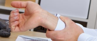
Another indicative test is the Adson test. On the affected side, the patient should raise the limb upward and tilt the head towards this shoulder. With SLM, the pulse in the arm will not be palpable. Specialists at the Manual Medicine Clinic “Galia Ignatieva MD” conduct physical examinations - examination, palpation, skin sensitivity tests of individual parts on the hand. During physical examinations, the doctor may measure temperature and pulse pressure in different arms and in different positions.
In addition to visual examination and physical examinations, the Manual Medicine Clinic “Galia Ignatieva MD” prescribes instrumental methods:
- electromyography (EMG), which allows you to assess the anatomical and functional characteristics of nerves and muscles;
- radiography - to identify anatomical abnormalities, the presence of an additional rib in the upper part of the chest;
- MRI – to visualize the condition of soft tissues, localization of compression of nerves and blood vessels;
- CT – for layer-by-layer visualization of changes in bone tissue;
- Ultrasound, Doppler mapping, angiography - to determine the presence of arterial stenosis and the speed of blood flow in them.
Specialists can also prescribe laboratory tests to identify inflammatory processes, assess blood sugar and hormone levels, which help in the differential diagnosis of SLM.
In the initial stage of SLM, instrumental diagnostic methods are uninformative and can give a false answer. Therefore, the most reliable method for diagnosing the syndrome is manual muscle testing. It allows you to conduct a comprehensive assessment of the condition and physiology, dynamics of movements, and identify the causes of disorders more quickly and informatively than other diagnostic methods.
Clinically Relevant Anatomy
The first area where vessels and nerves can be compressed is the most proximal and is called the interscalene triangle. This triangle is bounded by the anterior scalene muscle anteriorly, the middle scalene muscle posteriorly, and the medial surface of the first rib inferiorly. The presence of the scalene minor muscle and the fact that both the anterior and middle scalene muscles attach to the first rib can lead to narrowing of this space and therefore compression of the brachial plexus and subclavian artery nerves.
The second region is called the costoclavicular triangle, and is bounded anteriorly by the middle third of the clavicle, posteromedially by the first rib, and posterolaterally by the superior edge of the scapula. The subclavian vein, artery and nerves of the brachial plexus pass here, which then go into the subcoracoid process. Compression of these structures can occur as a result of congenital anomalies, trauma to the first rib or clavicle, or structural changes in the subclavian muscle or coracocostal ligament.
The last area is called the subcoracoid space or the space under the pectoralis minor muscle. This space is located under the coracoid process and the tendon of the pectoralis minor muscle. It is bounded above by the coracoid process, in front by the pectoralis minor muscle, and behind by 2-4 ribs. Shortening of the pectoralis minor muscle can result in a narrowing of this space and therefore compression of the neurovascular structures during hyperabduction.
Some anatomical abnormalities may also narrow the superior aperture. These include the presence of a cervical rib, congenital soft tissue anomalies, hypomobility of the clavicle, and functionally acquired anatomical changes. Soft tissue abnormalities may create a compressive effect on the neurovascular structures located in this location (eg, muscle hypertrophy or wider attachment of the middle scalene muscle to the 1st rib).
Treatment of scalene syndrome
After conducting research and establishing an accurate diagnosis, the doctor at the Manual Medicine Clinic “Galia Ignatieva MD” develops an individual SLM therapy plan. Treatment of scalene muscle syndrome is most often carried out using conservative methods:
- medicinal;
- physiotherapy;
- reflexology;
- exercise therapy;
- manual therapy.
Scalenus syndrome, the treatment of which requires symptomatic therapy, involves the use of the following groups of drugs:
- NSAIDs – medications eliminate inflammation and relieve pain;
- muscle relaxants – relax muscles, eliminate hypertonicity, spasm and, as a result, pain;
- opioid analgesics – to eliminate severe pain that is unresponsive to other types of painkillers;
- drugs for neuropathic pain and restoration of nerve structure.
After the severity of the symptoms of SLM has been reduced, physiotherapy methods can be added to the complex effect:
- shock wave therapy;
- magnetic therapy;
- laser therapy;
- DDT – diadynamic therapy;
- electrotherapy;
- vibration impact;
- myostimulation;
- phonophoresis;
- massage.
Physiotherapy eliminates inflammation, pain, activates blood circulation in soft tissues and normalizes trophic processes. In combination with medication, the effectiveness of treatment increases.
Exercise therapy is one of the important elements of the treatment program. Individually selected exercises allow you to:
- improve posture;
- eliminate muscle hypertonicity;
- correctly distribute the load;
- strengthen the muscle corset;
- restore range of motion in the neck and upper limbs.
Drug blockades are prescribed for the purpose of differential diagnosis and treatment of SLM, but due to the proximity of the nerve plexus, the procedure must be performed by a specialist with extensive experience.
Rice. 3. Manual treatment of SLM.
If a nerve in the neck is pinched, a vertebrologist can tell you what to do. The effectiveness of manual therapy at the Clinic of Manual Medicine “Galia Ignatieva MD” in relieving pain, restoring the anatomical and physiological characteristics of structures, eliminating muscle blocks and clamps has been proven by numerous clinical studies.
The specialist not only eliminates the signs and consequences of the pathology, but also acts on the root cause of SLM, since manual therapy has virtually no contraindications, it can be prescribed to patients with drug allergies, children and the elderly. Gentle manual techniques will restore mobility to the arm and neck, activate blood circulation, eliminate congestion, normalize metabolic processes and help remove metabolic products.
The use of surgical treatment is indicated only in exceptional cases - when there is a threat of arterial thrombosis, damage to nerve endings, the presence of an additional rib, etc. When the left neck hurts, the surgeon decides how to treat the anomaly.
Prevention of SLM
Specific methods for preventing SLM have not been developed. To prevent the occurrence of pathology, it is necessary to eliminate the causes of SLM:
- avoid hypothermia;
- eat rationally and control body weight;
- lead an active lifestyle - physical inactivity is one of the factors in the development of pathology;
- promptly identify and treat pathologies of the spinal column and musculotendinous system;
- avoid overstraining the muscles of the neck and shoulder girdle;
- train correctly;
- avoid prolonged fixed posture;
- the workplace must be ergonomic.
Anyone who has to spend a long time at a computer, desk, or operator console should know how to relieve neck muscle spasms with the help of special exercises. Examples of exercises can be found in specialized literature or shown by a specialist at the Manual Medicine Clinic “Galia Ignatieva MD”.
Manual therapy plays a great role in preventing relapse of SLM. A vertebrologist will show you how to relax your neck muscles. The patient can perform simple exercises and self-massage at home. In difficult cases, the help of a professional with extensive experience is necessary.