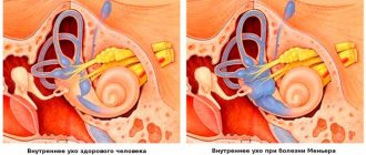
What is MRI of the brain based on?
The diagnostic effect of brain MRI is based on nuclear magnetic resonance. In response to the powerful radiation created by the generator, the hydrogen atomic nuclei contained in the tissues line up along the electromagnetic field lines and begin to vibrate. Each atom becomes like a spinning mini-spinning top, emitting energy waves.
Different structures emit different amounts of energy - some give it out more intensely, while others less so. The difference is recorded by a device that takes pictures (slices) in different projections.
To do this, the patient is placed inside a tomograph in which generators maintain a high-frequency electromagnetic field. Special radio emitters generate pulses, and coils record the energy sent by vibrating atoms.
The resulting slice images are combined into a three-dimensional matrix using a special computer program, in which dark or light unhealthy areas are visualized against a gray background.
Step-by-step scanning
The diagnostic procedure consists of several stages that take certain time periods:
- Preparatory stage before MRI of the brain. How long this stage lasts depends on the examination mode. If a session without contrast is planned, initial instruction, changing clothes, and positioning in the inside of the tomograph will take no more than 15 minutes.
- Examination. In normal mode - about half an hour. According to the contrast protocol - 50-60 minutes.
- Waiting for results. Typically, interpretation of the resulting photographic images will continue for 30-90 minutes. In particularly complex clinical situations, experts may need a day or more, since a consultation is expected to be convened. By agreement, you don’t have to wait for the pictures to be decrypted; you can leave an email address to which documents (descriptions and pictures) will be sent when ready.
Advantages of magnetic resonance imaging over other methods
An MRI examination gives results much more accurately than x-rays, echoencephalography (EchoEG), ultrasound and other diagnostic options. It allows you to obtain maximum data about existing tumors, diseases, post-traumatic and post-stroke changes. Unlike CT and X-rays, in this case the body is not irradiated.
Only soft tissues are visualized in the finished images. The bones of the skull are not visible, so they do not interfere with analysis and decoding.
The contrast agent used in MRI diagnostics is much less likely to cause allergic reactions compared to X-ray contrast agents used for radiography.
How to quickly find a diagnostic center?
How long an MRI of the brain takes can determine the technical base of a diagnostic medical institution. It is recommended to choose a tomography clinic in advance, based on its rating, prices for services, and the number of positive comments from clients. Piter-mrt provides a complete list of medical organizations located in different areas of the city. Each clinic has its own card with a description. Registration is made through the website; the telephone number of the consultation center is located at the top of the page. Time booking and consultation are provided free of charge.
How does the procedure work?
The patient removes all metal jewelry and takes out removable dentures containing metal.
The patient is placed on a movable table and secured with special belts. This measure is necessary because you will have to lie inside the tomograph, remaining motionless, for a long time.
A device equipped with wires that transmit and receive radio signals is placed on the head. The equipment is quite noisy and tires with constant clicks and whistles. Therefore, the patient’s ears are protected with earplugs. After this, the table slides into the device, and the specialist sits down at the computer, in which the transmitted data is analyzed and processed.
The technique takes pictures, the quality of which depends on the characteristics of a particular MRI scanner. The thinner the visual slices the equipment makes, the more accurate the final images will be. The duration of diagnosis is 20-30 minutes, and when contrast is used - up to an hour.
After MRI diagnostics, you can immediately return to your normal life. No side effects subsequently or during the MRI examination occur, with the exception of an extremely rare allergy to gadolinium salts.
Finished photographs are handed out printed or recorded on a magnetic medium - disk or flash card. It can be sent to email with SMS notification.
What to choose: CT or MRI for headaches?
Computed tomography (CT) and magnetic resonance imaging (MRI) are two ways of looking at the same problem differently.
MRI is a medical imaging technique, a way to look inside the human body without resorting to cuts or punctures. A study is carried out using a magnetic field. The magnetic field strength of the tomograph is many times higher than on Earth. Phones stop working near an MRI machine and bank cards become demagnetized. However, the magnetic field used is safe for humans. It makes it possible to look inside the human brain without X-rays and detect pathology at an early stage. CT differs from MRI in its operating principle based on X-ray radiation. A CT scanner is a modern version of an X-ray machine that can take a large number of scans from different angles and from them create a three-dimensional model of the area being examined. Magnetic resonance imaging is the dominant type of examination for head pathologies. Therefore, for headaches, it is best to start diagnostics with an MRI of the brain, rather than a CT scan, which is associated with radiation exposure to the body. Here, MRI of the brain will be the most informative and comprehensive type of diagnosis. However, there are situations when doctors will prefer to give a patient a CT scan of the brain. This happens in emergency cases. The main indication for a head CT scan will be traumatic brain injury. When doctors need to very quickly decide on surgical intervention, and they do not have 20-30 minutes for an MRI scan of the head, they will use computed tomography. If a patient is suspected of having a stroke, neurologists need to quickly differentiate whether the stroke is ischemic, associated with a blood clot, or hemorrhagic, which is caused by bleeding in the brain. Treatment for these two types of stroke is radically different. Therefore, if a person is admitted by ambulance, doctors will do a quick CT scan rather than an MRI if a stroke is suspected, and based on its results they will begin intensive therapy. Author: Varlaty Valentin Pavlovich
Neurologist, acupuncturist with 40 years of experience
Types of MRI of the brain
- Standard - done without the introduction of contrasting solutions, but at the same time provides a sufficient amount of information.
- With contrast, before which drugs containing gadolinium salts are injected into the vein - gadopentetic and gadoteric acids, Omniscan, Magnevist, etc. These solutions penetrate into the bloodstream and, once in the rays of the MRI scanner, highlight the resulting “picture”. At the same time, the changed areas become better visible, which simplifies decoding. The technique is most often used to detect vascular abnormalities, multiple sclerosis and tumor formations. The dose of contrast agent is selected individually, taking into account weight.
- MR angiography – is performed to assess the condition of blood vessels in atherosclerosis, aneurysms, blood clots and pre-stroke conditions. It is done with gadolinium contrast to show blood flow problems in detail.
- MR imaging of the pituitary gland, an appendage that is an endocrine gland. The pituitary gland secretes hormones responsible for reproductive function, tissue metabolism and the regulation of human growth. An examination is prescribed if an adenoma is suspected - a benign tumor that causes migraine-like pain, hormonal imbalances, gigantism, infertility, obesity and sexual dysfunction. The same method is used to identify malignant pituitary formations that have similar symptoms and are accompanied by a pronounced deterioration in health.
Are there differences in MRI and CT scan of the brain?
Many patients of our diagnostic center are interested in how CT differs from MRI of the brain. There are differences between these procedures, which boil down to the following MRI features:
- The ability to detect even minor disturbances in brain function during the development of a disease such as multiple sclerosis;
- Since the examination is performed in several projections, it allows one to obtain more informative data about the patient’s health status;
- It is used in cases where it is impossible to visualize some parts of the brain using computed tomography (for example, the brain stem, the cerebellum itself);
- Helps identify pathologies at the earliest stages of development. For example, the progression of ischemic stroke can be determined after 2 to 3 hours.
Therefore, between CT or MRI of the brain, doctors often prefer magnetic resonance imaging. After all, this type of diagnosis allows you to determine in detail and as accurately as possible the slightest changes and pathologies in the functioning of the brain and in all its structures.
Indications for general anesthesia for MRI
Intravenous or inhalational sedation is only necessary for patients who are unable to hold their body still for long periods of time. Main indications for general anesthesia:
- Claustrophobia is the fear of closed spaces. Such patients, while inside the device, experience panic, which negatively affects their health and makes MRI diagnostics impossible.
- Mental disorders accompanied by unpredictability of behavior and high excitability.
- Uncontrollable involuntary head movements (swaying, shaking, tics).
- Epilepsy and other types of convulsive readiness and seizures - anesthesia is given only intravenously due to the risk of provoking a seizure attack.
- Early childhood. Small children cannot lie still for a long time in an MRI scanner, so light mask anesthesia is indicated for them.
- Severe pain syndrome, in which prolonged stay in one position causes discomfort, cramps, pain and spasms.
Indications for use
Brain diagnostics are carried out in order to establish an accurate diagnosis or clarify the doctor’s suspicions regarding the development of complex pathologies.
MRI of the brain, how long does it take and in what cases is it prescribed? The study is prescribed when the following anomalies and pathologies occur:
- Cerebrovascular diseases;
- Tumors and neoplasms in the cerebellopontine ganglion;
- Paroxysmal states;
- Pituitary adenoma, epilepsy;
- Violations of visual and auditory activity;
- Sinusitis and multiple sclerosis;
- Pathologies at the base of the skull;
- Infectious diseases in the central nervous system (abscesses, meningitis, HIV infections);
- Bruises and other head injuries that led to internal hemorrhage;
- Pathologies in the vessels of the brain (thrombosis, development of aneurysm);
- Neurodegenerative diseases.
The procedure is also prescribed before and after surgery, including patients who have the following ailments:
- noise in ears;
- decreased memory and concentration;
- nosebleeds;
- impaired coordination of movements and sensitivity;
- migraine, headache and dizziness (if there is a violation of cerebrospinal fluid dynamics);
- mental disorders.
Indications for magnetic resonance imaging of the brain
- Neoplasms or their metastases. Diagnosis is prescribed for persistent migraine-like pain, sudden loss of vision and hearing, auditory, olfactory and visual hallucinations, attacks of confusion, sudden reading and writing disorders, often accompanying cancer pathologies.
- Epilepsy and other diseases manifested by fainting, confusion and convulsions.
- Suspicion of single or multiple cystic cavities filled with fluid, bloody or other contents.
- Possible presence of parasites (cysticercus and echinococci) carried through the vascular bed with the bloodstream into the head.
- Inflammations – meningitis, encephalitis, arachnoiditis, myelitis. Lesions caused by infections - measles, herpes, tuberculosis, toxoplasmosis, tick-borne encephalitis.
- Rehabilitation after stroke, traumatic brain injury and surgery. Using magnetic resonance diagnostics, the doctor evaluates the effectiveness of the treatment and predicts long-term results.
- The likelihood of developing multiple sclerosis, Alzheimer's disease and other degenerative processes.
- Children are examined for congenital pathologies and hydrocephalus.
With all these diseases, life and health directly depend on a timely diagnosis. Therefore, at the slightest suspicion of brain disorders in yourself or your child, you need to come to the clinic and be examined.
Contraindications to magnetic resonance imaging
MRI of the head is not performed if:
- exacerbation of serious mental pathologies;
- heart, kidney and liver failure;
- general serious condition;
- the presence of hearing and other implants, pacemakers, insulin pumps;
- with body weight over 250 kg.
Also, examination is not prescribed for severe damage to the respiratory system. MRI of the head is not recommended for pregnant women or children. However, if there is an urgent need for diagnosis, it can be carried out.
Important! Each case of MRI of the head is considered individually. The final decision to perform an MRI of the head is made by the attending physician. If necessary, the patient is first referred for consultation to specialists. If necessary, additional diagnostics are prescribed.
What the results show
MRI studies, especially those performed with contrast, reveal numerous pathological processes. In the sections, compactions, cystic cavities, and hematomas (collections of blood) are visible in detail. Scars, parasites and their cysts, foci of degeneration, sclerosis and inflammation are identified.
Vascular changes are diagnosed, manifested by impaired patency, narrowing or dilation of blood vessels, the appearance of aneurysms (protrusion of walls) and thrombosis.
The degree of tissue damage in traumatic brain injuries, hemorrhagic and ischemic strokes is determined. The affected areas look lighter and are visible even if they are small in size and have sparse neurological symptoms.
Congenital defects are determined - underdevelopment and hypertrophy of the organ, small and incorrectly located gyri, cysts, holoprosencephaly - lack of division into hemispheres. Hydrocephalus is detected - accumulation of fluid in the ventricles, which with this anomaly are greatly enlarged.
Pathological areas and neoplasms appear as dark or lightish spots of various sizes and shapes, standing out against a grayish background. Oncological seals, especially malignant ones, have blurred, uneven edges and surrounding areas of necrosis.
MR diagnostics is recommended periodically for everyone who has been treated for oncological pathologies of any location. It detects metastases, which usually accompany cancer recurrence.
Indications for MRI of the brain in adults
Typically, a referral for an MRI examination is issued by the attending physician. The following symptoms and conditions may serve as medical indications for scanning:
- suspicion of the presence of a benign or malignant tumor;
- injuries;
- persistent pain, migraine attacks;
- dizziness and loss of consciousness of unknown etiology;
- constant ringing in the ears;
- convulsions, epileptic attacks;
- sharp deterioration of memory, hearing, vision, speech disorders.
MRI of the brain is also used for dynamic monitoring of the patient’s condition after neurosurgery and to identify congenital developmental anomalies.
Contraindications
- Installed pacemakers and other electronic devices that are disrupted by the surrounding electromagnetic field.
- Fixed dentures with metal elements present in the mouth, crowns containing metal, braces and other orthodontic structures. The metal they contain is heated by a magnet and deteriorates, damaging surrounding tissue.
- Tattoos applied to the skin, made with metal-containing ink. Due to the heat caused by the electromagnets, burns may occur in these areas. In the absence of information about the pigment. used when applying a tattoo, it is better not to risk it and do a CT scan, ultrasound or x-ray. Examination is also prohibited for any metal piercing that cannot be removed.
- MRI examinations with contrast are not performed during pregnancy or if contrast agents are intolerant. Such an examination is not prescribed for severe kidney pathologies that make it difficult to eliminate gadolinium.
Magnetic resonance imaging is a safe and highly informative procedure that detects pathologies in the early stages. Therefore, in case of migraine-like phenomena, coordination problems, a sharp decrease in hearing and vision, fainting, or progressive memory deterioration, you must definitely go to the clinic and be examined. The price of MRI diagnostics is low and quite affordable for Muscovites and residents of Moscow Region.
Get an MRI of the brain >>>
Proper preparation for MRI diagnostics
Magnetic resonance imaging is one of the most highly informative examination methods. With its help, you can obtain comprehensive information about the structure and function of internal organs, as well as the state of the body’s support system. MRI diagnostics allows us to identify congenital developmental anomalies and post-traumatic scar deformations of organs, aneurysms of large vessels, malignant and benign tumors, pathological changes in the brain and parenchymal organs. Preparation for MRI is a set of measures aimed at reducing the risk of adverse consequences and increasing the information content of the examination.
When is contrast MRI of cerebral vessels prescribed?
An examination is prescribed in the presence of any manifestations of focal neurological deficit: dizziness, loss of consciousness, deterioration of visual ability, defects in sound perception, disturbances in the functioning of the vestibular apparatus. Before the examination, indications for MRI of the brain are determined, since the procedure requires the exclusion of metal objects and fear of closed spaces.
Contrast diagnostics of head vessels (MR angiography), based on the principle of nuclear magnetic resonance, is of great importance for identifying abnormalities in elderly patients.
The check should be carried out if the existence of tumors or swelling of the brain is suspected due to injuries received (participants in road accidents, household injuries). MRI of the vessels of the studied area makes it possible to promptly begin therapy that stops the development of pathologies of the nervous system and blood supply.
There may be more obvious indications for MR examination of the vessels of the head:
- Formation of blood clots in the cavities of veins/arteries, creating obstacles to free blood flow;
- Developmental defects;
- Tumors of a benign/malignant nature;
- Ischemic heart damage (myocardial infarction);
- Chronic vascular disease of the head associated with the deposition of cholesterol inside the lumens of the canals. Atherosclerotic plaques are detected, their size and persistence are determined;
- Impairment of cerebral blood supply, accompanied by damage to head tissue;
- Various inflammations of the vascular system;
- Protrusion of the walls of blood channels, disruption of the anatomical relationships between vessels of different sizes;
- Damage to the meninges, arteritis.
MRI of brain and neck vessels
In medical practice, several methods are used to diagnose the vessels of the brain and neck, but the most informative and reliable method is still magnetic resonance imaging. MRI of brain and neck vessels is performed for:
- dizziness;
- strokes;
- vegetative-vascular dystonia;
- cerebral vascular ischemia;
- constant headaches;
- vertebrobasilar insufficiency.
To ensure a clearer result, an MRI of the brain is performed with contrast.
MRI of the brain with contrast enhancement
This research method involves intravenous administration of a special dye. MRI of the brain with contrast is performed to diagnose vascular disorders and clarify the size and spread of tumors.
The contrast agent used for intravenous administration passes through the vessels and is retained in the tissues, which makes the resulting image clearer. Gadolinium containing contrast for MRI differs from contrast agents used in x-ray examination methods; it does not contain iodine, and allergic reactions occur extremely rarely.