Article for the “bio/mol/text” competition: Prion diseases are a phenomenon discovered in the twentieth century, and in it they began to play a big role: an increase in life expectancy in developed countries has led to the fact that more and more people began to live to see “their Alzheimer’s” "or "your Parkinson's." The nature of neurodegenerative diseases continues to remain vague, and scientists are still studying only certain aspects of them - for example, the reason for their development in old age or the ability to be transmitted from one species of living beings to another.
“Bio/mol/text”-2012
This article was submitted to the competition of popular science works “bio/mol/text”-2012 in the category “Best News Message”.
The competition is sponsored by the visionary Thermo Fisher Scientific.
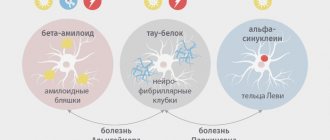
It all started when in the 20th century scientists became interested in the nature of unusual diseases in humans and animals: kuru, Creutzfeldt-Jakob, scrapie. The noticeable similarity in the pathology of these diseases gave rise to the hypothesis of their infectivity, which was subsequently confirmed experimentally. Then the question arose about the causative agent of these diseases. Before the answer was found, the extraordinary properties of the pathogens were revealed: they do not reproduce on artificial nutrient media, are resistant to high temperature, formaldehyde, various types of radiation, and the action of nucleases. Purification of the infectious material and its study made it possible to proclaim that “everything is to blame” for the protein, which 30 years ago received the name prion (from the English prion - protein infection).
Thus, famous American scientists - virologist and doctor D.K. Gaidushek, who discovered the infectious nature of prion diseases, in 1976, and biochemist S.B. Prusiner, who identified prions and developed the prion theory in 1997, were awarded Nobel Prizes. Their work became the impetus for subsequent research, thanks to which new types of prion infections were studied. But, even despite the undying interest in the “prion topic,” the formation of prions remains a mystery to this day.
Prion - neuron killer
A prion, also known in microbiology as infectious PrPSc, is a technical term that describes a defective form of a protein commonly found in mammalian brain tissue that promotes long-term memory function.
The development of prion disease in Wikipedia (eng.) is described as follows: “... These contagious molecules are responsible for the occurrence of transmissible spongiform encephalopathies or prionopathies. Their central manifestations are cerebellar disorders and extremely rapid dementia.”
Prions kill neurons very quickly, which provokes the rapid development of degenerative diseases.
According to the theory, we are talking about the source of many disorders of the central nervous system of animals (including humans). These proteins play a role in hereditary changes and evolution.
One of the regulatory factors of prion conversion is chaperones, whose function is to clean cells from pathogenic proteins. Both chaperones and prions belong to the group of cellular proteins.
Theories and hypotheses
S. B. Prusiner
The theory explaining why prions kill neurons was formulated by Professor S. B. Prusiner in 1982. He was also the first to use the word "prion" (originally intended to be used as a portmanteau of "proteinaceous" and "infectious", but it sounded better in the altered state).
Prusiner formulated the theory of prions as pathogenic proteins in connection with the search for the causative agent of Creutzfeldt-Jakob disorder. The agent he discovered was something new. Until 1982, it was believed that infectious diseases could only be caused by infectious organisms containing nucleic acid carrying genetic information. But the protein structure does not contain nucleic acid. It is a protein that multiplies by changing similar proteins in the body. In 1997, S. B. Prusiner received the Nobel Prize in this field.
Pathogenic prion proteins have the same primary structure (amino acid sequence), but differ in their conformational configuration. While wild PrPc has a strong α-helix predominance and about 5% β-sheet, pathogenic PrPSc (Sc – scrapie) has a β-sheet proportion of up to 40%. The reasons for the aberrant properties of PrPSc prions are still under investigation. Today there are several hypotheses.
- The viral hypothesis explains the effects of prions by virology. The involvement of RNA viruses in transmissible spongiform encephalopathies has been suggested. Both viroids and prions are small infectious pathogens; therefore, the result of exposure to the virus is a prion of an infectious nature.
- Multicomponent hypothesis . It is assumed that for the formation of infectious proteins as new pathogens, communication with polyanions and lipids is necessary.
- Heavy metal poisoning . Intoxication causes the development of infection when there is a lack or excess of copper in the body (an optimal amount of copper is necessary for healthy protein).
What is the toxicity of protein to a neuron?
Once the infectious protein appears, it can “imprint” its conformation onto healthy neurons. Toxic proteins can kill mitochondria in neurons, spreading disease to tissues.
Pathogenic proteins are extremely resistant to physical and chemical influences, which leads to difficulties in sterilization (attempts have been made to burn the brains of affected animals at 600°C; the ash subsequently infected approximately 1/3 of the animals). In accordance with what metabolic processes are disrupted by prions in the neuron, the most infectious are the tissues of the eye, brain and spinal cord.
Topic article: Signs of Alzheimer's disease
Biological essence of prions
Figure 1. The metaphor for neurodegenerative brain damage is that neural tissue becomes a sponge as a result of massive neuronal death.
The prion molecule is not something exotic: in a “normal” form it is present on the surface of the nerves of each of us. At the same time, we feel great, and our nerve cells are alive and healthy. However, this is all until our normal protein “degenerates” into an abnormal form. And if this happens, it will lead to terrifying consequences: the infectious form of prions has the ability to “stick together” with other molecules and, moreover, “convert” them into the same form, causing a “molecular epidemic”. As a result of this polymerization, toxic protein plaques appear on the nerve cell, and it dies [1]. In place of the dead cell, a void is formed - a vacuole filled with liquid. Over time, one neuron after another will disappear, and more and more “holes” will form in the brain, until finally the brain turns into a sponge (Fig. 1), which will inevitably be followed by death.
There is a simplified idea that polymerized prion fibrils “puncture” the neuron, causing its death. In fact, this is not entirely true: spherical prion aggregates preceding the fibrillar stage are also toxic (at least for Alzheimer's disease): “Alzheimer's neurotoxin: not only fibrils are poisonous.” - Ed.
But how can a normal natural protein (denoted PrPC) suddenly become pathological (PrPSc; Sc - from the word “scrapie”)? What is going to happen? As in the case of a “normal” infection, such transformation requires an encounter with an infectious prion molecule. There are two ways of transmitting this molecule: hereditary (due to mutations in the gene encoding the protein) and infectious. That is, the introduction of a prion can occur unexpectedly - for example, when eating insufficiently well-fried or cooked meat (which should contain the PrPSc form), during a blood transfusion, during organ and tissue transplantation, or when administering pituitary hormones of animal origin.
And then an amazing event occurs: normal protein molecules, in contact with pathological ones, themselves transform into them, changing their spatial structure (the mechanism of transformation remains a mystery to this day) [1]. Thus, the prion, as a real infectious agent, infects normal molecules, triggering a chain reaction that is destructive to the cell.
Infectious prions turned out to be non-toxic to nerve cells
Purified infectious prion proteins assembled into
Cassandra Terry
Prions that transmit neurodegenerative diseases and prions that damage neurons are different molecules, reports the Proceedings of the National Academy of Sciences
. This was revealed in experiments in which either an extract of purified prions (vectors of infection) or homogenized brain tissue of animals that showed symptoms of prion diseases, including neurodegeneration, were added to the nutrient medium of nerve cell cultures. In the first case, the cells in the cultures remained intact, in the second they were damaged.
Prions are protein infectious agents, and they differ from other proteins in their ability to take on very stable conformations and “infect” neighboring molecules of the same composition with them. As a result, proteins of a special, pathological conformation form clusters and deform the cells in which they are located. This occurs especially often in nerve cells, and many prion diseases affect the brain. The damage is very severe and irreversible, and the patient usually dies. There is no treatment for prion diseases yet.
Prion diseases can be hereditary if there are mutations in the genes of the corresponding proteins. They can also be contracted by eating the brains or meat of infected individuals (heat treatment and many digestive enzymes generally do not destroy prion clusters) or coming into contact with their body fluids. However, at the molecular level, the details of prion infections are still unclear.
Researchers from University College London, led by John Collinge, tested how various prions act on primary cultures of neurons (that is, “ready” nerve cells removed directly from the animal’s body) of the neocortex of the cerebral hemispheres and hippocampus of mice.
To the nutrient medium in which the same cultures were located, the researchers added purified rod-shaped clusters of molecules of the prion protein encoded by the Prnp
, isolated from the brains of mice with symptoms of prion neurodegenerative disease (from 107.7 to 108.1 infectious units per milliliter). In other cases, homogenized brain tissue from diseased mice was used instead of purified proteins and contained between 104.2 and 105.7 infectious prion units per milliliter. Finally, homogenates of the brains of healthy animals were introduced into some of the cultures, to which purified “infectious” prion proteins had previously been added. Proteins with a “healthy” conformation that were not capable of infecting cells were used as a control.
The state of neurons in culture—the number, length, and degree of branching of their processes, as well as whether they were alive—was observed using fluorescent microscopy: antibodies associated with fluorescent proteins were matched to molecules characteristic of different regions and functional states of the nerve cell. giving a glow of different colors, and looked at where and with what intensity it would appear.
The state of neurons in culture on different days after the addition of purified prions or homogenates of the brain of sick animals. The graphs show the lengths of the shoots depending on what and in what concentration the crops were treated.
Iryna Benilova et al. / Proceedings of the National Academy of Sciences, 2020
Share
The length of nerve cell processes and the number of processes on them decreased only in one case - when brain homogenate of mice with prion disease was added to the culture medium. Purified prion from the tissues of sick animals, including as part of the brain homogenate of healthy rodents, did not harm neurons, as did the purified “safely folded” prion protein from them. The neurotoxic effect of the homogenized brains of sick mice disappeared under the influence of two percent sodium lauroyl sarcosine, a substance with which homogenates were treated in some cases. At the same time, the number of molecules of the infecting prion did not decrease.
From these data, the researchers concluded that in prions, the function of infecting cells and the toxic effect on these cells are not strictly linked to each other. They are probably provided by different types of molecules and (or) forms of their clusters. Apparently, prions that form rod-shaped clusters are non-toxic to neurons, while proteins in clusters of some other shape, on the contrary, harm them. Poorly purified prions are known to damage nerve cells, but previous studies have not specified what shape groups of prions must be in order to be toxic. This is not easy to establish: in such mixtures there are prion clusters of various shapes and sizes. New research should be aimed at verifying this.
Prions that do not cause disease have already been found. In 2021, Italian scientists isolated PrPSc molecules from the brains of sheep suffering from scrapie, multiplied them, and “adapted” them to the structure of prions in bank voles ( Myodes glareolus
) and tried to infect rodents with these substances. For some reason, one of the varieties of PrPSc from sheep could not provoke scrapie in voles - probably due to the structure of the molecule.
Svetlana Yastrebova
Some information about prions
The researchers note:
- The prion protein includes 254 amino acid residues and weighs 33–35 kilodaltons [2];
- the gene encoding the PrP protein is found in humans, mammals, and birds [1];
- To completely destroy the prion protein, a temperature of at least 1000 degrees is required [1]!
- it is possible that prions take part in intercellular recognition and cellular activation [3];
- it is possible that the function of prions is to suppress age-related processes [3];
- with the development of clinical manifestations of prion diseases there are no signs of inflammation or changes in the blood;
- it is assumed that prions are involved in the development of schizophrenia and myopathy;
- The mechanism of action of prions and their transformation from a normal form to a pathological one remains unclear.
Conditions for the occurrence of diseases
The conditions for the occurrence of prion diseases are unique. They can form according to three scenarios: as infectious, sporadic and hereditary lesions. In the latter option, genetic predisposition plays a major role [2].
Renowned prion researcher Stanley Prusiner identifies two striking features of neurodegenerative diseases such as Creutzfeldt-Jakob disease, Alzheimer's disease, and Parkinson's disease. The first is that more than 80% of cases of the disease are sporadic (that is, random, occurring “by themselves”). Second: despite the fact that a large number of mutant proteins specific to a particular disease are expressed during embryonic development, the patterns of inheritance of these neurodegenerative diseases appear later. This suggests that certain processes occur during aging that release disease-causing proteins [5]. More than 20 years ago, the author argued that this process involves the random refolding of a protein into an incorrectly folded one, which corresponds to the transition to an infectious state - a prion.
Interesting facts about Alzheimer's disease: Chronic sleep deprivation may increase the risk of Alzheimer's disease ("New step in understanding Alzheimer's disease: sleep deprivation may be a risk factor"), and Alzheimer's neuropeptide itself (amyloid-β Aβ) may be part of the innate immune system ( "Perhaps Alzheimer's β-amyloid is part of the innate immune system." - Ed.
In the last decade, interest in this topic has renewed due to the possibility of developing diagnostics and effective therapy [5]. Many different explanations have emerged for age-related neurodegenerative diseases, such as oxidative modification of DNA, lipids and/or proteins; somatic mutations; altered innate immunity; exogenous toxins; DNA-RNA mismatches; disruption of chaperone function; absence of one of the gene alleles [5]. An alternative complex explanation is that different groups of proteins can form prions. Although small amounts of prions can be eliminated through protein degradation pathways, excessive accumulation over time allows prions to self-propagate throughout the body (Figure 2), resulting in central nervous system dysfunction [5].
Figure 2. Processes of neurodegeneration caused by prions. Top: The accumulation of a “normal” prion protein increases its likelihood of transitioning to a toxic conformation, which is described by a greater content of β-structure. Prions are most pathogenic in the form of oligomers; After fibril formation, toxicity decreases. Depending on which specific prion protein we are talking about, in a pathological state it can form plaques, tangles or inclusion bodies. Possible routes of drug intervention: (I) reduction of con precursor protein; (II) inhibition of the formation of the prion form; (III) destruction of toxic aggregates. Bottom: Hereditary senile neurodegeneration is explained by two events: the presence of a mutant form of the precursor and the formation of a prion from it, ready for oligo- and polymerization to form toxic forms.
[5]
Human prion diseases: symptoms and treatment prospects
The mechanisms of human diseases caused by prions (English from Pr otein (“protein”) and Infect ion (“Infection”; the word was first used by S. Prusiner [1] at the end of the 20th century) still remain poorly understood, despite There seems to be a large amount of research being carried out in this area. The purpose of this article is to summarize and clearly explain the currently available information regarding prions and their associated diseases.
For the purposes of this article, the following abbreviations are accepted: prion - proteinacious infectious particle; PrP - prion protein; PrPC , normal isoform of prion protein; PrPSc —infectious form of prion protein; P RNP is a gene encoding a prion protein.
Prion diseases (also known as transmissible spongiform encephalopathies, TSEs), which became known to man in the mid-18th century, are one of the most intriguing biological phenomena. Research into this phenomenon began in the 20th century with attempts to determine the biological essence of the causative agents of several specific diseases of animals and humans with similar symptoms. The hypothesis about their common etiology, put forward in the 1960s by scientists radiobiologist T. Alper and mathematician D. Griffith [2] and later supplemented and proven by physician S. Prusiner [3], gave impetus to subsequent research in this area. However, despite the deep interest of the scientific world, many aspects of the existence of prions remain unexplored to this day.
Human diseases associated with these specific protein infectious agents include Creutzfeldt-Jakob-Creutzfeldt disease (CJD) and its various variations, fatal insomnia (FFI/FSI), Gerstmann-Schraussler-Scheinker disease (GSS), kuru, variable protease-sensitive prionopathy, and prion disease associated with diarrhea and damage to the autonomic nervous system.
All of the above prion diseases remain fatal today, placing them in the category of the most dangerous diseases.
The essence of prions
After the process of translating genetic information, contained in the form of a nucleotide sequence of RNA (ribonucleic acid), into a specific amino acid sequence that forms the primary structure of all proteins, the newly synthesized proteins fold into certain structures. Prions are a type of misfolded protein molecule (Fig. 1), a defective form of the normal membrane protein PrP, which is expressed (manifests) primarily in cells of the central nervous system.
Rice. 1. Correctly “folded” (left) and defective (right) proteins [4]
Thanks to certain biological machinery, ordinary misfolded proteins are easily utilized and do not have a negative effect on the processes of human life. Prions are distinguished by their resistance to these mechanisms and the ability to convert their normal constituent proteins into similar ones. There are two hypotheses describing the mechanisms of this phenomenon. According to the first, heterodimeric model (Fig. 2) [5], the transformation occurs as follows: PrPSc joins the “healthy” PrP molecule and catalyzes a series of conformational changes leading to its transition to the prion form, after which the two abnormal proteins diverge and launch new rounds of this process. Moreover, the presence of an aggregated (“glued together”) form of the protein is not a necessary part of prion transformation.
Rice. 2. Heterodimeric model of prion replication [6]
An alternative hypothesis (Fig. 3) [5] - polymerization - states that catalysis of the conformational transformation of a normal protein into a pathological one can only occur through “nuclealization” followed by the formation of oligomeric or multimeric complexes. It is worth noting that recent research favors the second model.
Rice. 3. PrPSc replication cycle according to the polymerization hypothesis [7]
Exposure to PrPSc leads to a kind of “intracellular epidemic”: a lot of non-functional protein plaques are formed on the body’s cells, which is why it dies sooner or later.
Pathways of prion diseases
It is believed that there are only three ways of acquiring prion diseases: direct infection, hereditary transmission and sporadic occurrence by an unknown mechanism [8], but regardless of the origin, they can be transmitted by infectious means.
A highly conserved PRNP gene was detected, which carries information about the normal isoform of the PrP protein, located in the p-arm of human chromosome 20 [9]. PRNP has a length of 16 thousand nucleotide sequences and contains 2 exons. All hereditary prion diseases are associated with autosomal inheritance of mutations that occur in this gene.
Rice. 4. Localization of the PRNP gene on human chromosome 20 [10].
The main way prion disease occurs is spontaneous. According to one of the hypotheses explaining this process, a certain post-translational modification occurs in normal proteins [11]. Another hypothesis postulates that at some specific moment an indefinite number of cells in the body somatically (non-hereditarily) mutate and begin to produce the defective PrPSc protein [12].
A prion can enter an uninfected human body in various ways: by eating poorly prepared meat containing PrPSc, by blood transfusion from an infected person to a healthy person, by transplantation of infected organs and tissues.
Clinical picture of human prion diseases
Another important feature of prion proteins is the ability to take on a certain number of different conformations. This causes differences in the course and symptoms of prion diseases: different incubation periods, damage to different parts of the cerebral cortex, and disruption of various functions of the nervous system are possible [13]. Despite this, among all diseases associated with the action of prions, a series of common features can be traced: damage to the nervous system, the initial absence of an immune response to defective PrP proteins due to the constant presence of their “correct” isoform in the body [5], rapid progression of the disease after the end of the incubation period.
Creutzfeldt-Jakob disease ( CJD ) is a rare but best known human prion disease. There are several forms of CJD—hereditary, iatrogenic, sporadic, and variable—with the first three differing primarily in how they spread. The pathological pictures of hereditary, iatrogenic and sporadic CJD are similar: all cases show progressive cognitive impairment, cerebellar lesions and dysfunction, or a combination of these disorders; visual impairment up to blindness; myoclonic seizures. In the terminal stage, global cognitive impairment occurs, and death occurs 8–10 months after diagnosis of CJD [14]. Variable CJD has several deeper distinctive features: it affects young people under the average age of 30 years, its onset is characterized by behavioral changes, insomnia, depression; motor disorders appear approximately 6 months from the onset of the disease in the form of progressive ataxia, chorea, myoclonus; dementia occurs later than in the classic form, the patient is aware of his deteriorating condition. Variable CJD is characterized not only by onset at a younger age, but also by a median survival of more than 14 months [15].
Gerstmann-Straussler-Scheinker disease ( GSS ) is a disease that differs somewhat from CJD in several ways. This clinical form of TSE is caused by a mutation of the PNRP gene at codon 102, leading to the replacement of the amino acid proline with leucine [16]. The disease begins in middle age with the manifestation of cerebellar ataxia, speech disorders, dementia and changes in behavior. These symptoms may include diplopia, deafness, myoclonic seizures, and spasticity.
Kuru is a prion disease endemic to parts of Papua New Guinea. The main way this disease spread was ritual cannibalism. Symptoms include movement disorders (tremor, massive fasciculations, choreoathetosis, myoclonus). Death occurs approximately 2 years after the onset of the disease. In 2009, it was discovered that some members of one of the aboriginal tribes have innate immunity to kuru due to the appearance of a relatively new polymorphic modification of the PNRP gene [17].
Fatal insomnia is a rare TSE usually associated with inheritance of an autosomal dominant mutation (“familial” FFI). It is noted that there is also a sporadic form of this disease (“spontaneous”, FSI) [18]. In both cases, the following picture is observed: sleep disturbances, hallucinations, autonomic hyperactivation, motor disorders, and a sharp and progressive decline in cognitive abilities. The disease lasts from 8 to 72 months (the average is about 18 months), after which the patient dies. There is rapid death of neurons and astrogliosis of the anterior and medial thalamus and inferior olives, with subsequent damage to the cerebral cortex and cerebellum. The stages of the disease are described [18]: the initial form is characterized by the appearance of severe insomnia, panic attacks, pathological anxiety and phobias; at the second stage of the disease, the patient begins to experience hallucinations; at the penultimate stage, the patient loses the ability to sleep and rapidly loses weight; the terminal stage results in loss of speech and death.
In 2013, another clinical form of prion diseases , associated with damage to the autonomic nervous system [19]. The disease is associated with the appearance of a new mutation in the PRNP gene, leading to a shortening of the prion protein and subsequent disruption of its connection with cell membranes. In this case, the distribution of prion aggregates is not limited to the central nervous system. Presumably, PrPSc migrates to peripheral nerves and internal organs. A series of symptoms appear early in life: chronic diarrhea, autonomic failure and sensory polyneuropathy. In adulthood, damage to the central nervous system occurs, leading to the appearance of dementia and seizures among the symptoms.
Variable protease-sensitive prionopathy ( VPSPr ) is another new rare sporadic prion disease, first described in 2008. VPSPr is similar to GSS in terms of PrPSc features, however, no PRNP gene mutations were detected in the VPSPr prion [20]. The immunity of prion proteins that cause VPSPr to the action of proteases is significantly reduced. The disease is manifested by speech disorders (aphasia, dysarthria), cognitive impairment, and in some cases ataxia and parkinsonism.
Prospects for the treatment of prion diseases
Much research in this area provides evidence for the possibility of inhibiting prion replication and treating the diseases they cause.
As noted above, the body's immune response to PrPSc is absent. However, an experiment conducted with in vitro-derived prions proved that the use of antibodies produced against certain antigenic determinants of PrP induces inhibition of PrPSc proliferation [21], leading to a delay in the disease. Prion conversion can also be stopped using “β-structure blockers” - peptide sequences enriched with the amino acid proline and having a composition homologous to PrPC [5]. Another approach is based on the use of antisense oligonucleotides (ASO) - short fragments of nucleic acids that stop translation from messenger RNA due to the formation of loop-like regions on it. It is ASO that is currently the most effective method of inhibiting prion replication: experiments with the introduction of ASO into the cerebrospinal fluid of laboratory mice, conducted at Rocky Mountain Laboratories, led to a delay in the manifestation of prion diseases in experimental subjects by 113–135 days [22] . Astemizole, which belongs to the group of histamine H1 receptor (H1R) blockers, has a proven antiprion effect [23]. In addition, to prevent the multiplication of prions, it is possible to use mutations of the PNRP gene, leading to changes Q171R and E219K in the amino acid sequence of PrP: mutant prion proteins are unable to transform into a pathological form [24].
At the moment, treatment of prion diseases can only be symptomatic. The use of Brefeldin A, which destroys the Golgi apparatus and thereby slows down the proliferation of PrPSc, and NMDA receptor antagonists, which promote longer survival of infected cells, in the treatment of CJD has not achieved much success [25]. Attempts to use classical antiviral agents in the treatment of CJD and GSS also failed [25]. Traditional hypnotics have zero effectiveness in the treatment of FSI and FFI, although a case of delayed death has been reported with the simultaneous use of a number of potent drugs (diazepam, ketamine, nitric oxide) [26]. VPSPr therapy can be based on the use of the increased sensitivity of PrPSc to the action of proteases: delivery to the body of a mixture of specific immobilized cysteine proteases will probably delay the onset of the disease and prolong the life of the patient. Etiotropic treatment of major TSEs, apparently, should be based on the use of the above or similar methods of inhibition of prion replication.
Literature:
- Stanley B. Prusiner - Autobiography. NobelPrize.org.
- Alper T., Cramp WA, Haig DA, Clarke MC Does the agent of scrapie replicate without nucleic acid? //Nature. - 1967. - May (vol. 214, no. 5090). — P. 764–766.
- Taubes, Gary. The game of name is fame. But is it science? // Discover. - 1986. - December (vol. 7, no. 12). — P. 28–41.
- Mayo Foundation of Medical Education and Research.
- I. S. Shkundina, M. D. Ter-Avanesyan. Prions. Advances in biological chemistry, v. 46, 2006. - pp. 7–9.
- Nora Whisler. Lecture 34: PRION. — 2015. — P. 22.
- Dipendra Paj Pandeya, Nimish K. Acharya and Seong-Tshool Hong. Review: The Prion and its Potentiality. // Biomedical Research. — 2010.
- Groschup MH, Kretzschmar HA, eds. Prion Diseases Diagnosis and Pathogeneis // Archives of Virology - New York: Springer, 2001. - Vol. Suppl 16.
- Oesch B., Westaway D., Wälchli M., et al. A cellular gene encodes scrapie PrP 27–30 protein // Cell. - Cell Press, 1985. - Vol. 40, no. 4. - P. 735–746.
- Andreas Papassotiropoulus, Adriano Aguzzi, M. Axel Wollmer, Christoph Hock. The prion gene is associated with human long-term memory. // Human Molecular Genetics. — 2005.
- Prion Diseases (Transmissible Spongiform Encephalopathies) [Online].
- Prion Clinic: Sporadic Prion Disease [Online].
- N. N. Zavadenko, G. Sh. Khondkaryan, R. Ts. Bambeeva, A. A. Kholin, E. N. Saverskaya. Human prion diseases: modern aspects. // Journal of Neurology and Psychiatry. S. S. Korsakova. - 2018. - vol. 118, ed. 6. - pp. 91–92.
- ICTVdB Index of Viruses. US National Institutes of Health website [Online].
- Clinical and Pathologic Characteristics | Variant Creutzfeldt-Jakob Disease, Classic (CJD) [Online].
- Arata H, Takashima H, Hirano R, et al. Early clinical signs and imaging findings in Gerstmann–Sträussler–Scheinker syndrome (Pro102Leu). // Neurology. — 2006.
- Mead S, Whitfield J, Poulter M, Shah P, Uphill J, Campbell T, Al-Dujaily H, Hummerich H, Beck J, Mein CA, Verzilli C, Whittaker J, Alpers MP, Collinge J. A Novel Protective Prion Protein Variant that Colocalizes with Kuru Exposure. // New England Journal of Medicine. — 2009.
- R.Turner. Dying To Sleep: Fatal Familial Insomnia (FFI) [Online].
- Mead S, Gandhi S, Collinge J, Caine D, Gallujipali D, Carswell Ch, Hyare H, Joiner S, Ayling H, Lashley T, Linehan JM, Al-Doujaily H, Sharps B, Revesz T, Sandberg MK, Reilly MM, Koltzenburg M, Forbes A, Rudge P, Brandner S, Warren JD, Wadsworth JDF, Wood NW, Holton JL, Collinge J. A novel prion disease associated with diarrhea and autonomic neuropathy. // New England Journal of Medicine. — 2013.
- Gambetti P, Puoti G, Zou WQ. Variably protease-sensitive prionopathy: a novel disease of the prion protein. // Journal of Molecular Neuroscience. — 2011.
- M. Enari, E. Flechsig, C. Weissmann. Scrapie prion protein accumulation by scrapie-infected neuroblastoma cells abrogated by exposure to a prion protein antibody. // Proceedings of the National Academy of Sciences. — 2001.
- G. Raymond, et al. Antisense oligonucleotides extend survival of prion-infected mice. // JCI Insight. — 2021.
- Scripps Research Institute Scientists Identify First Potentially Effective Therapy for Human Prion Disease; Unique drug screening approach for prion diseases identifies tacrolimus and astemizole as antiprion agents // Proceedings of the National Academy of Sciences. — 2013.
- K. Kaneko et al. Evidence for protein X binding to a discontinuous epitope on the cellular prion protein during scrapie prion propagation. // Proceedings of the National Academy of Sciences. — 1997.
- M. Maniz, P. Kalakoti, M. Henry, J. Thakur, R. Menger, B. Guthikonda, A. Nanda. Creutzfeldt-Jakob disease: updated diagnostic criteria, treatment algorithm, and the utility of brain biopsy. // Neurosurgical Focus. — 2015.
- David Robson. The tragedy of people who stop sleeping: is it possible to help them? [Online] // BBC Future. — 2021.
Laboratory diagnosis and treatment
Diagnosis is based on intracerebral infection of mice or hamsters, which slowly (up to 150 days) develop the corresponding disease if the patient was sick [2]. Histological examination of the brains of dead animals is often carried out [2].
Unfortunately, to date, effective methods for treating prion diseases have not yet been developed, although attempts are being made to prevent the conformational transition of a normal protein into an abnormal one. Therefore, the most reliable way to prevent the development of infectious forms is prevention [2].
The solution to the “prion issue” is becoming especially relevant in connection with the growing threat of an epidemic through invasive medical operations and even when taking medications.
Lethargy
Iron deficiency
Diabetes
Atherosclerosis
Stroke
9977 04 May
IMPORTANT!
The information in this section cannot be used for self-diagnosis and self-treatment.
In case of pain or other exacerbation of the disease, diagnostic tests should be prescribed only by the attending physician. To make a diagnosis and properly prescribe treatment, you should contact your doctor. Lethargy: causes of occurrence, what diseases it occurs with, diagnosis and treatment methods.
Definition
The functioning of the nervous system is based on the reflex arc - the path along which irritation (signal) from the receptor passes to the effector organ. A reflex arc is a set of neural circuits consisting of sensory, intercalary and motor neurons that provide perception of a signal from the outside, conduct it to special centers of the brain, where the received information is analyzed and a response impulse is generated.
Thus, a high speed of impulse conduction and information processing is one of the necessary conditions for adequate speed of functioning of the nervous system.
Due to the fact that the structure of the nervous system is very complex, consisting of many cells connected to each other by nerve processes (nerve fibers), this system is subject to significant influence from the environment. Thus, changes in the composition of the fluid that washes the nerve cells, changes in temperature and other factors can significantly slow down its coordinated work.
Types of retardation
Retardation is manifested by a slowdown in the speed of certain nervous functions. In this case, both isolated changes can be observed (for example, a slowdown in motor activity, inhibition of perception), and complex ones - when all the main functions of the nervous system, including higher mental activity, slow down.
Retardation can develop acutely or gradually.
The more sudden it appears, the more attention must be paid to finding the reasons for its development. Possible reasons for the development of lethargy
There are many reasons for the slowdown of nervous processes, and one of them is
nervous exhaustion
. This term refers to the insufficiency of nerve tissue reserves to perform its functions, which develops during prolonged hard work of the nervous system.
The next condition associated with lethargy is oxygen starvation
central nervous system.
Lack of oxygen may be due to its insufficient supply from the outside (for example, staying in a poorly ventilated room), impaired gas exchange in the lungs due to various bronchopulmonary diseases
, impaired blood supply as a result of damage to the vessels supplying the central nervous system, or as a result of
heart failure
associated with poor blood supply to all organs and tissues.
Also, impaired oxygen delivery develops with anemia
- a decrease in the concentration of hemoglobin in the blood. Hemoglobin is a protein found in red blood cells that binds and carries oxygen from the lungs to every cell of our body.
Due to the fact that the nervous system's oxygen needs are high, oxygen deficiency quickly leads to disruption of its functioning.
Another cause of lethargy is a state of
hypoglycemia
- a decrease in blood glucose levels. It is observed when there is insufficient intake of glucose from food, during significant physical exertion (when glucose is predominantly spent by intensively working muscles), and when patients with diabetes mellitus overdose on insulin.
Lethargy also occurs due to toxic damage to the nervous system as a result of cerebral edema.
Diseases that cause lethargy
Among diseases of the cardiovascular system that may be associated with insufficient blood supply to the brain,
atherosclerotic vascular damage
, including against the background
of arterial hypertension, diabetes mellitus, heart failure
due to
myocardial infarction, myocarditis
and other diseases.
Bronchopulmonary diseases leading to insufficient oxygen saturation of the blood include pneumonia, chronic obstructive pulmonary disease, bronchial asthma
(especially at the time of an attack), etc.
Anemia
may develop due to a disorder of hematopoiesis (for example, iron deficiency anemia), destruction of red blood cells (hemolytic anemia), acute or chronic blood loss.
Hypoglycemia
observed not only in diabetes mellitus, but also in endocrine diseases, such as adrenal insufficiency, insulinoma, long-term severe liver diseases, etc.
Toxic damage to the central nervous system can be caused by the intake of certain toxic substances
from the outside, and by intoxication due to the development
of liver failure
(for example, with cirrhosis of the liver) or
renal failure
.
Intoxication syndrome accompanies most infectious diseases
, as well as many
malignant processes
.
Separately, it is worth noting life-threatening conditions that lead to cerebral edema and are manifested, among other things, by lethargy - these include stroke
, compression of the brain
by an intracranial tumor
,
traumatic brain injury
.
Which doctors should I contact if I feel lethargic?
Due to the fact that the cause of the development of lethargy can be diseases of a variety of organs and systems, if this symptom appears, you should consult a general practitioner, or. These specialists, after establishing the probable cause, can refer the patient to a nephrologist or hepatologist.
Diagnostics and examinations for lethargy
Diagnosis of diseases accompanied by lethargy begins with a survey and examination of the patient. Often this is enough to make a preliminary diagnosis. However, to confirm it, some additional research is necessary:
- a clinical blood test with a leukocyte count, which is necessary primarily to exclude anemia;
Prospects
Apparently, interest in prions will not fade until assumptions about them are fully confirmed and effective methods for treating prion diseases are found. The article [6] talks about the need for modern research, which requires careful consideration of foreign prions in extraneuronal tissues.
The authors used mice as model objects: two lines that transgenically expressed sheep prion protein, and one line that expressed human prion protein (Fig. 3). The goal was to compare the effectiveness of interspecies transmission of infection through brain and spleen tissue. Intracerebral infection with a foreign prion protein resulted in the absence or small amount of the infectious agent in the brains of these mice. However, infectious foreign prions were detected in the spleen at earlier stages of infection compared to when neurotropic prions were used, suggesting that lymphatic tissue may be more permissive to the spread of foreign prions compared with the brain.
Figure 3. The ability of the Sc237 hamster prion to infect and transmit when injected into the brain or spleen of transgenic mice having the sheep (tg338; white mice) or human (tg7; gray mice) PrP prion protein. The number of diseased/injected mice is shown in parentheses; Below is the average lifetime (in days).
[6]
What causes this preferential replication of prions in lymphatic tissues is still unknown. However, the findings indicate that humans may be more sensitive to foreign prions than previously thought based on the presence of prions in the brain, and for this reason, asymptomatic carriers of prion disease may not be recognized. This once again confirms that such a powerful biomolecule as the prion is fraught with many mysteries, the disclosure of which may help in understanding a number of insoluble problems of humanity...
Prion diseases (PDs), or transmissible spongiform encephalopathies (TSEs), are a group of neurodegenerative disorders characterized by rapidly progressive dementia and movement disorders. PZ can be inherited, occur sporadically, be acquired and contagious [1-4], have a long incubation period (years), but quickly progress after the manifestation of clinical symptoms. Among the first manifestations of mental illness are usually behavioral disturbances and personality changes, myoclonus, visual disturbances, and disturbances in the motor and coordination spheres. PDs do not have effective treatment; survival rate for sporadic and acquired forms in many cases does not exceed one year after the onset of the disease [1–4].
Since PZs are rare and begin with nonspecific symptoms that indicate unusual organ and tissue damage, they are difficult to diagnose. The practitioner has to carry out a differential diagnosis, which includes a wide range of pathological conditions of various etiologies, including other neurodegenerative, vascular, autoimmune, infectious, paraneoplastic, toxic, metabolic and iatrogenic syndromes [5].
In humans, prions cause Creutzfeldt-Jakob disease, Gerstmann-Straussler-Scheinker disease, fatal familial insomnia, and some others. In connection with outbreaks of disease in mammals, concerns have been raised about the possibility of transmission of TSE to humans, in particular bovine spongiform encephalopathy (mad cow disease), chronic wasting disease of deer and elk. A deeper understanding of the pathogenesis of PZ can lead to an expansion of the spectrum of these diseases due to forms that were not previously considered as such or had an unknown etiology, and at the same time allows us to count on the development of effective therapy for PZ.
A prion as an infectious agent does not introduce new genetic material into the body, but is a modified form (due to an abnormal three-dimensional structure) of one of the proteins present in it with self-sustaining properties, i.e. the ability to catalyze the conformational transformation of a normal cellular protein homologous to it into a similar one (prion) [1].
The term “prion” appeared in the middle of the 20th century. The nature of spongiform encephalopathies remained unknown for a long time. In the 60s, English researchers T. Alper et al. [6] and J. Griffith [7] hypothesized that some TSEs are caused by pathogens consisting solely of proteins. They discovered that the pathogen that causes scrapie in sheep
.: scrapie - pruritus) and Creutzfeldt-Jakob disease has unusual properties and exceptional resistance to ionizing radiation. This cast doubt on the assumption that these diseases are caused by a virus, although prions, like viruses, are able to increase their numbers using the functions of living cells. The dose of radiation required to destroy half of the particles of an infectious agent depends on their size: the smaller the particle, the less likely it is to be hit by a charged particle. On this basis, it was found that prions are significantly smaller in size than viruses.
Various opinions have been expressed regarding the composition of prions. The main hypothesis was about their protein composition, first put forward by J. Griffith [7] and substantiated in more detail by S. Prusiner [8, 9]. It was after this that the term “prion” was introduced (prion - proteinacious infectious particle, protein infectious particle, or PrP - prion protein, prion protein). In 1982, S. Prusiner [8] isolated this agent from the brain of sick animals and then studied its properties. The infectious agent consisted of a single protein. Based on the experimental data obtained, the concept of prion proteins was formulated [8, 9], which was initially met with great skepticism and then became generally accepted.
All mammalian PZs known to date are caused by the PrP protein. Its form with normal tertiary structure is designated PrPC .
.: common - ordinary or cellular - cellular).
The pathological form of the protein, which determines its infectivity, is called PrPSc (from English
: scrapie - sheep scrapie (scrapie), after the name of one of the first diseases with an established prion nature) or PrPTSE (from
English
: Transmissible Spongiform Encephalopathies - transmissible spongiform encephalopathies).
The main provisions of the concept of S. Prusiner [8, 9] are as follows [1]:
1. The infectious agent is the PrPSc protein, which replicates itself in the absence of nucleic acid.
2. The transformation of the protein from the normal PrPC form to the infectious PrPSc form occurs in the form of a conformational transition in different ways:
- spontaneously (sporadic forms);
- based on the entry from outside of the pathological form of PrPSc (acquired forms);
- as a result of mutations in the P RNP
, causing the formation of PrPSc from PrPC (hereditary forms).
Prions are the only known infectious agents whose reproduction occurs without the participation of nucleic acids. The infectious isoform of PrPSc is capable of converting the normal PrPC protein into the infectious isoform by changing its conformation (tertiary structure). The prions that appear as a result of this transformation begin to rearrange new protein molecules, and a chain reaction is started, during which a huge number of incorrectly “folded” molecules are formed. This in turn changes the interaction of PrPC with other proteins. Studies of the structure of PrPC have shown that it consists of approximately 40% α-helices and 3% β-sheets; in PrPSc, instead of α-helices, β-sheets predominate [1]. High β-sheet content correlates with PrPSc's resistance to enzymatic degradation and its infectivity.
It has been established that the normal and pathological forms of the prion protein are indistinguishable from each other in amino acid sequence, but have different conformations. The normal host protein PrPC is found on the surface of many cells, particularly neurons. When the structurally modified form of PrPSc enters healthy cells, it initiates a chain reaction and leads to the conversion of PrPC to PrPSc. The prion form of the protein is extremely stable and accumulates in the affected tissue, causing its damage and ultimately death. The stability of the prion form means that prions are resistant to denaturation by chemical and physical agents [1].
Following the determination of the primary structure of PrPС, the P RNP
, which is present in the genomes of all mammals, birds and fish.
In humans, this gene is localized in the short arm of chromosome 20 (20p) and has a length of 16 thousand bp. and contains 2 exons. The gene is highly conserved, and the highest levels of its expression are observed in neurons of the central nervous system. It encodes a polypeptide of 253 amino acid residues in length, which is shortened by cellular enzymes during maturation. The mature form of PrP consists of 208 amino acid residues and has a molecular weight of 35–36 kDa [10]. Genetic forms of PZ are caused by a mutation in the PRNP
. In Creutzfeldt-Jakob disease, more than 50 mutations have been described [11], most often in codons 178, 200 and 210; in Gerstmann-Straussler-Scheinker disease, mutations occur in codons 102 and 117; in familial fatal insomnia, in codons 129 and 178. In addition to mutations A number of polymorphism variants have been described [12].
Expressed P RNP
PrPC protein is a glycosylphosphatidylinositol anchored cell surface glycoprotein. PrPC (normal membrane protein of mammals) is part of cell membranes in various tissues - mainly in neurons of the central nervous system, lymphoreticular tissue, and cells of the immune system [1]. The functions of the PrPC protein are not fully understood. Apparently, it plays an important role in maintaining the safety of neurons and glia in relation to oxidative stress, is involved in the regulation of intracellular calcium (Ca+) content in neurons, copper metabolism, is involved in cell adhesion, the transmission of intracellular signals, ensuring the normal functioning of synapses and signal transmission in nervous tissue, as well as in maintaining circadian rhythms in cells, tissues, organs and the body as a whole [3, 13].
When the PrPC protein transitions to the prion state PrPSc, its α-helices transform into β-sheets [1, 4], and the conformation changes. These abnormal isoforms aggregate into highly structured amyloid fibers, which accumulate to form plaques. The end of each fiber serves as a kind of axis to which free protein molecules can attach, causing the fibril to grow. In most cases, only PrP molecules that are identical in primary structure to PrPSc can attach (therefore, prion transmission is usually species specific). However, cases of interspecies transmission of prions are also possible [14].
As mentioned above, there are three options for the occurrence of PZ: direct infection, hereditary and sporadic (occurring spontaneously) forms. In some cases, the development of PZ is determined by a combination of these factors (for example, infection and predisposition determined by the genotype) [1, 15]. In most cases (about 85%) PZs arise spontaneously for unknown reasons; hereditary forms account for approximately 10% of cases, the remaining cases are caused by prion infection [15].
Infection of both humans and animals is most often nutritional. From the intestine, prions are transported into the blood and lymph. Prions enter the central nervous system through the hematogenous route, breaking the blood-brain barrier, which is confirmed by the transmission of PZ through blood transfusion [16]. In addition to hematogenous spread, prions can enter the brain via the vagus and peripheral nerves (neuroinvasion) [17].
It was previously thought that the central nervous system was the only tissue in which prions accumulate, but it has been confirmed that prions enter the brain after they replicate in the spleen, appendix, tonsils and other lymphoid tissues. Although PDs are neurological diseases, critical events in their pathogenesis occur in peripheral lymphoid organs [18]. Lymphoid organs are affected in the early stages of PZ, and the spleen and lymph nodes are the primary sites of PrPSc replication after infection and are also significantly damaged. Experiments have shown that removal of the spleen and other effects on peripheral lymphoid structures delay the onset of clinical manifestations of PZ [18].
Under the influence of prions, fibrils are formed in the brain, amyloidosis (extracellular dysproteinosis, characterized by amyloid deposition with the development of tissue atrophy and sclerosis) and astrogliosis (proliferation of astrocytic neuroglia, hyperproduction of glial fibers) develop in the absence of infiltrative inflammatory reactions. As a result, the death of neurons, the formation of vacuoles, protein/amyloid aggregates, and sponge-like changes in the brain occur [19–21]. After infection and replication in the CNS, prions spread along peripheral nerves to other tissues, where secondary prion replication occurs [20, 21].
Prions infect different species of mammals, but the PrP protein is very similar in all species. But due to small differences between PrP in different species, transmission of PZ from one species to another is uncharacteristic, i.e., there is a species barrier [1, 22]. However, the human variant of Creutzfeldt-Jakob disease is caused by a prion that typically infects cows and causes TSE in cattle, which is transmitted through contaminated meat.
Clinical forms of PZ
Currently, the following clinical forms of human PD are known: Creutzfeldt-Jakob disease (CJD), the main type; variant CJD (vCJD); Gerstmann-Straussler-Scheinker disease; variable protease-sensitive prionopathy; fatal insomnia and fatal familial insomnia; kuru; PZ associated with diarrhea and autonomic neuropathy.
CJD
is the most common human PZ, accounting for about 85% of all cases of PZ. The leading clinical manifestation of CJD is rapidly progressive dementia, which occurs without obvious causes and is accompanied by behavioral changes (especially in younger patients), disorders of higher mental functions (aphasia, amnesia and attention deficit), myoclonus, and pyramidal disorders [23].
The classic clinical triad of CJD includes rapidly progressive dementia, myoclonus, and ataxia. Among movement disorders, myoclonic hyperkinesis is most often observed (occurs spontaneously or is provoked by auditory and tactile stimulation), but extrapyramidal disorders are also found: dystonia, choreoathetosis, tremor, hemiballismus, atypical parkinsonism [24].
Other neurological disorders are represented by visual disorders (diplopia, blurred or blurred vision, visual field defects, visual agnosia, possible formation of cortical blindness), epileptic seizures, behavioral changes, peripheral neuropathy, and in later stages - akinetic mutism. Psychiatric symptoms are observed in 80-90% of patients and occur early in the disease in 20-26%. These include insomnia, anxiety, depression, irritability, hallucinations, psychosis, and cognitive disorders [25].
Confirmation of CJD using laboratory methods is based on the results of magnetic resonance imaging (MRI) of the brain (diffusion-weighted MRI or FLAIR mode reveals characteristic signal changes at the level of the caudate nucleus, globus pallidus, cortical zones), electroencephalography (EEG) (typical periodic complexes of sharp waves on a slower and low-amplitude background), identification of protein markers of the pathogen in the cerebrospinal fluid (in particular, protein 14−3-3) using immunoblotting, a new method of vibration-induced conversion in real time (RT-QUIC), through which a minimal amount of prion protein is detected [21, 26].
The diagnosis is verified by histochemical examination of microslides from a brain biopsy, and hereditary cases and other genetic forms of PZ (fatal familial insomnia and Gerstmann-Straussler-Scheinker disease) can be established.
But since not all parts of the brain can show histological changes typical of CJD, confirmation of the diagnosis after a biopsy may give inconclusive results. Therefore, neurosurgeons strive to obtain samples for research from those areas of the brain in which there are the most significant changes according to MRI (usually deep subcortical structures) [15].
There are several forms of CJD: sporadic, genetic (familial), iatrogenic and vCJD. Regardless of the form, the course of the disease is characterized by rapid progression with a fatal outcome. Average survival usually ranges from 6 months to a year [12, 15].
Sporadic CJD
occurs much more often than other forms - in 85% of cases [15]. The incidence is 1 case per 1 million population per year. The neurodegenerative process starts spontaneously as a result of somatic gene mutation or random structural changes in PrP, leading to the formation of PrPSc. In contrast to vCJD, clinical and pathological manifestations are more heterogeneous, which may be due to the existence of different molecular phenotypes. The disease usually begins at the age of 60-70 years, about 90% of patients die within one year.
Genetic (familial) CJD. About 5-15% of cases of CJD are genetic (familial), with an autosomal dominant mode of inheritance and high penetrance associated with mutations in the PRNP
.
Familial CJD has similar clinical, neuroradiological and laboratory characteristics to the sporadic form. More than 50 PRNP
, and their frequency increases with age [11].
Iatrogenic CJD
can be transmitted by cadaveric cornea or dura transplantation, the use of stereotactic intracerebral electrodes, or the use of growth hormone prepared from human pituitary gland. The clinical picture is similar to that of sporadic CJD [12, 15]. The incubation period depends on the site of inoculation. When using contaminated electrodes that are applied directly to the brain, it is the shortest (16–28 months), and after peripheral administration of growth hormone it lasts from 5 to 30 years [27].
There are three case reports of CJD being transmitted as a result of blood transfusion from a donor who was diagnosed with vCJD during the outbreak in the UK [28].
vCJD
develops after eating beef contaminated with prions. In 1986, an epizootic of bovine TSE, also called “mad cow disease,” broke out in the UK, leading to the death of more than 160,000 cattle [29]. This new disease was caused by the use of meat and bone meal supplements when, due to poorly controlled animal by-product processing regulations, PrPSc from scrapie-infected sheep and other TSE-infected cattle ended up in cow feed. Typically, the technology for producing such flour after thorough grinding of the raw materials includes treatment with active fat solvents, as well as heat treatment at a temperature of 130 °C. However, in the late 70s, entrepreneurs, deciding to increase the nutritional value of meat and bone meal, reduced the heat treatment regime to 110 ° C, and also reduced the amount of fat extracting substances. It was these changes that contributed to the cattle epizootic [3].
It has been proven that TSE in cows has led to the emergence of a new type of CJD, called “variant CJD” [29]. The first cases of vCJD were reported in 1995, when the disease was diagnosed in 2 British adolescents [30, 31]. Due to the long incubation period, the link between the disease and contaminated meat in the UK was not established until the incidence of TSE in cows became an epidemic. The TSE epidemic was brought under control following massive slaughter of livestock and changes in production technology that dramatically reduced contamination of meat with nerve tissue components. In the UK, the annual number of new cases of vCJD, which peaked in 2000, has been steadily declining, with only 1 case confirmed in 2013 [15, 32].
The majority of vCJD cases are diagnosed in the UK, with approximately 200 cases reported in 2015, compared with approximately 60 cases in other countries [15, 32]. All patients developed vCJD after eating meat obtained from cattle affected by TSE. But despite the widespread occurrence of TSE, affecting hundreds of thousands of cattle, relatively few people who consumed meat from diseased animals developed vCJD [15, 33].
The incubation period (the time between eating contaminated beef and the onset of symptoms) was long: most patients were infected in the late 1980s, and the peak incidence occurred in the early 2000s, i.e., the incubation period was 11-12 years. In the most recently diagnosed cases, the incubation period ranged from 12 to more than 20 years [32, 33].
Clinical and pathomorphological manifestations of vCJD differ from other forms of CJD. The disease affects young people under the age of 30 on average, its onset is characterized by personality changes: the patient loses previous interests, begins to avoid loved ones, and develops anxiety, insomnia, and depression. Motor disturbances appear approximately 6 months from the onset of the disease in the form of progressive ataxia, chorea, and myoclonus. Dementia occurs later than in the classic form, and the patient becomes aware of his deteriorating condition. Quite quickly he loses the ability to self-service. vCJD is characterized not only by onset at a younger age, but also by a median survival rate exceeding 14 months [15, 32, 33]. It is likely that the differences in survival are due in part to the age of the patients.
Moreover, the development of vCJD is caused not only by exposure to a pathogen from contaminated meat, but also by genetic predisposition, one of the factors of which is polymorphism in codon 129 of the PRNP
, which determines the insertion of the amino acid valine or methionine in the corresponding position of the prion protein. In all confirmed cases, a homozygous state for this polymorphism with the incorporation of methionine was detected. It is assumed that the increased susceptibility to the development of PZ in homozygotes at codon 129 is due to the absolute homology of prion protein molecules, which facilitates intermolecular interaction and, consequently, the conversion of PrPC to PrPSc [29]. On the contrary, heterozygosity (valine/methionine) in codon 129 has a certain protective effect, which is confirmed by a later (10-20 years) onset of the disease in heterozygotes compared to homozygotes in some hereditary forms of PD [12, 33].
Gerstmann-Straussler-Scheinker disease
, or subacute spongiform encephalopathy, is inherited in an autosomal dominant manner and usually develops in middle age. The average life expectancy after onset (5 years) exceeds that for CJD. Compared to CJD, the prevalence of Gerstmann-Straussler-Scheinker disease is approximately 100 times lower [3, 12].
The disease begins with the development of cerebellar ataxia with difficulty walking and maintaining balance, dysarthria, which is accompanied by progressive personality changes and dementia. Double vision, gaze paresis, deafness, and pyramidal symptoms (spasticity) may occur. Myoclonus is much less common than in CJD. Life expectancy ranges from 2 to 10 years.
If characteristic symptoms and family history are present, Gerstmann-Straussler-Scheinker syndrome is more likely in young patients (under 45 years of age). The diagnosis is confirmed by genetic research data [3, 12, 33].
Variable protease-sensitive prionopathy
- a rare PZ, described in 2008, diagnosed in 2-3 cases per 100 million population [34]. Variable protease-sensitive prionopathy is similar to Gerstmann-Straussler-Scheinker disease in terms of accumulation of the abnormal prion protein PrPSc and patterns of changes in the brain, but unlike Gerstmann-Straussler-Scheinker disease, no mutations have been identified in the gene for this protein. The clinical manifestations differ from CJD, and PrPSc is less resistant to protease degradation, with some cases being more protease sensitive than others, as reflected in the name “variable protease-sensitive.” The disease manifests itself with mental and speech disorders (aphasia, dysarthria), and cognitive impairment. Ataxia and parkinsonism may develop. The average age of onset of the disease is 70 years, survival is about 2 years. Approximately 40% of patients have a family history of dementia [34].
Fatal insomnia
- a rare hereditary or sporadic PZ that causes sleep disturbances, movement disorders and leads to death. Fatal insomnia is usually associated with an autosomal dominant mutation. There are descriptions of about 40 families affected by this disease [33]. Genetic testing can confirm the diagnosis. The average age of onset is about 40 years (from 20 to 60 years), life expectancy is 8-72 (on average 18.4) months. A description of verified sporadic fatal insomnia in a 13-year-old adolescent was recently published [35].
Early symptoms of fatal insomnia include increasing difficulty falling asleep and staying asleep, as well as cognitive decline, ataxia, and psychiatric disturbances (behavioral changes, mood disorders). These are accompanied by symptoms of sympathetic hyperactivation: arterial hypertension, tachycardia, hyperthermia, sweating.
The staged progression of the disease has been described [33]. At the initial stage, the patient suffers from increasingly severe insomnia, panic attacks and phobias (lasting an average of 4 months). Later, panic attacks become severe and are accompanied by hallucinations (on average 5 months). Next comes a complete inability to sleep, accompanied by rapid loss of body weight (on average 3 months). Finally, the patient loses speech, becomes unresponsive to his surroundings, and then dies (average 6 months).
Kuru
- a rare PZ, endemic to the highlands of Papua New Guinea and the Fore tribe, spread through ritual cannibalism. Although the last rituals associated with cannibalism ended in the mid-20th century, between 1996 and 2004. 11 new cases of kuru were reported, suggesting that the incubation period of the disease may last more than 50 years [33].
The first symptoms of kuru are tremor (like shaking) and ataxia. Later, dementia and movement disorders develop (choreoathetosis, myoclonus, massive fasciculations). Death usually occurs within 2 years of the onset of symptoms and is caused by pneumonia or pressure ulcer infections.
In 2009, it was found that some members of the Fore tribe have innate immunity to kuru, thanks to a new polymorphism of the PRNP
[36].
PZ associated with diarrhea and autonomic neuropathy
, is a new clinical form described in 2013 in 11 members of a British family, manifested by symptoms of damage to the autonomic nervous system, rather than the central nervous system [37].
The disease is associated with a new Y163X mutation in the PRNP
, leading to a shortening of the prion protein, as a result of which it lacks the “anchor” that binds the protein to cell membranes; this probably creates the preconditions for its migration to other tissues. Therefore, the accumulation of prion amyloids is not limited to the central nervous system, but extends to peripheral nerves and internal organs. Peripheral symptoms predominate at the onset of the disease, and signs of central nervous system damage appear later.
This PZ shows how significantly a new mutation can change the tissues where abnormal proteins settle, causing symptoms of the disease. Diagnosis P.Z. should be excluded in patients with unexplained chronic diarrhea in combination with neuropathy, as well as in familial cases resembling amyloid polyneuropathy.
Symptoms begin in young adulthood and include chronic watery diarrhea, autonomic failure (eg, urinary retention, incontinence, orthostatic hypotension), and, most notably, sensory polyneuropathy. At the age of 40-50 years, the patient experiences a decline in cognitive function and seizures. The disease progresses over decades, and once symptoms appear, patients can live up to 30 years [37].
Prevention of PD
Today there are no treatments for P.Z. All known human diseases are fatal, and prescriptions are limited to maintenance therapy. Relatives of patients with a family history of PD are recommended to receive genetic counseling.
Special vaccines for animals are being developed, which in the future should help in creating a vaccine against PZ for humans [38]. Using genetic engineering methods, a cow was produced that lacked the gene necessary for the formation of prions, i.e., theoretically, it is immune to TSE [38].
Due to the lack of effective treatment, measures are needed to prevent transmissible forms of P.Z. According to modern research, the main way to acquire PZ is through the consumption of contaminated food. Prions are believed to be present in urine, saliva and other body fluids and tissues, and can also persist for long periods in the environment in soil and animal remains.
Infection with prions can occur through the use of unsterile surgical instruments. Prions are not susceptible to standard disinfection methods, so they can pose a danger to other patients and surgeons, pathologists or laboratory technicians who come into contact with contaminated tissue or instruments. Doctors who come into contact with biological fluids and tissues of patients with suspected PD should wear gloves and avoid contact of contaminated material with mucous membranes. If contaminated material comes into contact with the skin, it is first disinfected with a 4% sodium hydroxide solution for 5-10 minutes, then washed with running water [39].
Infection can be prevented by taking precautions when handling infected tissue and using appropriate techniques to clean contaminated equipment. To disinfect materials and instruments, autoclaving at 132 °C for 1 hour or sterilization in a 1N sodium hydroxide solution or 10% sodium hypochloride solution for 1 hour is recommended. Sterilization should lead to denaturation of prions (by hydrolysis or destruction of the tertiary structure) to a state in which they will be unable to change the configuration of other proteins [39]. Prions are resistant to proteases, heat, radiation, and formalin treatment, although these measures reduce their infectivity [40]. The effectiveness of ozone sterilization as a method of deactivating prions in contaminated water has been demonstrated [41].
Research is being conducted using genetically modified organisms, in particular yeast fungi, as well as a number of bacterial strains for the fermented biotransformation of organic food waste (including those containing a prion - the causative agent of scrapie sheep) with the effect of reducing prion activity and, as a result, preventing re-infection of animals and people [42].
In modern medicine, biological medicinal products are used, for the production of which animal tissue is used. Is there a potential risk of developing PZ in patients using them? In this regard, the issues of ensuring and assessing the safety of such drugs for humans are extremely relevant, which must be solved with the help of modern technologies.
The biological safety of biological products based on animal tissue from contamination by prions is ensured by specially developed procedures that meet the requirements of the European Medicines Agency (Guideline 410/01 Rev.2), as well as the requirements of the European Pharmacopoeia (Eur. Ph. 7.0, 01/2008: 50107, paragraph. 5.1.7 “Virus safety” and Eur. Ph. 7.0, 01/2008: 50208, paragraph 5.2.8. “Reducing the risk of infection of animals with the spongiform encephalopathy virus through human and veterinary drugs”) to minimize the risks of prion transmission through pharmacological products , intended for humans and veterinary use.
is a manufacturer of modern medications, including the peptidergic nootropic drug Cortexin. Geropharm purchases raw materials of animal origin used for the production of dry extract - a pharmaceutical substance. The raw materials are taken from young healthy animals, which undergo the most stringent selection according to geographical criteria and are used only after confirmation of viral safety and receipt of a veterinary certificate issued by the State Service for Veterinary Safety.
The production of the drug Cortexin is based on modern technological processes aimed at isolating low molecular weight water-soluble peptides from raw material tissues, the molecular weight of which is less than 10 kDa. According to the technology enshrined in the industrial regulations, the production of the drug includes aqueous extraction of a complex of hydrophilic polypeptides from a dry extract, followed by ultrafiltration through a hollow fiber filter with a cutoff capacity of 8 kD, as well as final filtration of the solution through membrane filters with pore sizes of 0.45 and 0.22 microns before freeze drying. Ultrafiltration makes it possible to completely exclude from the final preparation peptides with a molecular weight of more than 10 kDa, while, as is known, the molecular weight of prions is 33-35 kDa, and the molecular fragment resistant to proteolysis is 27-30 kDa.
It is important to emphasize that the substance of the drug (cortexin extract dry) is an intermediate product and is not directly used for the preparation of a sterile medicinal product, but is processed at additional technological stages, as a result of which the composition of the finished dosage form (lyophilisate) includes only water-soluble pharmacologically active polypeptides with molecular weighing less than 10 kDa.
Thus, based on the physicochemical properties of prions described above and taking into account the conditions for obtaining the peptide substrate used in the production of the drug cortexin, we can confidently exclude the possibility of the presence of not only PrPSc, but also fragments of the molecule of this protein in the solution used to obtain the drug . For each batch of finished products, as part of routine monitoring, the manufacturer performs an analysis for the absence of high molecular weight proteins using validated quality control methods, which confirms the high biological safety of Cortexin from prion contamination.
The study of prions and the diseases they cause is a relatively new and rapidly developing area of biomedical research. The ever-increasing interest in PZ is due, on the one hand, to the fact that prions represent a completely new type of infectious agent, the identification of which can be compared in its significance with the discovery of the world of single-celled microorganisms by Anthony van Leeuwenhoek and then with the discovery of the kingdom of viruses by Dmitry Iosifovich Ivanovsky. On the other hand, medicine currently does not have effective means of treating this pathology, and the long incubation period of PD with the development of neurodegenerative changes and inevitable death evokes mystical horror. Therefore, it is important to prevent infection with prions through both nutritional (consumption of contaminated meat products) and transmissible iatrogenic (use of medicinal biological products obtained from animal tissues) routes. The reliable and highly regulated production of peptidergic biologics provides optimism regarding the safety of these drugs along with their effectiveness.
The authors declare no conflict of interest.
*e-mail; https://orcid.org/0000-0003-0103-7422
Literature
- Abramova Z.I. Study of proteins and nucleic acids. Kazan: Kazan State University, 2006. - 157 pp.;
- Novikov D.K., Generalov I.I., Danyushchenkova N.M. Medical microbiology. Vitebsk: VSU, 2010. - 597 pp.;
- Prudnikova S.V. Microbiology with basics of virology. Krasnoyarsk: IPK SFU, 2008;
- Pozdeev O.K., Pokrovsky V.I. Medical microbiology. M.: Geotar-med, 2001. - 765 pp.;
- S. B. Prusiner. (2012). A Unifying Role for Prions in Neurodegenerative Diseases. Science
.
336 , 1511-1513; - V. Beringue, L. Herzog, E. Jaumein, F. Reine, P. Sibille, et. al.. (2012). Facilitated Cross-Species Transmission of Prions in Extraneural Tissue. Science
.
335 , 472-475; - Carolina Pola. (2012). Prion escape to spleen. Nat Med
.
18 , 360-360; - Elements: “10 facts about prions and amyloids”;
- Elements: “Geometry of protein bodies”;
- Charles Weissmann. (2012). Mutation and Selection of Prions. PLoS Pathog
.
8 , e1002582.