Basal ganglia and their functions
basal ganglia,
called ganglia by histologists of the last century, they are nuclear-type structures that are located in the thickness of the white matter of the forebrain closer to its base.
In mammals, the basal nuclei include the strongly elongated and curved caudate nucleus
and
the lentiform nucleus embedded in the thickness of the white matter.
It is divided into three parts by two white plates: the largest, laterally lying
shell,
and
the globus pallidus,
consisting of internal and external sections (Fig. 3.29).
| Rice. 3.29 Afferent and efferent connections of the basal nuclei 1 - caudate nucleus, 2 - putamen, 3 - substantia nigra, 4 - thalamus, 5, 5* - internal and external parts of the globus pallidus, 6 - subthalamic nucleus, 7 - sensorimotor cortex, 8 - motor cortex, 9 - paths to the brainstem; arrows indicate afferent pathways, efferent pathways, internuclear interactions. |
These anatomical formations form the so-called striopallidal system
(From Latin striatus - striped and pallidus - pale.)
,
which, according to phylogenetic and functional criteria, is divided into the ancient part paleostriatum and the new part neostriatum.
The paleostriatum
is represented by the globus pallidus, and
the neostriatum,
which appears for the first time in reptiles, consists of the caudate nucleus and putamen, which are collectively called
the striatum,
or striatum.
The caudate nucleus and putamen are anatomically connected and are characterized by alternating white and gray matter, which justifies the term striatum.
The subthalamic nucleus is also often included in the striopallidal system.
(Lewis body) and
the substantia nigra
of the midbrain, which form a functional unity with the basal ganglia. The striatum consists mainly of small cells, the axons of which are directed to the globus pallidus and the substantia nigra of the midbrain.
The striatum is a kind of collector of afferent inputs going to the basal ganglia. The main sources of these inputs are the neocortex (mainly sensorimotor), nonspecific nuclei of the thalamus, and dopaminergic pathways from the substantia nigra.
In contrast to the striatum, the globus pallidus
consists of large neurons and is the concentration of the output, efferent pathways of the striopallidal system. The axons of neurons localized in the globus pallidus approach various nuclei of the diencephalon and mesencephalon, including the red nucleus, where the red nucleus-spinal tract of the extrapyramidal motor regulation system begins.
Another important efferent pathway runs from the internal part of the globus pallidus to the anteroventral and ventrolateral nuclei of the thalamus, and from there continues to the motor areas of the cerebral cortex. The presence of this pathway determines a multi-link loop-like connection between the sensorimotor and motor areas of the cortex, which occurs through the striatum and globus pallidus to the thalamus. It is noteworthy that, as part of this striopallidothalamocortical pathway, the basal ganglia act as an afferent link in relation to the motor areas of the cerebral cortex. Numerous connections of the striopallidal system with various parts of the brain indicate its participation in integration processes, however, to date, much remains unclear in the knowledge about the functions of the basal ganglia.
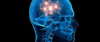
The basal ganglia play an important role in the regulation of movement
and
sensorimotor coordination.
It is known that when the striatum is damaged,
athetosis is observed -
slow worm-like movements of the hands and fingers.
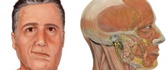
Degeneration of cells of this structure also causes another disease - chorea,
expressed in convulsive twitching of the facial muscles and muscles of the limbs, which are observed at rest and when performing voluntary movements.
However, attempts to elucidate the etiology of these phenomena in animal experiments did not yield results. Destruction of the caudate nucleus in dogs and cats did not lead to the occurrence of hyperkinesis,
characteristic of the diseases described above.
Local electrical stimulation of certain areas of the striatum causes so-called circulatory motor reactions in animals,
characterized by turning the head and torso in the direction opposite to the irritation. Stimulation of other areas of the striatum, on the contrary, leads to inhibition of motor reactions caused by various sensory stimuli.
The presence of certain discrepancies between experimental and clinical data apparently indicates the occurrence of systemic disturbances in the mechanisms of movement regulation during pathological processes in the basal ganglia. Obviously, these disorders are associated with changes in the function of not only the striatum, but also other structures.
As an example, we can consider the possible pathophysiological mechanism of parkinsonism.
This syndrome is associated with damage to the basal ganglia and is characterized by a complex of symptoms such as
hypokinesia -
low mobility and difficulty in the transition from rest to movement;
waxy rigidity,
or
hypertonicity,
independent of the position of the joints and the phase of movement;
static tremor
(shaking), most pronounced in the distal limbs.
All these symptoms are caused by hyperactivity of the basal ganglia, which occurs when the dopaminergic (most likely inhibitory) pathway that runs from the substantia nigra to the striatum is damaged. Thus, the etiology of parkinsonism is due to dysfunction of the striatum and midbrain structures, which are functionally combined into the striopallidal system.
To clarify the role of the basal ganglia in the implementation of movements, data from microelectrode studies are successfully used. Experiments on monkeys have shown a correlation between the discharges of neurons in the striatum and slow, side-to-side worm-like movements of the paw. As a rule, the neuron discharge precedes the onset of slow movement, and during fast “ballistic” movements it is absent. These facts allow us to conclude that striatal neurons are involved in the generation of slow movements that are subject to correction by sensory feedback. The basal ganglia represent one of the levels of a movement regulation system built on a hierarchical principle.
Receiving information from the associative zones of the cortex, the basal ganglia are involved in creating a program of targeted movements, taking into account the dominant motivation. Next, the relevant information from the basal ganglia enters the anterior thalamus, where it is integrated with information coming from the cerebellum. From the thalamic nuclei, impulses reach the motor cortex, which is responsible for implementing the program of purposeful movement through the underlying brainstem and spinal motor centers. So, in general terms, we can imagine the place of the basal ganglia in the entire system of motor centers of the brain.
Physiology
All subcortical nuclei are again conventionally combined into two systems. The first is called the striopallid system, which includes:
- pale globe;
- caudate nucleus of the brain;
- shell.
The last two structures consist of many layers, which is why they are grouped under the name striatum. Ballus pallidus is a brighter, lighter color and is not laminated.
The lenticular nucleus is formed by the globus pallidus (located inside) and the shell, which forms its outer layer. The amygdala and the amygdala are components of the limbic system of the brain.
Let's take a closer look at what these brain nuclei are.
Caudate nucleus
Paired component of the brain related to the striatum. The location is in front of the thalamus. They are separated by a strip of white matter called the internal capsule. Its anterior part has a more massive thickened structure, the head of the structure is adjacent to the lenticular core.
Structurally, it consists of Golgi neurons and has the following characteristics:
- their axon is very thin, and dendrites (processes) are short;
- nerve cells have reduced physical dimensions compared to normal ones.
The caudate nucleus has close connections with many other distinct brain structures and forms a very wide network of neurons. Through them, the globus pallidus and thalamus interact with sensory areas, creating pathways with closed circuits. The ganglion also interacts with other parts of the brain, and not all of them lie in its vicinity.
Experts do not have a consensus on what the function of the caudate nucleus is. This once again confirms the scientifically unfounded theory that the brain is a single structure, any of its functions can be easily performed by any part. And this has been repeatedly proven in studies of people injured due to accidents, other emergencies and diseases.
It is certainly known that it takes part in autonomic functions and plays an important role in the development of cognitive abilities, coordination and stimulation of motor activity.
The striatal nucleus consists of layers of white and gray matter alternating in a vertical plane.
Black substance
The component of the system that is most involved in the coordination of movements and motor skills, maintaining muscle tone and controlling postures. Participates in many autonomic functions, such as breathing, cardiac activity, and maintaining vascular tone.
Physically, the substance is a continuous strip, as was believed for decades, but anatomical sections have shown that it consists of two parts. One of them is a receiver that sends dopamine to the striatum, the second - a transmitter - serves as a transport artery for transmitting signals from the basal ganglia to other parts of the brain, of which there are more than a dozen.
Lenticular body
Its location is between the caudate nucleus and the thalamus, which, as stated, are separated by the external capsule. In front of the structure, it merges with the head of the caudate nucleus, which is why its frontal section has a wedge-shaped shape.
This nucleus consists of sections separated by a thin film of white matter:
- shell – darker outer part;
- pale ball.
The latter is very different in structure from the shell and consists of type I Golgi cells, which predominate in the human nervous system, and are larger in size than their type II. According to neurophysiologists, it is a more archaic brain structure than other components of the brain nucleus.
Other nodes
The fence is the thinnest layer of gray matter between the shell and the island, around which there is a white substance.
The basal ganglia are also represented by the amygdala, located under the shell in the temporal region of the head. It is believed, but not known for sure, that this part belongs to the olfactory system. It is also where the nerve fibers coming from the olfactory lobe end.
Lenticular nucleus
The lentiform nucleus (nucl. lentiformis) is located lateral and anterior to the thalamus. It is wedge-shaped with the apex facing the midline. Between the posterior edge of the lenticular nucleus and the thalamus is the posterior leg of the internal capsule (crus posterius capsulae internae). The anterior face of the lentiform nucleus below and in front is fused with the head of the caudate nucleus.
Two strips of white matter divide the lenticular nucleus into three segments: the lateral segment - the putamen , which has a dark color, is located on the outside, and the two ancient parts of the globus pallidus (globus pallidus) of a conical shape are facing the middle.
Caudate nucleus
The caudate nucleus (nucl. caudatus) is club-shaped and curved backwards.
Its anterior part is expanded, called the head (caput) and is located above the lenticular nucleus, and its posterior part - the tail (cauda) passes above and lateral to the thalamus, separated from it by the medullary stripes (stria medullaris). The head of the caudate nucleus participates in the formation of the lateral wall of the anterior horn of the lateral ventricle (cornu anterius ventriculi lateralis). The caudate nucleus consists of small and large pyramidal cells. Between the lentiform and caudate nuclei there is an internal capsule (capsula interna).
The internal capsule (capsula interna) is located between the thalamus, lentiform and caudate nuclei and is a layer of white matter formed by projection fibers on the way to the cortex and from the cortex to the underlying parts of the central nervous system.
On a horizontal section of the cerebral hemisphere at the level of the middle of the thalamus, the internal capsule is white and resembles the shape of an angle open outward. The internal capsule is divided into three sections: the anterior leg (crus anterius capsulae internae), the knee (genu capsulae internae) and the posterior leg (crus posterius capsulae internae).
Above the inner capsule, the fibers form a radiant crown (corona radiata). The short anterior leg of the capsule is formed by axons that arise from the cells of the frontal lobe cortex and go to the thalamus (tr.
frontothalamicus), into the red nucleus (tr. frontorubralis), to the cells of the bridge nuclei (tr. frontopontinus). In the knee of the internal capsule there is a corticonuclear tract (tr. corticonuclearis), connecting the cells of the motor cortex with the nuclei of the motor cranial nerves (III, IV, V, VII, IX, X, XI, XII). The posterior limb of the internal capsule is slightly longer than the anterior one and borders the thalamus and the lentiform nucleus. In its anterior part there are fibers emanating from the cells of the posterior sections of the frontal (motor) cortex and heading to the nuclei of the anterior columns of the spinal cord.
Somewhat posterior to the corticospinal tract are fibers running from the lateral nuclei of the thalamus to the posterior central gyrus, as well as from cortical cells to the nuclei of the thalamus. The posterior leg contains fibers passing from the cortex of the occipital and temporal lobes to the pontine nuclei. In the posterior section, auditory and visual fibers pass, starting from the internal and external geniculate bodies and ending in the temporal and occipital lobes.
Along the entire length of the internal capsule there are transverse fibers that connect the lentiform body with the caudate nucleus and the thalamus. Fan-shaped diverging fibers of all pathways forming the internal capsule form the corona radiata in the space between it and the cerebral cortex. Minor damage to small areas of the internal capsule due to the compact arrangement of the fibers causes severe disorders of motor functions and loss of general sensitivity, hearing and vision on the side opposite to the injury.
Striatum
The striatum receives afferent impulses mainly from the thalamus, partly from the cortex; sends efferent impulses to the globus pallidus.
The striatum is considered as an effector nucleus that does not have independent motor functions, but controls the functions of a phylogenetically older motor center - the pallidum a (globus pallidus).
The striatum regulates and partially inhibits the unconditioned reflex activity of the globus pallidus, i.e., it acts on it in the same way as the globus pallidus acts on the red nucleus. The striatum is considered the highest subcortical regulatory and coordination center of the motor apparatus.
In the striatum, according to experimental data, there are also higher vegetative coordination centers that regulate metabolism, heat generation and heat removal, and vascular reactions.
Apparently, in the striatum there are centers that integrate and unite unconditioned reflex motor and autonomic reactions into a single holistic act of behavior.
The striatum influences organs innervated by the autonomic nervous system through its connections with the hypothalamus. With lesions of the striatum, a person experiences athetosis - stereotypical movements of the limbs, as well as chorea - strong abnormal movements that occur without any order or sequence and involve almost all the muscles (“St. Vitus’s dance”).
Both athetosis and chorea are considered to be the result of a loss of the inhibitory influence that the striatum has on the pallidum.
Pale ball
The globus pallidus (globus pallidus), pale nucleus, is a paired formation that is part of the lenticular nucleus, which is located in the cerebral hemispheres and is separated by an internal capsule. The pallidum is the motor nucleus. When it is irritated, you can get a contraction of the neck muscles, limbs and the entire torso, mainly on the opposite side.
The pallid nucleus receives impulses via afferent fibers coming from the thalamus and closing the thalamo-pallidal reflex arc. The pallid nucleus, being effector-connected with the centers of the midbrain and hindbrain, regulates and coordinates their work.
One of the functions of the pallidum is considered to be inhibition of the underlying nuclei, mainly the red nucleus of the midbrain, and therefore, when the globus pallidus is damaged, a strong increase in the tone of the skeletal muscles is observed - hypertonicity, because the red nucleus is freed from the inhibitory influence of the globus pallidus. The thalamo-hypothalamo-pallidal system takes part in higher animals and humans in the implementation of complex unconditioned reflexes - defensive, orientation, food, sexual.
In humans, when stimulating the globus pallidus, the phenomenon of almost doubling the volume of short-term memory was obtained.
Investigating the spatiotemporal relationships between speech elements (vowel phonemes) and recorded impulse activity, a correlation was identified indicating the involvement of a particular structure in the process of auditory memory. In a number of cases, such relationships were obtained by studying the globus pallidus and the dorsomedial thalamic nucleus.
Amygdala nucleus
The amygdala nucleus (corpus amygdaloideum), or amygdaloid complex, is a group of nuclei and is localized inside the anterior pole of the temporal lobe, lateral to the septum of the perforated substance.
The amygdaloid complex is a structure included in the limbic system of the brain, which is characterized by a very low threshold of excitation, which can contribute to the development of epileptiform activity.
The complex contains both larger (pyramidal, pear-shaped) and medium-sized (multipolar, bipolar, candelabra-shaped) and small cells.
The amygdaloid complex is divided into a phylogenetically older - corticomedial - and a newer basal-lateral part. The group of corticomedial nuclei is characterized by low acetylcholinesterase (AChE) activity and is largely associated with olfactory function, forming projections to the paleocortex. The connection with sexual function is confirmed by the fact that stimulation of these nuclei facilitates the secretion of luliberin and folliberin.
Neurons of the basal lateral nuclei are characterized by higher AChE activity, give a projection to the neocortex and striatum, and also facilitate the secretion of ACTH and growth hormone. When the amygdaloid complex is stimulated, convulsions, emotionally charged reactions, fear, aggression, etc. occur.
Fence
The claustrum is a thin layer of gray matter separated by an outer capsule of white matter from the lenticular nucleus. The fence below is in contact with the nuclei of the anterior perforated substance (substantia perforata anterior).
They assume participation in the implementation of oculomotor reactions of tracking an object.
Structure and location
The basal ganglia of the brain is a collection of gray matter in the white matter, located at the base of the brain and part of its anterior lobe. As we can see, the gray matter not only forms the hemispheres, but is also located in the form of separate clusters called ganglia. They have a close connection with the white matter and cortex of both hemispheres.
The structure of this region is based on a slice of the brain. It includes:
- amygdala;
- striatum (composed of the caudate nucleus, globus pallidus, putamen);
- fence;
- lenticular nucleus.
Between the lenticular nucleus and the thalamus there is a white substance called the internal capsule, and between the insula and the fence is the external capsule. Recently, a slightly different structure of the subcortical nuclei of the brain has been proposed:
- striatum;
- several nuclei of the midbrain and diencephalon (subthalamic, pedunculopontine and substantia nigra).
Together they are responsible for motor activity, motor coordination and motivation in human behavior. This is all that can be said for sure about the function of the subcortical nuclei. Otherwise, they, like the brain as a whole, are poorly understood. Absolutely nothing is known about the purpose of the fence.
Functions of the basal ganglia
The main structures of the basal ganglia (
rice.
66 )
The basal nuclei are the caudate nucleus (
nucleus caudatus
), putamen (
putamen
) and globus pallidus (
globulus pallidus
);
some authors attribute the claustrum to the basal ganglia
.
All these four nuclei are called the striatum ( corpus striatum
).
There is also a striatum (s triatum
) is the caudate nucleus and putamen.
The globus pallidus and shell form the lentiform nucleus ( nukleus lentioris
). The striatum and globus pallidus form the striopallidal system.
Rice. 66. A - Location of the basal ganglia in the volume of the brain. The basal ganglia are shaded red, the thalamus is shaded gray, and the rest of the brain is blank. 1 – Globus pallidus, 2 – Thalamus, 3 – Putamen, 4 – Caudate nucleus, 5 – Amygdala (Astapova, 2004).
B – Three-dimensional image of the location of the basal ganglia in the volume of the brain (Guyton, 2008)
Functional connections of the basal ganglia.
The basal ganglia
do not have input from the spinal cord, but do have direct input from the cerebral cortex
.
The basal ganglia are involved in motor functions, emotional and cognitive functions
.
Excitatory pathways
They go mainly to the striatum: from all areas of the cerebral cortex (directly and through the thalamus), from the nonspecific nuclei of the thalamus, from the substantia nigra (midbrain) (Fig. 67).
Rice. 67. Connection of the basal ganglia circuit with the corticospinocerebellar system for the regulation of motor activity (Guyton, 2008)
The striatum itself has a mainly inhibitory and, partially, excitatory effect on the globus pallidus.
From the globus pallidus the most important path goes to the ventral motor nuclei of the thalamus, from them the excitatory path goes to the motor cortex of the cerebrum. Some fibers from the striatum go to the cerebellum and to the centers of the brain stem (RF, red nucleus and then to the spinal cord.
Braking paths
from the striatum they go to
the substantia nigra
and, after switching, to the nuclei of the thalamus (Fig. 68).
Rice. 68. Nerve pathways secreting various types of neurotransmitters in the basal ganglia. Ax – acetylcholine; GABA – gamma-aminobutyric acid (Guyton, 2008)
Motor functions of the basal ganglia
In general, the basal ganglia, having bilateral connections with the cerebral cortex, thalamus, and brainstem nuclei, are involved in the creation of programs of targeted movements, taking into account the dominant motivation. In this case, neurons of the striatum have an inhibitory effect (transmitter - GABA) on neurons of the substantia nigra. In turn, neurons of the substantia nigra (transmitter - dopamine) have a modulating effect (inhibitory and excitatory) on the background activity of striatal neurons.
When dopaminergic influences on the basal ganglia are disrupted, movement disorders such as parkinsonism are observed, in which the concentration of dopamine in both nuclei of the striatum drops sharply. The most important functions of the basal ganglia are performed by the striatum and the globus pallidus.
Functions of the striatum
.
Participates in turning the head and body and walking in a circle
, which are part of the structure of indicative behavior.
Defeat
the caudate nucleus in diseases and when destroyed in experiments leads to violent, excessive movements (hyperkinesis: chorea and athetosis).
Functions of the globus pallidus
.
Has a modulating effect
to the motor cortex, cerebellum, RF, red nucleus. When stimulating the globus pallidus in animals, elementary motor reactions predominate in the form of contraction of the muscles of the limbs, neck and face, and activation of eating behavior.
Destruction of the globus pallidus
is accompanied by a decrease in motor activity -
adynamia
(pallor of motor reactions), and it (destruction) is accompanied by the development of drowsiness, “emotional dullness”, which
makes it difficult to carry out
existing
conditioned reflexes
and worsens
the development of new ones
(impairs short-term memory).
Symptoms of basal ganglia dysfunction
The subcortical ganglia, along with other brain structures, are subject to various primary and secondary pathologies. And since their functionality mainly consists of maintaining the autonomic nervous system, the general condition of a person directly depends on the healthy functioning of the nerve connections.
Symptoms of the pathology increase gradually and quite often patients do not pay attention to them at the initial stage. Calcification of the basal ganglia is considered a sluggish process, since a long period of time is required for the accumulation of calcifications on the surface of the nucleus.
Progressive pathologies include corticobasal degeneration. The clinical picture increases gradually, with an increase in the number of self-destruction of nerve cells.
All clinical manifestations of pathology of the basal ganglia are divided into two groups: hyper or hypokinetic disorders.
General symptoms:
- Various movement disorders - tremor, impaired sense of balance, atony or hypertonicity of muscles, erratic movements, pathological postures;
- Changes in the emotional sphere - apathy or depression;
- Feeling tired (broken);
- Disturbances in the functioning of vital centers (blood circulation, breathing);
- Speech center disorder;
- Poor facial expressions;
- Inadequate assessment of one's behavior.
Symptoms can also be supplemented by general cerebral manifestations, since the basal ganglia of the brain function in conjunction with other structures and systems.
Pathological states of nuclei
The causes of nuclear pathology in the brain are numerous infections and traumatic brain injuries, and congenital anomalies cannot be ruled out.
In patients with pathologies of the basal ganglia of the brain, there are signs of irritation - the damaged nervous tissue does not respond to various stimuli (external or internal), as a result of which spontaneous outbreaks of irritation occur in the cerebral cortex without an obvious source.
The most common diseases:
- Cortical paralysis - the striopallidal system is affected, resulting in convulsive syndrome. A characteristic feature is the proboscis symptom and involuntary head movements;
- Parkinson's disease is a progressive degenerative disease where neurons die and dopamine production stops. The muscles become rigid, as a result of which the patient suffers from various movement disorders;
- Hethington's disease is a genetic disease that, in addition to muscular manifestations, is combined with mental personality disorders.
To treat diseases, complex therapy is selected, which also includes correctional work with a psychologist and speech therapist. Thanks to complex work, the irritation of the nervous system is restored and the correct physiological reaction is formed.