Treatment of the disease
Treatment of torsion dystonia is conservative and surgical.
- Surgery:
- Stereotactic operations are indicated in cases where the disease rapidly progresses and deprives the patient of the ability to work and the ability to self-care. Stereotactic operations are aimed at destroying brain structures and are responsible for pathological impulses. Preparation for such an operation takes place in several stages. First, a special stereotactic frame is attached to the patient's head. A magnetic resonance or computed tomography scan of the brain is performed. Then, using special calculations using a tomogram and stereotaxic landmarks, the coordinates of the target structure are calculated. The surgeon accesses the required part of the brain and destroys the required structures in various ways. The duration of the disease and its severe course in the later stages are not contraindications to surgical intervention. After stereotactic operations, hyperkinesis and muscle rigidity in most cases decrease or disappear;
- it is possible to perform surgical interventions on peripheral nerves (neurotomy, radiotomy) for local forms of torsion dystonia. At the same time, their pathological effect on muscle groups is eliminated;
- in the later stages of torsion dystonia with joint deformities and muscle contractures, orthopedic operations are sometimes performed, so patients may also need the help of an orthopedic surgeon to correct pathological processes in the joints.
- Electrical stimulation of subcortical structures is the latest technique, which consists in the fact that electrodes producing electrical discharges are implanted into the area responsible for pathological impulses. These impulses suppress the activity of the pathological area of the brain. The operation is also performed using stereotactic guidance. After determining the coordinates of the target structure, the surgeon performs surgical access under intraoperative X-ray control and installs electrodes in certain subcortical nuclei. The neurostimulator itself, which produces electrical impulses, is a small device that is implanted into the subcutaneous fatty tissue of the subclavian region. After surgery, high-frequency electrical impulses are constantly sent to the patient's brain. In addition, the patient can adjust the parameters of the neurostimulator depending on his state of health.
- Drug therapy consists of prescribing certain groups of medications:
- anticholinergics;
- benzodiazepines;
- neuroleptics;
- anticonvulsants;
- muscle relaxants;
- Botox injections;
- GABAergic drugs;
- tranquilizers;
- glucocorticoids;
- adrenergic blockers;
- B vitamins.
Local forms of torsion dystonia (spasm of the hand, torticollis, adducted feet) are treated with botulinum toxin. It blocks the transmission of nerve impulses in those muscles in which spasm occurs.
Therapeutic gymnastics - anaerobic physical activity has a positive effect on the course of torsion dystonia. Hydrotherapy is also indicated.
Torsion dystonia
As a rule, torsion dystonia debuts with periodically occurring tonic focal spasms, observed mainly when the muscle group affected by dystonia is loaded. For example, at the beginning of its development, writer's cramp appears only during writing. Involuntary spastic contractions correspond to uncontrolled motor acts (hyperkinesis). The latter can be athetoid, choreatic, myoclonic, tonic, hemiballic, tic-like or tremor-like in nature. In the distal parts of the limbs they are less pronounced than in the proximal ones. Rotational movements of the torso or limbs along their longitudinal axis are typical.
Pathognomonic is a change in the intensity of spastic postures and involuntary motor acts in accordance with the functional activity and position of the body, as well as the emotional state of the patient. Corkscrew-like movements of the torso are observed mainly during walking, hyperkinesis of the limbs - when trying to perform a purposeful action. During sleep, the disappearance of all tonic pathological manifestations and hyperkinesis is observed. The ability of patients to adapt to the emerging movement disorders, temporarily reduce the severity of hyperkinesis, maintain self-care and perform complex motor acts (for example, dancing) is noted.
Frequent muscle contractions can cause the development of their hypertrophy, long-term spasm is a connective tissue replacement of muscle tissue with the formation of shortening of the muscle and a persistent decrease in its ability to stretch. Long-term forced position of a limb during muscle spasm leads to degenerative processes in articular tissues and the formation of joint contractures. Tonic spasms of the back muscles cause curvature of the spine: lumbar lordosis, scoliosis or kyphoscoliosis. In later stages, spasms of the trunk muscles can cause respiratory problems.
In some cases, torsion dystonia begins with local forms, which gradually transform into a generalized version. The latter is characterized by an elaborate gait with swaying, periodic adoption of an abnormal pose and freezing in it. In some patients, the disease has a stable course with the preservation of isolated local manifestations and without generalization of the dystonic process. A similar course is observed mainly in cases of late onset (in the period from 20 to 40 years).
Diagnosis of the disease
The diagnosis is made based on clinical data. Usually it is not in doubt if there are already cases in the family.
Methods of analysis and diagnostic tests indicated for this pathology:
- X-ray examination.
- Electromyography - allows you to identify disturbances in the activity of electrical potentials of muscles.
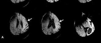
- Magnetic resonance imaging of the brain and spinal cord - to assess areas of damage.
- Electroneuromyography - allows you to evaluate the electrical activity of skeletal muscles and peripheral nerves.
- Electroencephalogram - helps to identify disturbances in the bioelectrical activity of the brain.
Differential diagnosis is carried out with torsion-dystonic syndromes, especially those caused by the chronic form of epidemic encephalitis and hepatocerebral dystrophy. Differential diagnostic significance for epidemic encephalitis is the acute development of the disease, sleep disturbance, diplopia, convergence insufficiency, gaze convulsions, autonomic disorders and, in later stages, symptoms of parkinsonism. Hepatocerebral dystrophy, in contrast to torsion dystonia, is characterized by a low level of ceruloplasmin in the blood, the presence of a Kayser-Fleischer ring (deposition of a greenish-brown pigment containing copper on the periphery of the cornea of the eyes), and cirrhosis of the liver.
Other diseases of the extrapyramidal system that occur with torsion-dystonic syndromes differ from torsion dystonia in that they do not progress and undergo reverse development (to varying degrees) with a decrease in the frequency and severity of hyperkinesis. In their clinical picture, along with hyperkinesis, there are other symptoms of brain damage that are not observed with torsion dystonia.
Diagnostics
Due to the very clear clinical picture and the hereditary nature of the disease, most often there are no problems with diagnosis.
Objectively, the doctor can determine the rigidity of some muscles, while sensitivity and muscle strength are not reduced, and reflexes are preserved. No intellectual impairments are observed.
In some cases, it is necessary to conduct instrumental examinations. The following methods are used:
- electromyography;
- electroencephalography;
- computed or magnetic resonance imaging (MRI);
- electroneuromyography.
Differential diagnosis is carried out with the following diseases:
- secondary torsion-dystonic syndromes;
- hepatocerebral dystrophy;
- epidemic encephalitis;
- Huntington's chorea.
Prices
| Disease | Approximate price, $ |
| Prices for diagnosing migraine | 7 060 — 8 260 |
| Prices for diagnosing childhood epilepsy | 3 100 — 4 900 |
| Prices for brain shunting for hydrocephalus | 33 180 |
| Prices for treatment of Parkinson's disease | 58 600 |
| Prices for migraine treatment | 9 680 |
| Prices for the diagnosis of amyotrophic lateral sclerosis | 6 550 |
| Prices for diagnosing epilepsy | 3 520 |
| Prices for rehabilitation after a stroke | 78 300 — 82 170 |
| Prices for treatment of childhood epilepsy | 3 750 — 5 450 |
| Prices for treatment of multiple sclerosis | 4 990 — 17 300 |
Treatment and observation of the patient
For mild to moderate severity, conservative treatment is indicated. In this case, appropriate medications, physiotherapy, water procedures, and physical therapy are prescribed. Teaching the patient emotional self-regulation is very important, as this significantly reduces the frequency of attacks.
The following groups of drugs are indicated for the treatment of torsion dystonia:
antiparkinsonian (levodopa);- muscle relaxants (mellictin, baclofen);
- neuroleptics;
- tranquilizers (Elinium, Seduxen)
- anticholinergics (bromocriptine, triperiden);
- vitamins (B1, B6);
- centrally acting antispasmodics (diphenyltropin).
If conservative therapy is ineffective, as well as in the last stages of the disease (for example, when the patient can no longer fully care for himself), surgery is indicated.
As a radical treatment, stereotactic surgery is performed to remove the ventrolateral nucleus of the thalamus. In some cases, destruction of the subthalamic zone is indicated. In the local form, the intervention is performed on the side opposite to those parts of the body where disturbances are observed. In case of generalized lesions, two operations are indicated every 6-8 months.
In any case, after surgical treatment, the patient is prescribed appropriate medication support. This is necessary in order to reduce the likelihood of relapses. You should periodically contact a neurologist to monitor the disease. The same is recommended for the patient’s closest relatives.
Diagnostic measures for torsion dystonia
In the neurological status, the following symptoms allow one to suspect the development of torsion dystonia:
- rigidity of certain muscle groups;
- preservation of tendon reflexes;
- no reduction in muscle strength;
- absence of sensory disorders.
The neurologist, first of all, must differentiate idiopathic torsion dystonia from secondary torsion-dystonic syndromes. Secondary syndromes, in most cases, are characterized by symptoms of damage to the nervous system, sleep disturbances, and autonomic disorders. As part of differential diagnosis, patients are referred to:
- EEG;
- Echo-EG;
- Doppler ultrasound of head vessels or REG;
- MRI or CT scan of the brain.
Torsion dystonia can be distinguished from hepatocerebral dystrophy by the fact that the patient does not have pigment deposits in the cornea. There is also no liver damage and a decrease in blood ceruloplasmin is observed. The difference with epidemic encephalitis is the absence of an acute period. A patient with torsion dystonia does not have sleep disorder, diplopia, convergence disorder, or autonomic dysfunction.