About other diseases starting with the letter “B”: Basilar impression, Basilar migraine, Beriberi, Bettolepsy, Amyotrophic lateral sclerosis, Alzheimer's disease, Wilson's disease, Hallerwarden-Spatz disease, Hamstorp disease, Hippel-Lindau disease, Canavan disease, Creutzfeldt disease- Jacob's disease, Lafora's disease, Machado-Joseph disease, Moya-moya disease, Morton's disease, Parkinson's disease, Pick's disease, Refsum's disease, Fahr's disease.
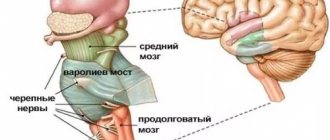
Hallervorden-Spatz disease is a progressive, inherited disorder caused by iron deposits in the brain. It is expressed by Parkinson's disease, a disruption in mental perception and in the state of consciousness, the manifestation of a convulsive symptom, contraction of muscle groups, and visual disturbances.
An important sign of the presence of this pathology is the “eye of the tiger” pattern in the form of a round or linear zone in the forebrain, which is visible during magnetic resonance imaging.
Therapy is aimed at eliminating the symptoms of the disease with drugs used to treat movement disorders: drugs to increase domofin levels, medications against epilepsy. Treatment is also aimed at stopping seizures and suppressing the patient’s depression. Nowadays, the disease is incurable and all actions are aimed exclusively at reducing negative manifestations.
Hallervorden-Spatz disease was characterized in 1922 by German brain research specialists, and the disease was named after the names of these morphologists. The most common clinical manifestations of this anomaly are considered to be cumulative symptoms that manifest themselves in convulsions, muscle contractions and tics. Also a distinctive feature are signs of parkinsonism, mental retardation, pathology of the optic nerves, and damage to the retina of the eye.
This pathology is quite rare. After the development of pronounced pathological phenomena after an asymptomatic course, three forms are noted: childhood, youth and adult.
Before the advent of the necessary equipment, the diagnosis was made exclusively by specialists in the field of pathological anatomy. After the introduction of the necessary equipment into medical organizations, it became possible to make a diagnosis during life. The cause of iron accumulation became known only in 2001. It turned out that the reason for everything is damage to the gene, which causes disruption of the brain. After the results of these studies, the disease was renamed in reference books to pantothenate kinase-associated neurodegeneration.
Causes of the disease
Hallervorden-Spatz disease appears due to a disruption in the functioning of genes. It can be both hereditary and accidentally acquired due to certain mutations. The genetic basis is considered to be a deviation in the pantothenate kinase gene. The genetic substrate is disturbances in the gene for photosynthesis enzymes, indicated on the map of the 20th chromosome 20 p 12.3 - p 13.
About 50 mutations have been identified. Ultimately, this genetic damage leads to a decrease in pantothenate kinase, which leads to a concentration of sulfur acid. This amino acid establishes a strong connection with iron and has a detrimental effect on proteins. As a result of the reaction, oxidation occurs, which programs cell death. In place of destroyed cells, glial cells multiply. This inexplicable reaction most often affects the paired structure of the forebrain and the substantia nigra, located in the midbrain. It is there that concentrated iron is found, which is colored brown. Also, as a result of research, formations were found that were located in the medulla, cerebral cortex and in the peripheral nervous system.
Features of genetic failure
Hallervorden-Spatz disease is a hereditary genetic pathology.
The disease can be either familial or sporadic (random), but it is always inherited. The disease belongs to the autosomal recessive type of inheritance. To transmit the disease, both parents must be heterozygous carriers of the disease and have one mutated allele.
The disease is caused by a mutation in the pantothenate kinase gene (PANK2), which is located on chromosome 20 at the 20p12.3-p13 locus. The pantothenate kinase gene is responsible for encoding the protein pantothenate kinase 2, which in turn inhibits the accumulation of the amino acids cysteine and panthetheine.
As a result, chemical compounds with iron ions are formed, which have a negative effect on proteins and trigger peroxidation processes, which leads to programmed cell death of neurons. Instead of dead neurons, glial tissue (auxiliary cells of nervous tissue) grows.
Pathological processes occur in the globus pallidus, red nuclei and substantia nigra (substantia nigra). A greenish-brown substance that contains iron accumulates in them. In addition, spheroid neuroaxonal formations (extensions of axons with proliferation of tubular and membranous structures) are observed in the white matter of the brain, cerebral cortex, peripheral nerve trunks and spinal cord.
Symptoms of the disease
The most famous type of Hallervorden-Spatz disease begins in childhood between 4 and 10 years of age. The most common signs of the disease are:
Problems with muscle tone, especially in the legs. It also happens that the patient takes positions that look unnatural. In such cases, it is difficult for the child to walk.- The process then affects the torso, face and throat.
- Also with the above manifestations is dystonia, which is observed in several parts of the body. Local dystonia, involuntary spasm of the eye muscle, facial spasm, and spasmodic torticollis sometimes occur.
- Increased muscle tone, in which muscle tissue becomes dense and a state of low motor activity occurs with a limited number of movements.
- Episyndrome.
- Mental disorders such as decreased memory and alertness. Sometimes mental retardation, called oligophrenia, develops in the future. The behavior of such patients may become aggressive and deviate from moral standards.
- Impaired pronunciation and vision as a result of nerve atrophy can be identified as an independent symptom in some patients.
A diagnosis made in childhood usually leads to complete immobility after 10-15 years.
The juvenile form of Hallervorden-Spatz pathology appears at the age of 10 years and has a slow course. There are also problems with muscle tone, involuntary contractions appear, which are localized in the limbs, muscles of the mouth and jaw. There are also typical behavioral characteristics associated with intelligence and psyche.
The adult form of Hallervorden-Spatz disease is established after adulthood. It is also expressed by neurological symptoms, which are characterized by tremor and muscle tone. Insufficient motor activity, painful muscle condition, tremors and loss of coordination of movements are revealed. Loss of coordination can easily be identified using the Tavenard test - a standing patient is pushed forward, thereby throwing him off balance. The doctor is usually behind you.
The main feature of the course of the disease is considered to be a combination of neurological signs with other deviations of the muscle group. The category of impairment of mental processes can vary from the preservation of all functions to progressive dementia.
Features of the clinical picture
Signs of this pathology may vary in each specific case, and the manifestation of symptoms depends on the form of the disease in the individual.
The childhood form of Hallervorden Spatz appears between the ages of 5 and 10 years. It is considered a classic form of the disease.
In the vast majority of cases (90%), the disease begins with torsion dystonia, which affects the leg muscles. The patient experiences muscle contraction, difficulty walking, and changes in gait. Then the muscles of the face, pharynx and trunk are affected.
The presence of blepharospasm, hand spasm, spasmodic torticollis, and facial hemispasm may be present. A third of patients have muscle rigidity, hypokinesia, and tremor (signs of Parkinsonism syndrome).
Typical signs of the childhood form of the disease also include:
- epileptic syndrome;
- aggressiveness;
- mental retardation (occurs due to deterioration of memory and attention);
- asociality;
- visual disturbances (optic atrophy, retinal degeneration);
- mental disorders.
The childhood form of the disease progresses quickly, and complete loss of the ability to move occurs 10-15 years after the onset of the disease.
The teenage form develops and progresses more slowly. At the beginning of the disease the following appear:
- focal torsion dystonia (most often affects the muscles of the limbs and maxillo-oral muscles);
- neuropsychological disorders;
- behavioral disorders;
- intellectual disorders.
The atypical form of the disease is the rarest (15% of cases), and proceeds differently from the first two forms. The most typical symptoms for this form are:
- Parkinsonism syndrome with the inability to maintain balance while standing;
- involuntary movements in different parts of the body, dystonia, chorea and athetosis (slow and jerky movements), hemiballismus (sweeping movements), myoclonus (non-prolonged muscle spasms);
- dementia;
- epileptic syndrome;
- depression, aggression;
- pathological reflexes that arise as a result of destructive processes in the neurons of the brain.
Diagnostics
Due to the manifestation of various symptoms, it is not always possible for a neurologist to establish an accurate diagnosis of Hallervorden-Spatz disease. The dominant feature is considered to be age up to 30 years, a complex of motor activity disorders, an increase in the number of symptoms, as well as the presence of a characteristic sign on MRI images.
Additional signs of this disease may include pathological reflexes, decreased mental abilities, episyndrome, blurred vision, retinal atrophy, as well as a family history of this disease.
For diagnostic purposes, additional studies are carried out, in particular, an electroencephalogram is performed on the patient. To make a diagnosis, they rely on data from the neurological status and examination, which is based on the pattern of total electrical activity of the brain.
If there is obvious deterioration in vision, the help of an ophthalmologist is required to make a diagnosis, who will conduct a study to determine visual acuity and examine the fundus of the eye with special instruments.
A geneticist determines the presence of mutations when compiling a family tree, and also performs DNA diagnostics. Also, with the help of additional PET, reduced metabolism is detected in the formation of gray matter in the subcortical part of the brain.
There are a number of other diseases with similar pathologies, but their clinical picture does not include a number of features of Hallervorden-Spatz disease. Similar diseases in terms of symptoms: Huntington's disease, Wilson's disease, Machado-Joseph disease and other diseases associated with genetic brain damage.
When making a diagnosis, doctors rely on data obtained from MRI. All patient images show a pigmented area with iron. An image with an unambiguous appearance of this disease is called “Eye of the Tiger.”
The darker tissues of this zone look like an eye, and the lighter, bright ones look like a pupil. The moment of appearance of the “Eye of the Tiger” has not yet been determined by scientists. According to some, it appears even before certain symptoms, according to others, some time after the clear manifestation of the disease.
Diagnostic criteria and techniques
Making a diagnosis of Hallervorden-Spatz disease can be problematic for neurologists due to the polymorphism of symptoms.
Main criteria for diagnosis:
- onset of illness before age 30;
- typical picture in MRI results;
- constant development of symptoms;
- extrapyramidal syndromes;
- pyramid signs;
- epileptic seizures;
- cognitive impairment;
- retinal pigmentary degeneration;
- presence of the disease in a family history.
“Eyes of the tiger” on MRI with contrast is a typical picture of Hallervorden Spatz disease.
The disease must be differentiated from other diseases that have a similar clinical picture: Huntington’s chorea, Wilson-Konovalov disease, choreoacanthocytosis, Machado-Joseph disease.
For diagnosis, the patient is referred for the following examinations:
- Study of neurological status by a neurologist, neuropathologist.
- Electroencephalography.
- Consultation with an ophthalmologist . Direct ophthalmoscopy and visual acuity testing are possible.
- Genetic studies to determine the type of inheritance.
- DNA studies (detection of mutations in the PANK2 gene).
- Positron emission tomography of the brain . It is carried out to detect decreased blood circulation in the frontoparietal lobes, striatum, and globus pallidus.
- Magnetic resonance imaging of the brain . Required to detect the "eye of the tiger" - a hyperintense oval area in the hypointense zone that occurs due to iron accumulation. There is no single theory about the development of the “eye of the tiger”; there are assumptions about its occurrence before the onset of clinical manifestations of the disease, and about its appearance years after the onset of the disease. MRI can also detect spheroid neuroaxonal formations.
Course of the disease
The usual onset of the disease is in childhood, sometimes with severe symptoms already in the first year of life. Manifestation in adulthood is rarely possible. First, extrapyramidal motor disorders occur, especially gait disturbances with a tendency to fall or leg dystonia, and less commonly, mental disorders. Subsequently, movement disorders (dystonia, choreoathetosis, tremor) with neck rigidity, hyperreflexia and mental disorders (as a rule, intellectual disorder in the form of progressive dementia). Dysarthria and dysphagia are also found. Retinal pigmentation or optic nerve atrophy may be detected. In adults, parkinsonism-plus syndrome predominates - with dementia, hyperreflexia and prominent dystonia. The course of the disease is progressive, the neurological condition of patients worsens gradually.
Death usually occurs before the age of 30, in a state of complete mental collapse of the personality.