Diagnosis and treatment of cerebellar disorders
The neurologist examines and tests superficial and deep reflexes.
Electronystagmography and vestibulometry are performed. A general blood test is prescribed. A lumbar puncture is performed to detect infection in the cerebrospinal fluid, as well as markers of stroke or inflammation. The head is held. The condition of the cerebellar vessels is determined using Dopplerography. Treatment of cerebellar diseases in ischemic stroke is carried out using thrombus lysis. Fibrinolytics (streptokinase, alteplase, urokinase) are prescribed. To prevent the formation of new blood clots, antiplatelet agents (aspirin, clopidogrel) are used.
For ischemic and hemorrhagic strokes, metabolic drugs (Mexidol, Cerebrolysin, Cytoflavin) improve metabolism in brain tissue. To prevent recurrent strokes, drugs that lower blood cholesterol are prescribed, and in case of hemorrhagic hemorrhage, antihypertensive drugs are prescribed.
- Causes of cerebellar ataxia
- Causes and signs of weak development of reflexes
- How does a “normal” cerebellum work?
- Cerebellar ataxia with acute onset
- Diagnostics
- Consequences of the disease and prevention
- Cerebellar diseases
- Palmar chin reflex Marinescu Radovici
- Study of oral automatism reflexes in neurology
Neuroinfections (encephalitis, meningitis) require antibiotic therapy. Pathologies of the cerebellum caused by intoxications require detoxification therapy, depending on the nature of the poisons. Forced diuresis, peritoneal dialysis and hemodialysis are performed. In case of food poisoning - gastric lavage, administration of sorbents.
For oncological lesions of the cerebellum, treatment is carried out in accordance with the type of pathology. Chemotherapy, radiation therapy, or surgical treatment are prescribed. If the outflow of cerebrospinal fluid is disrupted, causing cerebellar syndrome, an operation is performed with craniotomy and shunting of the pathways for the outflow of cerebrospinal fluid.
Conclusion
Damage to the cerebellum, the consequence of which can be disability, the patient’s need for care, requires timely and thorough treatment, as well as care and rehabilitation of the patient. If there is a sudden disturbance in gait or speech disorder, it is necessary to visit a neurologist.
Coordination motor disorder caused by cerebellar pathology. Its main manifestations include gait disorder, disproportionality and asynergy of movements, dysdiadochokinesis, and changes in handwriting such as sweeping macrography. Typically, cerebellar ataxia is accompanied by scanned speech, intention tremor, postural tremor of the head and torso, and muscle hypotonia. Diagnosis is carried out using MRI, CT, MSCT, MAG of the brain, Dopplerography, cerebrospinal fluid analysis; if necessary, genetic research. Treatment and prognosis depend on the causative disease that caused the development of cerebellar symptoms.
Balametrix balancing board exercises
To work on cerebellar stimulation, a specially designed device is used - the Bilgow balancing board (Belgau). The exercises are somewhat similar to physical therapy.
The child stands on the surface of the board, which, in turn, is fixed on a round base, which forces him to balance in an attempt to maintain his balance. The base of the structure is very similar to the base of children's rocking toys. The difference is that here the child does not have to sit and rock, but, on the contrary, balances in a standing position and performs various exercises.
The surface of the board has special markings, and the angle of the platform is adjusted by rockers that change the radius from 5 to 50 degrees. Depending on the level of inclination, the level of difficulty of the exercises changes.
Classes are conducted under the strict supervision of a specialist, both for safety reasons and for full effectiveness. Despite the fact that the exercises themselves do not seem unusual or difficult (throwing a ball, tossing a ball or soft pillow between a child and an assistant, naming numbers or objects in a certain sequence), the effect of the exercises will only occur with the correct selection of exercises.
Children enjoy practicing on the balance board. Such “lessons” do not cause them fear, negativity or fear of not completing the task.
Causes of cerebellar ataxia
Ataxia may be a consequence of:
- severe head injury (traumatic brain injury);
- accumulation of fluid in the cavities of the brain, spinal canal (hydrocephalus);
- congenital malformation of the brain/skull;
- infectious disease of the brain (encephalitis);
- cerebrovascular accidents (ischemic stroke);
- cerebral palsy;
- epilepsy in children;
- malignant neoplasms;
- abscesses.
Sensitive ataxia syndrome develops as a result of damage to:
- posterior brain stems,
- peripheral nodes,
- posterior nerves, optic thalamus,
- parietal lobe of the brain.
Cerebellar ataxia is a consequence of damage to the cerebellar vermis, its peduncles and hemispheres. Occurs in encephalitis, sclerosis.
The vestibular form of the disease is caused by damage to any part of the vestibular apparatus - nuclei in the brain stem, cortical center in the temporal lobe of the brain, vestibular nerve, labyrinth.
Cortical ataxia occurs when the frontal lobe of the brain is damaged, which is caused by disturbances in the functioning of the fronto-pontine-cerebellar system.
The best doctors for the treatment of ataxia
8.4 13 reviews
NeurologistHirudotherapistDoctor of the highest category
Zanegin Dmitry Andreevich Experience 14 years 8.4 7 reviews
NeurologistCardiologistGastroenterologistArrhythmologistTherapistDoctor of the highest category
Karmanov Sergey Sergeevich Experience 12 years Candidate of Medical Sciences9 2 reviews
VertebrologistReflexotherapistNeurologistDoctor of the highest category
Sbitneva Natalya Georgievna Experience 39 years 8.6 7 reviews
Neurologist
Bondareva Anastasia Nikolaevna Experience 9 years 8.2 2 reviews
NeurologistDoctor of the first category
Kubyshta Svetlana Mikhailovna Experience 14 years 8.7
NeurologistNeurophysiologistFunctional diagnostics doctor First category doctor
Seleznev Fedor Alekseevich Experience 9 years Candidate of Medical Sciences8.8 41 reviews
NeurologistManual therapist
Podryadov Nikolay Evgenievich Experience 34 years 8.8 124 reviews
Neurologist
Gadaborsheva Tamara Magomedovna Experience 41 years 9.5 76 reviews
ReflexologistNeurologistManual therapistDoctor of the highest category
Shnigirist Alexander Ilyich Experience 27 years Candidate of Medical Sciences8.8 14 reviews
Neurologist Chiropractor Hirudotherapist Reflexotherapist Doctor of the first category
Derzhavina Irina Nikolaevna Experience 35 years
Most often, the main cause of the development of acute cerebellar ataxia is ischemic stroke, which is caused by atherosclerotic occlusion or embolism of the cerebral arteries that supply the cerebellum. A hemorrhagic stroke, compression of the cerebellum under the influence of an intracerebral hematoma or traumatic damage to the cerebellum resulting from a head injury cannot be excluded.
Late cerebellar ataxia of the subacute form often occurs as a symptom of an intracerebral tumor, which is located in the cerebellum. These are astrocytoma, hemangioblastoma, medulloblastoma, ependymoma. It can also develop as a result of normal pressure hydrocephalus arising from subarachnoid hemorrhage.
One of the reasons is meningitis and brain surgery. In addition, this form of ataxia can occur with a lack of vitamins, an overdose of anticonvulsants, an endocrine disorder, for example, with hyperparathyroidism or hypothyroidism. It may be a paraneoplastic syndrome for malignant tumors that are localized outside the brain. This could be lung cancer, ovarian cancer, non-Hodgkin's lymphoma and some other forms of oncology.
Why do children need cerebellar stimulation?
Very often, children with speech and mental disorders experience awkwardness in movements, clumsiness, and lack of coordination. It is these characteristics that are a sign that the child has problems in the functioning of the cerebellum and brainstem.
A disrupted connection between the cerebellum and the frontal lobes of the brain leads to a slowdown in the formation of speech, intellectual and mental processes.
The Cerebellar Stimulation Program improves:
- level of concentration;
- all types of memory;
- development of speech - both oral and written, as well as reading skills (read more about how to teach a child to read);
- mathematical and logical abilities;
- analysis and synthesis of information;
- ability to plan;
- work of the emotional-volitional sphere.
Thus, this method is indicated for improving learning - both at school and in special corrective classes conducted by speech therapists, psychologists and defectologists.
It has been proven that exercises to stimulate the cerebellum, carried out before the start of corrective measures, significantly increase their effectiveness!
Causes and signs of weak development of reflexes
It is advisable to study information about what reflexes a newborn should have; in some cases, they may not manifest themselves to an insufficient extent.
The reasons for this situation may be the following:
- received birth injuries;
- dysfunction of the nervous system;
- premature birth of a child;
- spine pathologies;
- previous asphyxia.
A sign of weakening is insufficient expression or one-sided manifestation of symmetrical reflexes. In some cases, the baby is not ready to respond to the stimulus; for example, if the child has no desire to eat, the sucking reflex will not be pronounced.
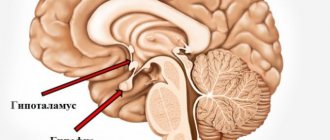
How does a “normal” cerebellum work?
Before approaching the description of diseases of the cerebellum, it is necessary to briefly talk about how the cerebellum is structured and how it functions.
The cerebellum is located at the bottom of the brain, under the occipital lobes of the cerebral hemispheres.
It consists of a small middle section, the vermis, and hemispheres. The vermis is an ancient section, and its function is to ensure balance and statics, and the hemispheres developed along with the cerebral cortex, and provide complex motor acts, for example, the process of typing this article on a computer keyboard.
The cerebellum is closely connected to all the tendons and muscles of the body. They contain special receptors that “tell” the cerebellum what state the muscles are in. This sense is called proprioception. For example, each of us knows, without looking, in what position and where his leg or arm is, even in the dark and at rest. This sensation reaches the cerebellum via the spinocerebellar tracts that ascend in the spinal cord.
In addition, the cerebellum is connected with the system of semicircular canals, or the vestibular apparatus, as well as with the conductors of the joint-muscular sense.
The olivocerebellar pathway passes through the inferior cerebellar peduncle, which connects it with unconscious movements. The reverse, efferent pathway is the path from the cerebellum to the red nuclei.
It is this path that works brilliantly when, having slipped, a person “dances” on the ice. Without having time to figure out what is happening, and without having time to get scared, the person restores his balance. This triggered a “relay” that switched information from the vestibular apparatus about changes in body position immediately through the cerebellar vermis to the basal ganglia, and then to the muscles. Since this happened “automatically”, without the participation of the cerebral cortex, the process of restoring balance occurs unconsciously.
The cerebellum is closely connected to the cerebral cortex, regulating conscious movements of the limbs. This regulation occurs in the cerebellar hemispheres
Read also
Noise in ears
Tinnitus cannot be called an independent disease.
It would be more accurate to designate it as a hearing disorder, which manifests itself as a hum, ringing or buzzing in the ears, then, as the real source of sound... Read more
Meniere's disease
Meniere's disease is a disease in which the leading symptoms are systemic dizziness, accompanied by hearing loss, a feeling of fullness and tinnitus. Paroxysmal dizziness,…
More details
Benign positional paroxysmal vertigo
How great it is when you feel dizzy with happiness, but unfortunately there are people who are afraid of this symptom because this symptom greatly interferes with their quality of life. Dizziness is not a diagnosis, it is a symptom...
More details
Vestibular paroxysmia
Attacks of systemic dizziness lasting several seconds, less often minutes, and are accompanied in most cases by tinnitus and hearing loss. Seizures often occur during hyperventilation...
More details
Videonystagmography
Videonystagmography (VNG) is the latest non-invasive method for qualitative and quantitative assessment of vestibular disorders. This method is used to study ocular nystagmus. Any true...
More details
Cerebellar ataxia with acute onset
Stroke appears to be the most common cause of acute ataxia in clinical practice. Lacunar infarctions in the area of the pons and supratentorial can cause ataxia, usually in the picture of ataxic hemiparesis. Ischemia in the area of the thalamus, posterior limb of the internal capsule and corona radiata (area of blood supply from the posterior cerebral artery) can manifest as cerebellar ataxia. At the same time, “silent” lacunar infarcts are often found in the cerebellum. Cerebellar infarction may also present with isolated dizziness. Cardiac embolism and atherosclerotic occlusion are the two most common causes of cerebellar stroke.
Hemiataxia with hemihypesthesia are characteristic of strokes in the thalamus (branch of the posterior cerebral artery). An isolated ataxic gait sometimes occurs when the penetrating branches of the basilar artery are affected. Hemiataxia involving certain cranial nerves develops with damage to the upper parts of the pons (superior cerebellar artery), inferolateral parts of the pons and lateral parts of the medulla oblongata (anterior inferior and posterior inferior cerebellar arteries), usually in the picture of brainstem alternating syndromes.
Extensive cerebellar infarctions or hemorrhages are accompanied by the rapid development of generalized ataxia, dizziness and other brainstem and cerebral manifestations, often in connection with the development of obstructive hydrocephalus.
Cerebellar tumors, abscesses, granulomatous and other space-occupying processes sometimes manifest themselves acutely and without severe symptoms (headaches, vomiting, mild ataxia when walking).
Multiple sclerosis sometimes develops acutely and rarely occurs without cerebellar symptoms. There are usually other signs (clinical and neuroimaging) of multifocal damage to the brainstem and other parts of the nervous system.
Guillain-Barré syndrome occurs as a rare form of lesion involving the cranial nerves and ataxia. But even here, at least mild hyporeflexia and an increase in protein in the cerebrospinal fluid are detected. Miller Fisher syndrome occurs acutely with the development of ataxia, ophthalmoplegia and areflexia (other symptoms are optional) followed by good recovery of impaired functions. These manifestations are very specific and sufficient for clinical diagnosis.
Encephalitis and post-infectious cerebellitis often involve the cerebellum. Cerebellitis in mumps is especially common in children with premorbid cerebellar anomalies. Chickenpox can cause cerebellitis. Epstein-Bar virus causes infectious mononucleosis with secondary acute cerebellar ataxia. Acute post-infectious ataxia is especially common among the consequences of childhood infections.
Intoxication is another common cause of acute ataxia. As a rule, there is an ataxic gait and nystagmus. If ataxia is detected in the limbs, it is usually symmetrical. The most common causes: alcohol (including Wernicke encephalopathy), anticonvulsants, psychotropic drugs.
Metabolic disorders such as insulinoma (hypoglycemia causes acute ataxia and confusion) are fairly common causes of acute ataxia.
Hyperthermia in the form of prolonged and intense heat stress (high fever, heat stroke, neuroleptic malignant syndrome, malignant hyperthermia, lithium toxicity hyperthermia) can affect the cerebellum, especially in the rostral region around the vermis.
Obstructive hydrocephalus, which has developed acutely, is manifested by a whole complex of symptoms of intracranial hypertension (headache, drowsiness, stupor, vomiting), among which acute cerebellar ataxia often occurs. With the slow development of hydrocephalus, ataxia can manifest itself with minimal cerebral disorders.
[], [], [], [], [], []
Features of children with cerebellar dysfunction
If we talk specifically about cerebellar dysfunctions, then in isolated form, they are extremely rare.
Cerebellar dysfunction can be a consequence of congenital or acquired pathologies of the cerebellum.
Symptoms of cerebellar dysfunction usually include ataxia. In addition, cerebellar dysfunction may include the following characteristics:
- disturbances of motor functions, tone (impaired coordination, slower reactions when changing one type of movement to the opposite, increased or decreased muscle tone, disorder of uniformity of movement, dysarthria, etc.);
- disorders of higher mental functions; we can highlight the following features of cognitive processes in children with cerebellar dysfunction: decreased neurodynamic processes,
- decreased attention and memory functions,
- violation of spatial and “quasi-spatial” perception;
- violation of control and programming of mental processes, decreased self-regulation,
- speech disorders (speech becomes chanted, slow, loses smoothness).
Diagnostics
When diagnosing ataxia, the following methods are used:
- MRI of the brain (detects atrophy of the brain stems and spinal cord, the upper parts of the vermis);
- electroencephalography of the brain (diagnoses reduction of alpha rhythm, diffuse theta and delta activity);
- electromyography (reveals axonal demyelinating damage to sensory fibers of peripheral nerves);
- DNA diagnostics (used to determine hereditary types of ataxia). By conducting indirect DNA diagnostics, doctors determine whether the ataxia pathogen can be inherited by other children in the family;
- magnetic resonance angiography (allows you to assess the integrity and patency in the cranial cavity and identify brain tumors).
Additional diagnostic procedures include consultation with a neurologist, ophthalmologist, and psychiatrist. Laboratory diagnostics for ataxia show a violation of amino acid metabolism - a reduced concentration of alanine and leucine, a decrease in their excretion in the urine.
Since the etiology of this disease involves a fairly wide range of diseases and its congenital nature cannot be ruled out, diagnosing the disease involves consulting the following specialists:
- neurosurgeon;
- neurologist;
- traumatologist;
- oncologist;
- endocrinologist
The diagnostic program may include the following activities:
- spinal puncture and cerebrospinal fluid analysis;
- functional tests;
- CT and MRI of the brain;
- Dopplerography of the brain;
- electronystagmography;
- PCR studies;
- MSCT;
- DNA diagnostics.
In addition, genetic counseling may be needed.
Due to the fact that the etymology of cerebellar pathologies is very diverse, diagnosis is carried out by several specialists with a narrow focus, namely a traumatologist, neurosurgeon, oncologist, geneticist, and endocrinologist. A thorough neurological examination allows not only to determine the cerebellar nature of the ataxia, but also to identify approximately the affected area.
In order to exclude vestibular disorder, it is necessary to conduct a study of the vestibular analyzer. A neurologist may prescribe stabilography, vestibulometry, and electronystagmography. If there is a suspicion that ataxia has arisen as a result of an infectious lesion, a blood sterility test or PCR test is required.
DNA testing and genetic analysis can detect the hereditary nature of ataxia in the cerebellum. The treatment is presented below.
When conducting a full range of examinations, making a diagnosis does not cause difficulties.
A functional test, assessment of tendon reflexes, and blood and cerebrospinal fluid testing are required.
A computed tomography and ultrasound scan of the brain is prescribed, and an anamnesis is collected.
Consequences of the disease and prevention
Treatment of cerebellar ataxia will be successful only if the true cause of the development of the disease is correctly established. The disease can be completely cured only in rare cases. Most often, patients remain with symptoms characteristic of the disease, which periodically worsen. In cases where it was possible to start therapy for the underlying disease in a timely manner, the prognosis will be as positive as possible. The patient’s age, general health, and the form of manifestation of cerebellar ataxia have a significant influence.
There is no specific prevention against the development of ataxia. Experts recommend timely treatment of infectious and viral pathologies, vaccination against serious diseases, and monitoring the functioning of the thyroid gland and cardiovascular system. Before planning a pregnancy, you should definitely consult a geneticist if there are cases of this disease in the family.
Cerebellar diseases
Violations in the functioning of the organ are manifested by a diverse clinic. But problems associated with impaired coordination, gait, and muscle tone predominate.
Congenital pathologies appear from the first minutes of life. They are associated with underdevelopment of the cerebellum or one of its parts. The lesion becomes noticeable as motor skills develop. Ataxia almost always appears.
Hereditary problems are associated with a gene defect. They are transmitted from parents in an autosomal dominant and autosomal recessive manner. Among these pathologies are:
- Friedreich's ataxia;
- Ataxia-telangiectasia;
- Ataxia with vitamin E deficiency;
- Abetalipoproteinemia.
One type of ataxia is spinocerebellar disorders. They include a whole range of diseases with a hereditary transmission mechanism. In addition to the main symptoms, disturbances in walking, tone, and coordination dominate.
Acquired diseases of the cerebellum arise as a result of circulatory disorders in the organ, viral and bacterial infections. The toxic effects of alcohol, heavy metal salts, lithium, and anticonvulsants also lead to cerebellum problems. Atrophic phenomena in the organ appear from a lack of vitamin E and B12, with hypothyroidism.
Tumors develop in the cerebellum. They manifest themselves as symptoms of compression and loss of coordination. The most common types of tumors that develop are astrocytomas and meduloblastomas.
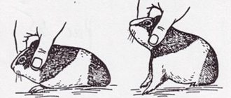
Palmar chin reflex Marinescu Radovici
The Marinescu-Radovici reflex (palmomental) is a typical reflex of oral automatism. It occurs normally in children up to 12-18 months, ensuring the cooperative performance of two different functions: grasping the mammary gland with the hands and simultaneous sucking of milk.
Determining this reflex during a neurological examination allows us to understand the cause of the disease and determine the location of the pathological process in the brain. The reflex was first described in 1920 by two neurologists Marinescu and Radovici.
Phenomena of oral automatism
The Marinescu-Radovic reflex refers to the reflexes of oral automatism. These are pathological reflexes on the face that occur during the phenomena of central (spastic) paralysis or paresis of the facial muscles, which are innervated by the cranial nerves.
The appearance of these reflexes indicates bilateral supranuclear damage to the brain stem, subcortical structures or cerebral cortex.
These reflexes, including Marinescu-Radovic, normally appear in children up to 18 months of age. This is due to the immaturity of brain structures and the ongoing differentiation of parts of the central nervous system.
https://youtube.com/watch?v=45bMF78O-iQ
Oral automaticity reflexes include:
- Marinescu-Radovic reflex, or palmomental reflex. It can be caused by streaking movements with a blunt object on the skin of the palm at the base of the thumb. In response to this, a slight upward displacement of the skin of the chin occurs on the patient’s face, which is associated with contraction of the mentalis muscle.
- Nasolabial (nasolabial) reflex. It is caused by tapping the back of the nose with a neurological hammer. In response to this, the person extends his lips slightly anteriorly, which is mediated by contraction of the orbicularis oris muscle.
- Proboscis reflex. Completely identical in appearance to nasolabial, but is caused by tapping on the upper or lower lip.
- The sucking reflex is manifested by sucking movements of the lips when touching or stroking the lips.
- Distance-oral Karchikyan syndrome. If you slowly bring a hammer closer to the patient’s lips, they will stretch out in the form of a “proboscis” without contact with the object.
All these reflexes are observed in newborns and children in the first year and a half of life, but then fade away and can occur in adults against the background of damage to brain structures.
Information about the Marineski-Radovici reflex
The reflex was first described in the 20s of the twentieth century by two Romanian neurologists Marinescu and Radovici. Observing this phenomenon in their patients, they established that the appearance of the reflex is associated with damage to the brain stem and is a reflection of the rudimentary synergy of grasping and sucking, or chewing.
The reflex consists of contraction of the mental muscle on the face with a gentle stroke of irritation of the palm in the area of the base of the thumb. As was revealed later, the nuclei of the seventh pair of cranial nerves are involved in this reflex.
A characteristic feature of this reflex is the one-sidedness of the reaction: when the skin on the right is irritated, the response is observed on the right half of the face, despite bilateral brain damage.
Use in diagnostics
As mentioned above, the palmar-chin reflex occurs in adults and children over one and a half years old with damage to brain structures: the brain stem, subcortical formations and the cerebral cortex.
Its appearance is associated with damage to the central motor neuron or its axon, and the occurrence of spastic or central paralysis in a certain muscle group in the patient. In this case, the inhibitory effect of the cerebral cortex neuron on the muscles disappears, as a result of which the reflex readiness of the muscles increases and pathological reflexes arise that are normally absent.
Most often this occurs as a result of the following diseases:
The appearance of any phenomena of oral automatism, incl. and the Marinescu-Radovic reflex, a serious warning sign indicating irreversible brain damage. Therefore, if such symptoms appear, you must immediately contact a medical facility to receive qualified medical care.
Symptoms of cerebellar ataxia
It is customary to distinguish two types of cerebellar ataxia: static (static-locomotor) and dynamic. Static cerebellar ataxia develops with damage to the cerebellar vermis, and dynamic ataxia develops with pathology of the cerebellar hemispheres and its connections. Each type of ataxia has its own characteristics. Cerebellar ataxia of any type is characterized by a decrease in muscle tone.
Static-locomotor ataxia
This type of cerebellar ataxia is characterized by a violation of the antigravity function of the cerebellum. As a result, standing and walking become too much of a strain on the body. Symptoms of static-locomotor ataxia may include:
- inability to stand straight in the “heels and toes together” position;
- falling forward, backward or swaying to the sides;
- the patient can stand only by spreading his legs wide apart and balancing with his hands;
- unsteady gait (like a drunk);
- when turning, the patient “slides” to the side, and he may fall.
To identify static-locomotor ataxia, several simple tests are used. Here are some of them:
- standing in the Romberg pose. The pose is as follows: toes and heels pushed together, arms extended forward to a horizontal level, palms facing down with fingers spread wide. First, the patient is asked to stand with his eyes open, and then with his eyes closed. With static-locomotor ataxia, the patient is unstable both with his eyes open and with his eyes closed. If no deviations are detected in the Romberg position, then the patient is asked to stand in a complicated Romberg position, when one leg must be placed in front of the other so that the heel touches the toe (maintaining such a stable position is possible only in the absence of pathology on the part of the cerebellum);
- the patient is asked to walk along a conventional straight line. With static-locomotor ataxia, this is impossible; the patient will inevitably deviate in one direction or another, spread his legs wide apart, and may even fall. They are also asked to stop abruptly and turn 90° to the left or right (with ataxia the person will fall);
- The patient is asked to walk with a side step. Such a gait with static-locomotor ataxia becomes as if dancing, the torso lags behind the limbs;
- "star" or Panov test. This test allows you to identify disorders in mildly expressed static-locomotor ataxia. The technique is as follows: the patient needs to successively take three steps forward in a straight line, and then three steps back, also in a straight line. First, the test is carried out with open eyes, and then with closed ones. If with open eyes the patient is even more or less able to perform this test, then with closed eyes he inevitably turns around (a straight line does not come out).
In addition to impaired standing and walking, static-locomotor ataxia manifests itself as a violation of coordinated muscle contraction when performing various movements. This is called in medicine cerebellar asynergies. Several tests are also used to identify them:
- the patient is asked to sit up sharply from a lying position with his arms crossed over his chest. Normally, the muscles of the trunk and the posterior group of thigh muscles contract synchronously, and the person is able to sit down. With static-locomotor ataxia, synchronous contraction of both muscle groups becomes impossible, as a result of which it is impossible to sit without the help of hands, the patient falls back and one leg rises at the same time. This is the so-called Babinski asynergy in the supine position;
- Babinsky's synergy in a standing position is as follows: in a standing position, the patient is asked to bend back, throwing back his head. Normally, for this, a person will have to involuntarily bend his knees slightly and straighten his hip joints. With static-locomotor ataxia, neither flexion nor extension occurs in the corresponding joints, and an attempt to bend ends in a fall;
- Orzechowski's test. The doctor extends his hands, palms up, and invites the standing or sitting patient to rest his palms on them. Then the doctor suddenly moves his hands down. Normally, the patient's lightning-fast, involuntary muscle contractions cause him to either lean back or remain motionless. This will not work for a patient with static-locomotor ataxia - he will fall forward;
- phenomenon of absence of reverse shock (positive Stewart-Holmes test). The patient is asked to forcefully bend his arm at the elbow joint, and the doctor counteracts this, and then suddenly stops the opposition. With static-locomotor ataxia, the patient's hand is thrown back with force and hits the patient's chest.
Dynamic cerebellar ataxia
In general, its essence lies in the disruption of smoothness and proportionality, accuracy and dexterity of movements. It can be bilateral (with damage to both hemispheres of the cerebellum) and unilateral (with pathology of one hemisphere of the cerebellum). Unilateral dynamic ataxia is much more common.
Some of the symptoms of dynamic cerebellar ataxia overlap with those of static-locomotor ataxia. For example, this concerns the presence of cerebellar asynergies (Babinski asynergia lying and standing, Orzechowski and Stewart-Holmes tests). There is only a slight difference: since dynamic cerebellar ataxia is associated with damage to the cerebellar hemispheres, these tests predominate on the affected side (for example, if the left hemisphere of the cerebellum is damaged, “problems” will occur with the left extremities and vice versa).
Dynamic cerebellar ataxia also manifests itself:
- intention tremor (tremor) in the limbs. This is the name for trembling that occurs or intensifies towards the end of the movement being performed. At rest, no trembling is observed. For example, if you ask a patient to take a ballpoint pen from a table, then at first the movement will be normal, but by the time the pen is actually taken, the fingers will begin to tremble;
- missing and missing. These phenomena are the result of disproportionate muscle contraction: for example, the flexors contract more than necessary to perform a particular movement, and the extensors do not relax properly. As a result, it becomes difficult to perform the most common actions: bring a spoon to your mouth, fasten buttons, lace your shoes, shave, and so on;
- handwriting disorder. Dynamic ataxia is characterized by large, uneven letters and a zigzag direction of the writing;
- chanted speech. This term refers to the intermittency and jerkiness of speech, the division of phrases into separate fragments. The patient’s speech looks as if he were speaking from the podium with some slogans;
- nystagmus. Nystagmus is involuntary shaking movements of the eyeballs. Essentially, this is the result of incoordination of contraction of the eye muscles. The eyes seem to twitch, this is especially pronounced when looking to the side;
- adiadochokinesis. Adiadochokinesis is a pathological movement disorder that occurs during the rapid repetition of multidirectional movements. For example, if you ask a patient to quickly turn his palms against their axis (as if screwing in a light bulb), then with dynamic ataxia the affected hand will do it more slowly and awkwardly compared to the healthy one;
- pendulum-like nature of knee reflexes. Normally, a blow with a neurological hammer under the kneecap causes a single movement of the leg of varying degrees of severity. With dynamic cerebellar ataxia, the leg oscillates several times after one blow (that is, the leg swings like a pendulum).
To identify dynamic ataxia, it is customary to use a series of tests, since the degree of its severity does not always reach significant limits and is immediately noticeable. With minimal lesions of the cerebellum, it can only be detected by testing:
- finger-nose test. With the arm straightened and raised to a horizontal level with a slight abduction to the side, with eyes open and then with eyes closed, ask the patient to touch the nose with the tip of the index finger. If a person is healthy, he can do this without much difficulty. With dynamic cerebellar ataxia, the index finger misses, and when approaching the nose, an intention tremor appears;
- finger-finger test. With eyes closed, the patient is asked to touch each other with the tips of the index fingers of slightly spaced hands. Similar to the previous test, in the presence of dynamic ataxia, no hit occurs, trembling may be observed;
- finger hammer test. The doctor moves the neurological hammer in front of the patient’s eyes, and the patient must place his index finger exactly on the elastic band of the hammer;
- test with hammer A.G. Panova. The patient is given a neurological hammer in one hand and is asked to alternately and quickly squeeze the hammer with the fingers of the other hand, either by the narrow part (handle) or by the wide part (elastic band);
- heel-knee test. It is performed in a supine position. It is necessary to raise the straightened leg approximately 50-60°, hit the knee of the other leg with the heel and, as it were, “ride” the heel along the front surface of the shin to the foot. The test is carried out with eyes open and then with eyes closed;
- test for redundancy and disproportion of movements. The patient is asked to stretch his arms forward to a horizontal level with his palms up, and then, at the doctor’s command, turn his palms down, that is, make a precise 180° turn. In the presence of dynamic cerebellar ataxia, one of the arms rotates excessively, that is, more than 180°;
- test for diadochokinesis. The patient should bend his arms at the elbows and seem to take an apple in his hands, and then quickly make twisting movements with his hands;
- Doinikov finger phenomenon. In a sitting position, the patient places his relaxed hands on his knees, palms up. On the affected side, flexion of the fingers and rotation of the hand is possible due to an imbalance in the tone of the flexor and extensor muscles.
Such a large number of tests for dynamic ataxia is due to the fact that it is not always detected with only one test. It all depends on the extent of the damage to the cerebellar tissue. Therefore, for more in-depth analysis, several samples are usually carried out simultaneously.