In this article you can find out what the head areas are, how this part of the body is structured and why did it appear during evolution in the first place? The article begins with the simplest thing - basic information about the organization.
What is meant by the skeleton of the head or, more simply, the skull? This is a collection of many bones, paired or not, spongy or mixed. The skull contains only two large sections:
- cerebral (the cavity in which the brain is located);
- facial (this is where some systems, such as the respiratory or digestive, originate; in addition, more sensory organs can be found here).
As for the brain region, it is worth mentioning that this area is divided into two:
- cranial vault;
- its foundation.
Evolution
It is important to know that vertebrates did not always have such a large head. Let's dive a little into the past. This part of the body appeared in ancient vertebrates during the fusion of the first three segments of the spine. Before this phenomenon, the same segmentation was observed. Each vertebra had its own pair of nerves. The nerves of the first vertebra were responsible for smell, the second for vision, and the third for hearing. Over time, the load on these nerves increased, it was necessary to process more and more information, which led to the thickening of these segments responsible for these sense organs. So they merged into the brain, and the union of the vertebrae formed the brain capsule (like a skull). Note that even the modern human head is still divided into the segments from which it was formed.
What is the average size of an adult's head? Length - 17-22 cm, width - 14-16 cm, height - 12-16 cm, circumference - 54-60 cm. The length of the head is usually greater than the width, so it is not round, but elliptical. It is also very interesting that the numbers (length, width and height) are not constant, they either increase or decrease. And all this depends on the location of the person.
Crown in babies: how to care for it (video)
All newborns, as well as children under the age of 1-2 years, have a soft crown or fontanel. This small area on the baby's head almost always causes concern for parents. Especially those who became mothers and fathers for the first time.
They are afraid to even touch the crown of the baby, let alone take care of such a vulnerable place, in their opinion. What if they harm the baby’s brain? After all, here he is not protected by the bones of the skull. It's like a hole in the skull, covered only by skin. But is this really so?
Or does the visible softness of the fontanel only create the illusion of its vulnerability? Surely Mother Nature couldn’t have made such a mistake? And leave the brain of a newborn child without a reliable protective shell. Surely she had her reasons for this. Let's try to understand what's going on here.
Crown in babies (or fontanel)
And, for starters, it won’t hurt you to know that, it turns out, your baby has more than one fontanel. There are six similar holes on a child’s head.
But the four temporal lobes heal while the baby is still in the womb (in rare cases, they can be felt within a couple of days after birth).
The small occipital fontanel usually closes when the child reaches one month of age.
And the most famous and largest - the parietal - can remain open for up to two years.
What are fontanelles
Fontana are membranous tissue in places of contact between the bones of the skull, or rather its remains (during the process of their formation, the bones of the skull go through two stages: membranous, then bone).
It is separated from the child’s brain by three structural membranes (dura, arachnoid, vascular). Between these membranes there is cerebrospinal fluid (CSF).
So, if you press the baby’s crown with your finger, nothing bad will happen. You won't be able to damage your baby's brain. But you will feel the pulsation.
Fontana can easily be called natural shock absorbers. They connect the bones of the skull and have their own functionality.
Their condition can tell about how the birth proceeded, about increased intracranial pressure in the child, or about the development of rickets.
Therefore, during the next scheduled examination, the pediatrician, the neurologist, and other doctors pay special attention to the baby’s crown.
Why are fontanelles needed?
- During childbirth, it is thanks to the fontanelles that the baby's head can significantly change its shape. Which greatly simplifies the birth process itself. Moreover, both for the woman in labor and for the baby. Passing through the birth canal, some bones of the newborn’s skull seem to overlap each other. This becomes possible because there are elastic connecting membranes between them.
- During the first year of his life, the child develops very actively. And his brain is growing especially rapidly. The soft crown provides the space necessary for this.
- Thanks to the existence of fontanelles called “windows” on the baby’s head, it is possible to examine the baby’s brain without causing him any harm or inconvenience. With the help of neurosonography, doctors can identify problems in the formation of brain structures and brain pathologies at the earliest stages of their development.
- The crown takes a fairly active part in the thermoregulation of the child’s body. Through its membrane, spontaneous, natural cooling of the meninges occurs.
- When a baby hits his head on a hard surface, the fontanel acts as a shock absorber, significantly softening the consequences of the impact.
- The size of the soft crown, its tension and pulsation provide doctors with important information about the health of the baby and the state of its nervous system.
Where are the fontanels located in newborns?
- The four lateral fontanels (also called temporal fontanelles) close, more often before the baby is born, less often - within a month after the birth of the child. The temporal fontanelles are located on both sides of the skull.
- The small fontanel is already closed by the time the child is born. It is felt more often in premature babies. But even in babies born on time, the presence of a small fontanelle is considered the norm if other indicators of the baby’s development are without deviations. The small fontanel is located on the back of the head (at the junction of the parietal and occipital cranial bones). It measures 0.5 cm in length and width. And it closes no later than 2-3 months after the birth of the baby.
- The large fontanel (anterior or soft crown) is located on the crown. At the junction of the parietal and frontal bones of the baby’s skull. It has the shape of a slightly elongated diamond, size - 2x2 cm, and is known more than others because it is the last to be tightened.
That is why, when we talk about the fontanel, we usually mean the soft crown (or the large fontanel). All newborns have it and it heals no sooner than a year later, or even later.
When does it overgrow?
Actually, you received the answer to this question a little higher. Indeed, most children's fontanel closes as they prepare to celebrate their first birthday. But this depends on the individual characteristics of each child.
If the baby’s crown tightens earlier or later (for example, at 8-9 months or 1.2-1.5 years), then parents should not worry, provided that the rest of the baby’s developmental characteristics are normal.
But there are also significant deviations from the norm. If a baby's fontanelle has already closed at 6 months, this can interfere with the normal growth of the child's brain (microcephaly). Here you need to immediately contact a pediatric neurologist.
But if after one and a half years the baby’s crown is still as soft, we can assume that he has developed rickets (a disorder of calcium-phosphorus metabolism). This disease must be treated in order to avoid irreversible changes in the child’s bone tissue.
Size of a large fontanel
The size of the crown of a newborn can tell a lot to the attending physician. Normally, its value ranges from 1 to 3 cm in different children.
If the baby's crown is larger, this may be evidence of:
Carefully observe the baby's behavior and well-being. If he sleeps restlessly, often burps, has digestive problems, lags behind his peers in development, etc., be sure to contact your local pediatrician and explain to him the reasons for your concern.
What does pulsation mean?
But you don’t need to worry about the fact that your little one’s fontanelle is pulsating. This is a completely natural phenomenon. This pulsation is a visible part of the general blood circulation. It repeats the rhythm of the baby's heart.
And the pulsation is visible due to the close passage of the crown of the middle cerebral artery to the membrane. And also due to the resonating effect of intracranial fluid.
Since the heart of any person is capable of changing its rhythm depending on physical activity, for example, or emotional shocks, the fontanel in a child can pulsate with different intensity.
But if you notice that your baby’s soft crown is pulsating strongly even in a calm state, consult a neurologist for advice. This may be evidence of high intracranial pressure in the baby.
Small or sunken crown: what does it mean?
In relation to the bones of the child’s skull, the fontanelle, in a calm state, should be neither retracted nor swollen. If the baby screams, the crown of the head may tense and swell slightly.
But a sunken crown is a real cause for alarm. As a rule, this is one of the symptoms of dehydration in the baby’s body. This condition can develop with severe digestive upset (diarrhea, vomiting). Which in itself is a reason to see a doctor.
How to care for your baby's crown
On the crown of the child’s head there is exactly the same skin as on his entire body. It also needs to be washed and dried. And, sometimes, moisturize with baby cream or oil to avoid the formation of parietal crusts or gneiss on the fontanel. And it's completely safe.
Don't be afraid to brush your baby either. The protective cavity of the skull, called the membrane and the fontanel located under the skin, is so strong that not a single comb can damage it.
What you really shouldn’t do, especially with a large crown in newborns, is to immerse the baby headfirst under water (dive). Through the large fontanel, the baby's brain can be affected by the difference in pressure under water and atmospheric pressure.
But a light massage of the child’s scalp will only benefit him. It will increase blood circulation and stimulate hair growth on a small head.
Video (Komarovsky)
The soft crown of a baby is a physiologically justified phenomenon and necessary for its successful birth and ensuring the subsequent intensive growth and development of the child.
This means that you need to treat the fontanel in the same way as you treat the baby’s head, arms, legs, back and tummy - provide care and carefully monitor the condition of the crown.
After all, as it turned out, its size and shape, as well as the time of overgrowth, can tell a lot about the state of the baby’s health.
React sensitively to any changes in the baby’s body. Then he will delight you with his good health and blooming appearance, generously giving his mom and dad the most sincere love in the world...
- Author: Natalya Galushko
zhdumalisha.ru
Brain
Before moving on to studying the areas of the head, it is worth saying that the head is considered the most important part of the body for a reason. After all, this is where they are located:
- brain;
- organs of vision;
- hearing organs;
- olfactory organs;
- taste organs;
- nasopharynx;
- language;
- chewing apparatus.
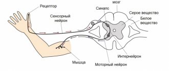
Now we will learn a little more about the brain. What is it and how does it work? This organ is formed from nerve fibers. Neurons (these are brain cells) are able to control the functioning of the entire human body by generating an electrical impulse. In total, twelve pairs of nerves can be observed that control the functioning of organs. Signals sent by the brain reach their destination through the spinal cord.
The brain is kept in fluid all the time, which prevents it from contacting the skull when the head moves. In general, our brain has pretty good protection:
- hard connective tissue;
- soft connective tissue;
- choroid;
- cerebrospinal fluid
The liquid in which our brain “floats” is called cerebrospinal fluid. The pressure of this fluid on the organ is considered to be intracranial pressure.
It is also important that the work of the brain and organs located on the head requires large energy costs. For this reason, we can observe intense blood circulation in this area. This:
- Nutrition: carotid and vertebral arteries.
- Outflow: internal and external jugular veins.
So, at rest, the head consumes about fifteen percent of the body’s total blood volume.
Changes in the fontanel
In some serious diseases, the state of the fontanelle changes. A bulging or, conversely, sunken fontanel becomes an “indicator” of pathology and indicates the severity of the disease. Thus, assessing the condition of the fontanel is an important diagnostic sign.
Protruding fontanelle in a newborn
Most often, a bulging fontanel accompanies meningitis, encephalitis, and intracranial hemorrhage. All these diseases are characterized by high intracranial pressure, which is why the fontanel bulges.
You should not make hasty conclusions and panic ahead of time. Brain diseases cannot be characterized only by bulging fontanel. But if there are accompanying threatening symptoms, you should urgently consult a specialist.
Alarming symptoms that, in combination with a bulging fontanel, threaten the child’s life:
- high temperature, which is difficult to break down and soon rises again;
- nausea and vomiting in a child;
- loud cry, irritability or, conversely, lethargy, drowsiness of the baby;
- convulsions, loss of consciousness;
- if the fontanel began to bulge after the baby fell or was injured;
- the appearance of strabismus, eye symptoms.
Sunken fontanel
If the soft crown has become sunken, this is a symptom of dehydration of the baby. The fontanelle changes, drops below the bones of the skull and indicates an acute lack of fluid for the baby. With repeated vomiting, diarrhea, and high fever, significant fluid loss occurs. Dehydration affects the entire body. The skin becomes dry, cracks may form on the lips, and the child’s well-being may be impaired.
It is necessary to give the child something to drink and arrange for the baby to be fed, if possible. And immediately consult a doctor for proper treatment and replenishment of lost fluid.
Skull and muscles
The skeleton of the head (skull) has an equally complex structure. Its main function is to protect the brain from mechanical damage and other external influences.
The entire human skull is formed by 23 bones. They are all motionless except for one - the lower jaw. As mentioned earlier, two departments can be distinguished here:
- cerebral;
- facial.
Bones related to the facial section (there are 15 in total) can be:
- paired - upper jaw, palatine bone, lacrimal, inferior nasal concha;
- unpaired - lower jaw, vomer, hyoid.
Paired bones of the medulla:
- parietal;
- temporal
Unpaired:
- occipital;
- frontal;
- wedge-shaped;
- lattice.
The entire brain section consists of a total of eight bones.
The cervical region, to which the skull is attached, allows the head to move. Movement is provided by the muscles of the neck. But on the head itself there are also muscle fibers that are responsible for facial expressions, one exception is the masticatory muscles, which are considered the strongest in this area.
Fontana. Physiology
New parents know that newborn babies have a special place on their heads that requires careful care - this is the fontanel. What is it? What is its function? What size is considered normal - large or small? When does the fontanel overgrow? Is there a danger of injuring him and disrupting his brain function? How to properly care for it?
The fontanelle is soft fibrous membrane tissue that connects two or more bones of the skull. Based on its location in the crown area, it is also called the crown.
Newborn babies have 6 fontanelles:
- Front
- Rear
- Two nipple-like
- Two wedge-shaped
When examining a newborn by pediatricians, a neonatologist, pediatrician and neurologist feel the size of the crown and monitor the rate of overgrowth of the anterior (large) fontanel throughout the year. It is the most visible, and it is by the large crown that pediatricians assess the normal development of the child. The last 5 close almost immediately or are invisible due to their small size.
The large crown should be diamond-shaped, and its normal size is from 0.5 to 4 cm. It is located in the middle of the crown (on the crown). Normally, the fontanel overgrows when the baby is one to one and a half years old. The skin on the soft part of the head pulsates to the rhythm of the baby’s heart. It's quite normal.
Function of the large fontanelle:
- Facilitating the passage of the baby through the mother's birth canal. With the help of fontanelles, the skull is elastic and tends to shrink. In the first weeks after birth, the baby's head has a slightly elongated shape.
- A child's brain grows and develops very rapidly. The fontanelles provide normal spatial conditions for this (so that there is room to grow)
- The crown is a kind of thermostat on the baby's head. When the body temperature rises above 38 C, the cooling mode “turns on” on the surface of the fontanel, thereby cooling the brain tissue.
- The crown is a kind of shock absorber that protects the baby’s brain from injury during falls and impacts.
It is advisable for all children under one year of age to undergo neurosonography - ultrasound of the brain. The procedure is painless and safe, but very informative, so it is recommended for all children, even in the absence of complaints and neurological symptoms.
Head areas
The entire head is conventionally divided into 13 regions. There they also distinguish between paired and unpaired. And so, six of them are classified as unpaired regions.
- The frontal area of the head (attention is focused on it in the next section of the article).
- Parietal (detailed information will be presented to your attention later).
- Occipital (discussed in more detail in a separate section of the article).
- Nasal, which completely matches the contour of our nose.
- Oral, also corresponds to the contour of the mouth.
- The chin, which is separated from the mouth by the geniolabial groove.
Now we move on to listing the seven paired areas. These include:
- The buccal region is separated from the nose and mouth by the nasolabial groove.
- Parotid-masticatory (contours of the parotid gland and muscles responsible for the chewing reflex).
- The temporal region of the head (the contours of the scales of the temporal bone, located below the parietal region).
- Orbital (outline of the eye sockets).
- Infraorbital (below the eye sockets).
- Zygomatic (cheekbone contour).
- Mastoid (this bone can be found behind the auricle, which, as it were, covers it).
Frontal region
Now we move on to a detailed examination of the frontal region of the head. The boundaries of the anterior section are the nasofrontal suture, the supraorbital edges, the posterior section is the parietal region, the sides are the temporal region. This section even covers the scalp.
As for the blood supply, it is carried out through the following arteries:
- supratrochlear;
- supraorbital.
They arise from the ophthalmic artery, which is a branch of the carotid artery. A well-developed venous network is observed in this area. All vessels of this network form the following veins:
- supratrochlear;
- supraorbital.
The latter, in turn, partially flow into the angular and then into the facial veins. And the other part goes into the eye.
Now briefly about the innervation in the frontal region. These nerves are branches of the ophthalmic nerve and have names:
- supratrochlear;
- supraorbital.
As you might guess, they pass together with the vessels of the same name. Motor nerves are branches of the facial nerve called temporal.
Fontana. Physiology
New parents know that newborn babies have a special place on their heads that requires careful care - this is the fontanel. What is it? What is its function? What size is considered normal - large or small? When does the fontanel overgrow? Is there a danger of injuring him and disrupting his brain function? How to properly care for it?
The fontanelle is soft fibrous membrane tissue that connects two or more bones of the skull. Based on its location in the crown area, it is also called the crown.
Newborn babies have 6 fontanelles:
- Front
- Rear
- Two nipple-like
- Two wedge-shaped
When examining a newborn by pediatricians, a neonatologist, pediatrician and neurologist feel the size of the crown and monitor the rate of overgrowth of the anterior (large) fontanel throughout the year. It is the most visible, and it is by the large crown that pediatricians assess the normal development of the child. The last 5 close almost immediately or are invisible due to their small size.
The large crown should be diamond-shaped, and its normal size is from 0.5 to 4 cm. It is located in the middle of the crown (on the crown). Normally, the fontanel overgrows when the baby is one to one and a half years old. The skin on the soft part of the head pulsates to the rhythm of the baby’s heart. It's quite normal.
Function of the large fontanelle:
- Facilitating the passage of the baby through the mother's birth canal. With the help of fontanelles, the skull is elastic and tends to shrink. In the first weeks after birth, the baby's head has a slightly elongated shape.
- A child's brain grows and develops very rapidly. The fontanelles provide normal spatial conditions for this (so that there is room to grow)
- The crown is a kind of thermostat on the baby's head. When the body temperature rises above 38 C, the cooling mode “turns on” on the surface of the fontanel, thereby cooling the brain tissue.
- The crown is a kind of shock absorber that protects the baby’s brain from injury during falls and impacts.
It is advisable for all children under one year of age to undergo neurosonography - ultrasound of the brain. The procedure is painless and safe, but very informative, so it is recommended for all children, even in the absence of complaints and neurological symptoms.
Parietal region
This area is limited by the contours of the bones of the crown. You can imagine it if you draw projection lines:
- in front - coronal suture;
- posterior - lambdoid suture;
- sides - temporal lines.
Blood supply is facilitated by arterial vessels, which are branches of the parietal branches of the temporal artery. The outflow is the parietal branch of the temporal vein.
Innervation:
- in front - the terminal branches of the supraorbital and frontal nerves;
- sides - auriculo-vesical nerve;
- posterior - occipital nerve.
Occipital region
The occipital region of the head is located below the parietal region, and is limited to the posterior region of the neck. So, the boundaries:
- top and sides - labdoid suture;
- bottom - the line between the tops of the mastoid processes.
Arteries contribute to blood supply:
- occipital;
- posterior ear.
The outflow is the occipital vein, and then the vertebral vein.
Innervation is carried out by the following types of nerves:
- suboccipital (motor);
- greater occipital (sensitive);
- lesser occipital (sensitive).
Nervous system
The article briefly talks about the nervous system of some areas of the human head. From the table you will find out more detailed information. In total, the head contains 12 pairs of nerves, which are responsible for sensations, the secretion of tears and saliva, the innervation of the muscles of the head, and so on.
| Nerve | Brief Explanation |
| Olfactory | Affects the nasal mucosa. |
| Visual | It is represented by a million (approximately) tiny nerve fibers, which are the axons of the neurons of the retina. |
| Oculomotor | Acts as muscles that move the eyeball. |
| Block | Dealt with irritation of the oblique muscle of the eye. |
| Trigeminal | This is the most important nerve located on our head. It innervates:
|
| Abductor | Innervation of the rectus muscle of the eye. |
| Facial | Innervation:
|
| vestibulocochlear | It is a conductor between the receptors of the inner ear and the brain. |
| Glossopharyngeal | Innervates:
|
| Wandering | It has the most extensive area of innervation. Innervates:
|
| Additional | Motor innervation of the pharynx, larynx, sternocleidomastoid and trapezius muscles. |
| Sublingual | Thanks to the presence of this nerve, we can move our tongue. |
Circulatory system
When studying the anatomy of the head, one cannot ignore such a complex but very important topic as the circulatory system. It is she who provides blood circulation to the head, thanks to which a person can live (eat, breathe, drink, communicate, and so on).
The functioning of our head, or rather the brain, requires a lot of energy, which requires a constant flow of blood. It has already been said that even at rest, our brain consumes fifteen percent of the total blood volume and twenty-five percent of the oxygen that we receive when breathing.
Which arteries supply food to our brain? Mainly:
- vertebrates;
- sleepy.
Its outflow from the bones of the skull, muscles, brain, and so on should also occur. This occurs due to the presence of veins:
- internal jugular;
- external jugular.
Medical Internet conferences
Introduction. The parietal foramen is found in 33–87% of skulls (about 60% in most populations), more often on the right [1–4 ] , demonstrating a weak connection with other non-metric characters [5]. The diameter of the parietal foramen usually does not exceed 2 mm [1, 6 – 8], in 70–78% of cases it ranges from 0.5 to 1.0 mm, in 19–23% – from 1.1 to 1.5 mm, in 3.7% it is more than 1.5 mm [9, 4], and the size of the right hole prevails over the size of the left one [4]. The diameter and number of parietal foramina are subject to pronounced age-related variability [9-13]. Through the parietal foramen, venous blood flows from the soft tissues of the calvarium and its periosteum into the superior sagittal sinus [14], and arterial anastomoses pass through the parietal foramen [7, 3, 15]. Various forms of diploe – emissary communications have been described [16].
Purpose: a comprehensive description of age, gender, topographic, bilateral and individual variability of the parietal foramen.
Methods. The study material included 819 cranial vaults. The material was divided into age groups in accordance with the age periodization scheme adopted at the 7th All-Union Conference on Problems of Age-Related Morphology, Physiology and Biochemistry of the Academy of Pedagogical Sciences of the USSR. Due to the fact that at least 30 skulls must be examined when analyzing nonmetric features, some age groups were combined (Table 1).
The frequency of parietal foramina was calculated both per skull and per side of the skull, taking into account the frequency of unilateral and bilateral foramina. The outer and inner diameters of the parietal foramina were measured using an MBS-2 binocular magnifier with an ocular ruler insert. The angle of inclination of the parietal foramen in the sagittal plane was determined using a protractor as the angle between the probe inserted into the hole and the outer surface of the post-obelion part of the parietal bone (obelion is a point in the median plane at the intersection of the line connecting the parietal foramina, or on the perpendicular restored from the hole) . The position of the hole on the outer surface of the skull was determined as the distance from the center of the hole to the obelion (using an MBS-2 binocular loupe with an ocular ruler insert) and from the obelion to the lambda (lambda is the connection point of the sagittal and lambdoid sutures of the skull) using a millimeter tape.
When statistically processing the primary data, methods of variation statistics were used and, to identify patterns of age-related changes, correlation and regression analysis was used. In this case, the age of the skull was taken as an independent variable, and the resulting mathematical models were considered as the basis for judging the patterns of age dynamics of the studied parameters of the parietal foramen. To determine the strength and reliability of the influence of gender and age factors on the frequency of parietal foramina, analysis of variance of two-factor complexes was used.
Results. Age-related changes in the frequency of the parietal foramina, regardless of the gender of the skull, are presented in Table 2 and are approximated by a parabolic function with one maximum, reflecting the pattern of increase in frequency until adulthood and its decrease in old and senile age:
p = 0.217 + 0.207 (x + 1) – 0.024 (x + 1)²,
where p is the overall frequency of the parietal foramina (on the right, left and on both sides of the skull), x is the serial number of the age group (Table 2).
In all the material studied, the overall frequency of the parietal foramina is 0.578±0.017. The frequency of the parietal foramen on one side is 0.334±0.017, on both sides – 0.244±0.015. The difference between these values reaches the level of statistical significance at the highest level of error-free judgment (t=3.97; p<0.001). In the unilateral position, the frequency of the right parietal foramen (0.195±0.014) is significantly (t=3.04; p<0.01) higher than the left one (0.139±0.012). Of all 624 detected holes, 54.6±2.0% were right, and 45.4±2.0% were left; the differences between these indicators are also statistically significant (t=3.20; p<0.01).
In the skulls of fetuses, out of 66 detected holes, 40 are located in the bone, and 26 are located in the membrane of the posterior fontanel, while the parietal holes occur with a frequency of 0.430±0.050, which is significantly (t=3.18; p<0.01) less than the frequency parietal foramina in the postnatal period (0.599±0.018). In fetal skulls, the differences in the frequencies of the parietal foramina on one side (0.200±0.040) and on both sides (0.230±0.042) are not significant (t=0.52; p>0.05) (even with a slight predominance of skulls with bilateral foramina positions) , as well as the differences in frequencies of the right and left holes.
Statistical significant differences were revealed between the maximum frequency of parietal foramina (in the second period of adulthood - 0.679±0.036) and in the first age group (0.545±0.045, t=2.06; p<0.05), as well as in old age (0.509 ±0.047, t=2.87; p<0.01). In old age, the differences between the frequencies of the parietal foramen on one side and on both sides are significant (t=2.31; p<0.05), while the significance of the differences between the frequencies of the right and left parietal foramina becomes statistically insignificant (t=1.47 ; p>0.05). In old age, the differences between the frequencies of the parietal foramina on one and both sides, as well as the frequencies of the right and left foramina, are statistically insignificant (t=0.78 and t=0.98, respectively; p>0.05).
In old and senile age, in the obelion section of the vault, 12% of skulls have holes with a diameter of 0.2–0.5 mm, which are not through. X-rays of the corresponding sections of the vaults will allow us to regard these openings as the mouths of the diploic canals. At the same time, the typical location and size indicate that they may be traces of parietal foramina obliterated from the inside.
To identify sexual dimorphism in the frequency of parietal foramina, 362 male and 236 female skulls of people aged 8–90 years were examined (Table 3).
Parietal foramina are significantly more common in female skulls than in male ones. These differences are most pronounced in the skulls of the second period of adulthood, mainly due to the high frequency of parietal foramina on both sides in female skulls. In male skulls, the frequency of parietal foramina on the right and both sides decreases with age, whereas the frequency of foramina on the left increases to a maximum in the skulls of older people. In female skulls, the frequency of parietal foramina on the right and left increases towards adulthood and decreases in old age.
Discussion. The total frequency of the parietal foramen found on all studied material (on the right, left and on both sides of the skull) is 57.8±1.7% (in the postnatal period - 59.9±1.8), while the parietal foramen is more often found on one side (33.4±1.7%) than on both (24.4±1.5%), mainly on the right, which corresponds to the information given in the literature (about 60%) [1 – 4]. A significantly higher frequency of parietal foramina in the postnatal period (59.9±1.8%) compared to the frequency of parietal foramina in fetuses (43.0±5.0%) suggests that the formation of foramina continues after birth. The absence in the skulls of fetuses of differences in the frequencies of the parietal foramina on one and two sides (with a certain predominance of skulls with a bilateral position of the foramina), as well as differences in the frequencies of the right and left foramina, may indicate the formation of bilateral variability of the parietal foramina (the predominance of skulls with a unilateral position of the foramen and predominance of holes on the right) in postnatal ontogenesis. In elderly and senile age, a decrease in the frequency of the parietal foramina occurs mainly due to the holes located on the right, due to which the characteristic right-sided predominance of the frequency of the parietal foramina is smoothed out. In this case, obliteration coming from the internal compact plate mainly affects the parietal foramina of small diameter. Parietal foramina are more common in female skulls; these differences are most pronounced in the skulls of people in the second period of adulthood. The age-related dynamics of the frequency of parietal foramina is different in male and female skulls.
Arteries
As already mentioned, the vertebral and carotid arteries, which are presented in pairs, supply food to the human head. The carotid artery is the basis of this process. It is divided into 2 branches:
- external (enriches the outer part of the head);
- internal (passes into the cranial cavity itself and branches, providing blood flow to the eyes and other parts of the brain).
Blood flow to the muscles is carried out by the external and internal carotid arteries. About 30% of the brain's nutrition is provided by the vertebral arteries. Basilar provides work:
- cranial nerves;
- inner ear;
- medulla oblongata;
- cervical spinal cord;
- cerebellum.
The blood supply to the brain varies depending on a person's condition. Mental or psychophysiological overload increases this indicator by 50%.
ZONES AND STAGES OF BALDING IN MALE ALOPECIA
However, alopecia does not always start from the crown and, unfortunately, is not always limited to this area.
Trichologists distinguish several stages of baldness: mild, moderate and severe - depending on the degree of hair loss, as well as several forms. The forms of alopecia depend on where a man’s hair falls out first (in the forehead, temples, crown).
When visiting a trichologist, the doctor, having examined the patient and combining information about the form and stage of baldness, will identify the pattern of alopecia. For representatives of the stronger sex, this model is determined by the Norwood-Hamilton scale.
Norwood-Hamilton scale (table)
So, for example, if a man’s hair on the top of his head has just begun to thin and small bald patches have appeared in the forehead area (the so-called “widow’s triangle”), this is a mild, second degree. And if only the hair on the back of the head reminds you of your former thick head of hair, the degree of baldness is severe and in this case only a hair transplant can help.
This is interesting
Recently, brutality is increasingly associated with a clean-shaven skull. Remember Vin Diesel, Jason Statham or Gosha Kutsenko - this is the personification of masculinity. Moreover, this identification has scientific roots - often baldness is associated with increased production of the most important male sex hormone - dihydrotestosterone.
WHY DO MEN HAVE A BALD SPACE ON THE TOP OF THEIR HEAD?
Androgenic (caused by hormonal factors) alopecia is the most common cause of baldness in the crown area. Up to 90% of all cases are associated with androgenic causes. The situation is further complicated by the fact that the predisposition to this type of baldness is often inherited (then we are talking about androgenetic alopecia). Treatment for this disease has not yet been invented.
Determine your cause of hair loss
(function(t, p) {window.Marquiz ? Marquiz.add([t, p]) : document.addEventListener('marquizLoaded', function() {Marquiz.add([t, p])})})( 'Inline', {id: '60d4194bb806c900493b4f8f', buttonText: 'Take the test', bgColor: '#e37557', textColor: '#ffffff', rounded: true, shadow: 'rgba(227, 117, 87, 0.5)' , blicked: true, buttonOnMobile: true})
Other causes of hair loss include:
- severe diseases, especially those related to the impact on the immune system: diabetes, oncology, formation of kidney stones, gall bladder;
- severe stress, vitamin deficiency;
- loss due to surgery of any organs of the digestive system, for example, the adrenal glands. As a result, the body loses the ability to synthesize vitamins, which are also necessary to nourish hair follicles);
- seborrhea (with this disease, the secretion of the sebaceous glands is disrupted, both increased and decreased sebum secretion may occur), which indirectly negatively affects the development and growth of hairs;
- radioactive exposure;
- burns and injuries. It is worth noting that most often with mechanical and burn damage, the hair follicles are not just damaged, but die, so it is almost impossible to restore hair using any methods other than transplantation.
Since the problem of baldness has been facing the entire male population of the globe for a long time and is ever acute, there are many methods to combat alopecia.
HOW TO GET RID OF BALD SPACE: POPULAR METHODS OF COMBATING BALD LOSS IN MEN
Conventionally, all methods can be divided into medical and non-medical. Of course, trichologists advise paying attention, first of all, to the first ones, but we will briefly look at everything.
Wig and bouffant
This may sound rather naive, but to this day many men (both old and young) try to hide their emerging bald spot by combing hair over it from “unaffected” places, such as the back of the head. And when hair loss becomes catastrophic, wigs are used (often expensive, made from natural hair). Some people try to disguise their bald head with hats, bandanas, etc. However, no matter how high-quality the wigs are, no matter how beautiful the headdresses are, sooner or later the secret becomes clear, and you have to either deal with the problem using other methods or come to terms with it.
Folk remedies and methods
In the arsenal of traditional medicine there are many remedies for stopping hair loss and enhancing hair growth, and some of these recipes have successfully migrated to classical medicine: for example, masks and rubbing with extracts of burdock root, nettle, and red pepper. However, there is one “but” here: in the case of androgenetic alopecia, all these methods are powerless, because they can only strengthen and “awaken” hair follicles affected by stress or vitamin deficiency, but folk remedies cannot defeat the hormonal causes of baldness.
Cosmetical tools
All kinds of shampoos, serums, masks, balms, advertised and not so much, sold both in regular supermarkets and exclusively in pharmacies. Here, some manufacturers emphasize fidelity to folk recipes, while others emphasize the use of the latest formulas and technologies. However, as in the previous case, almost no cosmetic product can fight androgenetic alopecia, which, as we said, is the most common cause of baldness in men. By the way, modern cosmetologists have found an alternative solution: a special powder that imitates short stubble recently went on sale.
Cosmetology procedures
Beauty salons offer a wide range of procedures designed to stimulate hair growth and stop hair loss: iontophoresis, plasma lifting, mesotherapy, myostimulation and the very exotic cryo- and ozone therapy. The effectiveness of all these procedures has not yet been fully proven clinically.
The greatest effect, according to various sources, is shown by:
- mesotherapy - the introduction of a special complex of drugs and vitamin “cocktails” into the scalp;
- myostimulation - exposure to pulsed current.
It is impossible not to notice that in the fight against androgenetic alopecia, all these methods will be useless.
Vitamin complexes and microelements
Strengthening the immune system is useful in any case, so you should not neglect taking multivitamins. If alopecia was caused by a weakened immune system, vitamin deficiency, or the body’s inability to synthesize this or that vitamin, external help will come in handy, including the hair.
Medications
Modern pharmacology offers two drugs that are effective in the treatment of androgenetic alopecia: Finasteride (which, by the way, is prescribed only to men) and Minoxidil, a product for external use that can partially stop hair loss and stimulate new growth. Both medications should be taken only on the recommendation of a trichologist.
The American remedy Rogaine is extremely popular. According to user reviews, it really helps. However, the drug has a lot of disadvantages. Firstly, it must be used regularly, otherwise the hair will begin to fall out even more. Secondly, you can only order the drug in an online store, and often it involves delivery from the USA (do not forget about sanctions). Thirdly, Rogaine is a very expensive pleasure.
Transfer
Unfortunately, most often androgenetic alopecia leads to a situation where no medications, cosmetics or other means can help. Even if the cause of baldness has been eliminated, it is almost impossible to restore already lost hair. Then the doctor may suggest hair transplantation. To do this, healthy hair follicles are taken from the patient from the donor area (usually the back of the head) and transplanted to the bald area. I can carry out transplantation in three ways: the Strip method, the FUE method and, finally, the HFE method.
- The Strip technique involves surgical intervention: the doctor uses a scalpel to cut out a flap of skin from the donor area, then divides it into small grafts, which are implanted into prepared incisions in the recipient area. This method is quite traumatic, scars remain after the operation, and repeated transplantation is impossible.
- The FUE (Follicular Unit Machine Extraction) method is less invasive: here, a punch with a diameter of up to 5 mm is used to remove grafts, leaving small round scars on the donor area. There will also be scars on the transplant area as incisions are made to implant follicles.
- The most advanced method is HFE (Hand Follicular Extraction), in which each graft is removed separately and manually using microsurgical instruments. No incisions are made in advance on the recipient area: the follicles are implanted directly into the skin, and the puncture is made with a special implanter. This procedure is almost non-invasive and minimally injures the skin of the donor area. HFE allows you to achieve almost 100% effectiveness in hair survival in the bald area. If necessary, you can re-transplant.
In Russia today, only one clinic practices the HFE transplantation technique. This is the HFE Clinic, where specialists with extensive experience in the field of hair transplantation work. The best trichologists consult here, and operations are performed by certified transplantologists. In our clinic, each client will be helped to achieve thick, beautiful hair.
Vienna
When considering the anatomy of the human head, it is difficult to ignore a very important topic - the venous structure of this part of the body. Let's start with what venous sinuses are. These are large veins that collect blood from the following parts:
- skull bones;
- head muscles;
- meninges;
- brain;
- eyeballs;
- inner ear.
You can also find another name for them, namely, venous collectors, which are located between the sheets of the lining of the brain. Leaving the skull, they pass into the jugular vein, which runs next to the carotid artery. You can also distinguish the external jugular vein, which is slightly smaller and located in the subcutaneous tissue. This is where blood collects from:
- eye;
- nose;
- mouth;
- chin
Generally speaking, everything listed above is called superficial formations of the head and face.
What influences closure
- Heredity. The size of the fontanelle and the time it closes in a baby primarily depend on genetic characteristics. If the parents had late or early overgrowth, then the baby will have the same thing. But provided there are no diseases that affect the time of crown overgrowth.
- The gestational age at which the baby was born. A premature child under 2-3 years of age will lag behind peers born at term in physical development. And accordingly, the period of closure of the fontanel is longer.
- A deficiency of calcium and vitamin D in a baby's body can cause prolonged overgrowth. And excessive content, on the contrary, causes early closure of the fontanel. Usually the reason for this is not a diet, but a metabolic disorder.
- Some diseases.
Muscles
To put it very briefly, all the muscles of our head can be divided into several groups:
- chewable;
- facial expressions;
- cranial vault;
- sense organs;
- upper digestive system.
You can guess the functions performed by their names. For example, chewing ones make the process of chewing food possible, but facial ones are responsible for human facial expressions, and so on.
It is very important to know that absolutely all muscles, regardless of their main purpose, are involved in speech.
ZONES AND STAGES OF BALDING IN MALE ALOPECIA
However, alopecia does not always start from the crown and, unfortunately, is not always limited to this area.
Trichologists distinguish several stages of baldness: mild, moderate and severe - depending on the degree of hair loss, as well as several forms. The forms of alopecia depend on where a man’s hair falls out first (in the forehead, temples, crown).
When visiting a trichologist, the doctor, having examined the patient and combining information about the form and stage of baldness, will identify the pattern of alopecia. For representatives of the stronger sex, this model is determined by the Norwood-Hamilton scale.
Norwood-Hamilton scale (table)
So, for example, if a man’s hair on the top of his head has just begun to thin and small bald patches have appeared in the forehead area (the so-called “widow’s triangle”), this is a mild, second degree. And if only the hair on the back of the head reminds you of your former thick head of hair, the degree of baldness is severe and in this case only a hair transplant can help.
This is interesting
Recently, brutality is increasingly associated with a clean-shaven skull. Remember Vin Diesel, Jason Statham or Gosha Kutsenko - this is the personification of masculinity. Moreover, this identification has scientific roots - often baldness is associated with increased production of the most important male sex hormone - dihydrotestosterone.
WHY DO MEN HAVE A BALD SPACE ON THE TOP OF THEIR HEAD?
Androgenic (caused by hormonal factors) alopecia is the most common cause of baldness in the crown area. Up to 90% of all cases are associated with androgenic causes. The situation is further complicated by the fact that the predisposition to this type of baldness is often inherited (then we are talking about androgenetic alopecia). Treatment for this disease has not yet been invented.
Determine your cause of hair loss
(function(t, p) {window.Marquiz ? Marquiz.add([t, p]) : document.addEventListener('marquizLoaded', function() {Marquiz.add([t, p])})})( 'Inline', {id: '60d4194bb806c900493b4f8f', buttonText: 'Take the test', bgColor: '#e37557', textColor: '#ffffff', rounded: true, shadow: 'rgba(227, 117, 87, 0.5)' , blicked: true, buttonOnMobile: true})
Other causes of hair loss include:
- severe diseases, especially those related to the impact on the immune system: diabetes, oncology, formation of kidney stones, gall bladder;
- severe stress, vitamin deficiency;
- loss due to surgery of any organs of the digestive system, for example, the adrenal glands. As a result, the body loses the ability to synthesize vitamins, which are also necessary to nourish hair follicles);
- seborrhea (with this disease, the secretion of the sebaceous glands is disrupted, both increased and decreased sebum secretion may occur), which indirectly negatively affects the development and growth of hairs;
- radioactive exposure;
- burns and injuries. It is worth noting that most often with mechanical and burn damage, the hair follicles are not just damaged, but die, so it is almost impossible to restore hair using any methods other than transplantation.
Since the problem of baldness has been facing the entire male population of the globe for a long time and is ever acute, there are many methods to combat alopecia.
HOW TO GET RID OF BALD SPACE: POPULAR METHODS OF COMBATING BALD LOSS IN MEN
Conventionally, all methods can be divided into medical and non-medical. Of course, trichologists advise paying attention, first of all, to the first ones, but we will briefly look at everything.
Wig and bouffant
This may sound rather naive, but to this day many men (both old and young) try to hide their emerging bald spot by combing hair over it from “unaffected” places, such as the back of the head. And when hair loss becomes catastrophic, wigs are used (often expensive, made from natural hair). Some people try to disguise their bald head with hats, bandanas, etc. However, no matter how high-quality the wigs are, no matter how beautiful the headdresses are, sooner or later the secret becomes clear, and you have to either deal with the problem using other methods or come to terms with it.
Folk remedies and methods
In the arsenal of traditional medicine there are many remedies for stopping hair loss and enhancing hair growth, and some of these recipes have successfully migrated to classical medicine: for example, masks and rubbing with extracts of burdock root, nettle, and red pepper. However, there is one “but” here: in the case of androgenetic alopecia, all these methods are powerless, because they can only strengthen and “awaken” hair follicles affected by stress or vitamin deficiency, but folk remedies cannot defeat the hormonal causes of baldness.
Cosmetical tools
All kinds of shampoos, serums, masks, balms, advertised and not so much, sold both in regular supermarkets and exclusively in pharmacies. Here, some manufacturers emphasize fidelity to folk recipes, while others emphasize the use of the latest formulas and technologies. However, as in the previous case, almost no cosmetic product can fight androgenetic alopecia, which, as we said, is the most common cause of baldness in men. By the way, modern cosmetologists have found an alternative solution: a special powder that imitates short stubble recently went on sale.
Cosmetology procedures
Beauty salons offer a wide range of procedures designed to stimulate hair growth and stop hair loss: iontophoresis, plasma lifting, mesotherapy, myostimulation and the very exotic cryo- and ozone therapy. The effectiveness of all these procedures has not yet been fully proven clinically.
The greatest effect, according to various sources, is shown by:
- mesotherapy - the introduction of a special complex of drugs and vitamin “cocktails” into the scalp;
- myostimulation - exposure to pulsed current.
It is impossible not to notice that in the fight against androgenetic alopecia, all these methods will be useless.
Vitamin complexes and microelements
Strengthening the immune system is useful in any case, so you should not neglect taking multivitamins. If alopecia was caused by a weakened immune system, vitamin deficiency, or the body’s inability to synthesize this or that vitamin, external help will come in handy, including the hair.
Medications
Modern pharmacology offers two drugs that are effective in the treatment of androgenetic alopecia: Finasteride (which, by the way, is prescribed only to men) and Minoxidil, a product for external use that can partially stop hair loss and stimulate new growth. Both medications should be taken only on the recommendation of a trichologist.
The American remedy Rogaine is extremely popular. According to user reviews, it really helps. However, the drug has a lot of disadvantages. Firstly, it must be used regularly, otherwise the hair will begin to fall out even more. Secondly, you can only order the drug in an online store, and often it involves delivery from the USA (do not forget about sanctions). Thirdly, Rogaine is a very expensive pleasure.