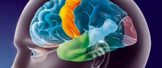
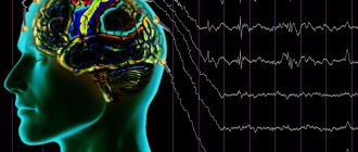
What are the rhythms?
The rhythms generated by the brain differ into five types depending on the amplitude and frequency of vibrations:
- The alpha rhythm is recorded in 95% of healthy patients at the moment of relaxation, when they close their eyes. Best expressed in the occipital regions. α-wave frequency 8-12 Hz. With visual stimuli, there is a deficiency of this radiation. At the same time, in people with congenital blindness or optic nerve atrophy, the α rhythm is absent.
- Beta rhythms are the fastest brain oscillations in the range of 12-40 Hz. They are associated with the processes of learning and concentration. Therefore, they are formed in a child during the development of logical thinking. Normally, this process ends by age five. Beta waves are generated naturally when you are awake. Under stress, their level increases, but a deficiency, on the contrary, is associated with absent-mindedness syndrome, depression and emotional disorders. Without β-activity, no meaningful human activity is possible.
- The gamma rhythm is clearly visible when solving complex problems. These are the highest frequency rhythms generated during an active thought process. Completely disappears in the deep sleep phase.
- The delta rhythm is characteristic of the recovery period and natural sleep. These are the slowest waves. They are formed in the prenatal period. Asynchronous delta waves appear during coma or drug intoxication.
- Theta activity manifests itself in the fetus already at 2-3 months and predominates in children under three years of age. They represent patterns of electrical activity in the range of 4-8 Hz. Normally, theta waves of an adult appear in the twilight state, during the transition from sleep to wakefulness. A significant number of them can be observed in a confused state, with mental disorders. A large amount of θ rhythm can signal a state of chronic stress.
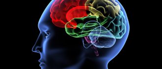
Types of bioelectric activity
Bioelectrical activity, depending on the scale (from a single neuron to the coordinated activity of large neural ensembles), manifests itself in different forms.
➥ Main article: Functional systems, structure and organization of the brain
| Micro level of the brain | Meso level of the brain | Macro level of the brain | |
| Regular bioelectrical activity | spike trains; Regular pattern of subthreshold membrane potential oscillations (SMPO) | Oscillations of neural populations | Brain rhythms or waves generated by the largest neural ensembles |
| Irregular bioelectrical activity | thermal noise; Channel noise; burst noise; Synaptic noise; Spike | 1/f noise | Disorganized EEG 1/f noise |
| Transient bioelectrical activity | Irregular subthreshold membrane potential oscillations; Spindle-like subthreshold membrane potential oscillations | Non-epileptiform transient activity (SWR); Epileptiform transient activity (interictal epileptiform activity, IED) | Non-epileptiform (eng. nonepileptiform) transient activity; Epileptiform transient activity (eng. epileptiform discharges); Event-related potential (ERP) |
Rhythmic activity
Regular subthreshold oscillations of membrane potential
➥ Main article: Rhythmic activity of the brain
Rhythmic or regular bioelectrical activity is a repeating frequency pattern in the central nervous system. In other words, these are the same type or self-similar oscillatory events that repeat at equal intervals.
Neural oscillations generated by the bioelectrical activity of the brain are observed throughout the central nervous system at all levels of its organization and include spike trains/bursting, a regular pattern of subthreshold oscillations of the membrane potential, high-amplitude oscillations of the local field potential and large-scale oscillations (brain waves).
EEG is one of the most accessible electrophysiological methods for recording rhythmic activity. Since the electric field created by a single neuron or a collection of closely located neurons is so small that it cannot be recorded from the surface of the head, EEG is a method that reflects the total activity of a large pool of neurons, the magnitude of which is sufficient to generate a potential difference on the surface of the scalp. It should be emphasized that the electrical activity must be as powerful as is required for the signal to pass through the bones of the skull and skin, which have noticeable resistance to electric current. Again, based on the above necessary and sufficient conditions: the activity recorded from the scalp is due to the relatively powerful total activity of the neuronal pool, it logically follows that the total EEG is primarily due to the activity of cortical neurons as those closest to the skin surface heads - place of registration.
Rhythmic processes play a key role in the functional activity of the brain.
Irrhythmic activity
Irrhythmic or irregular activity refers to chaotic or stochastic changes in the bioelectrical activity of the brain.
At the micro level
Irregular brain activity can be divided into molecular, synaptic and 1/f noise,3 and spiking activity by mechanisms.
Spikes
(English spikes) can also be extremely irregular, both during constant, spontaneous activity, and during evoked activity at a high frequency of impulse emission. There is uncertainty about the source of this irregularity, ranging from intrinsic sources of noise in neurons to collective effects in large-scale cortical networks. Cortical interneurons show highly irregular spike timing in response to direct current injection in vitro. This is in sharp contrast to cortical pyramidal cells, which exhibit irregular bioelectrical activity in vivo but regular activity in vitro. In vitro recordings and computational models have shown that this is associated with the rapid activation kinetics of interneuronal K ion currents. In this case, an arrhythmic spike can contribute to the irregular activity of the entire cortical network.45
Above is a normal EEG, below is an EEG of a patient with hypsarrhythmia.
At higher levels of organization, nonrhythmic activity can manifest itself in the form of disorganization of the bioelectrical activity of the brain. Types of disorganization:
- Disorganization of bioelectrical activity with a predominance of alpha activity. On the EEG, alpha activity is the main one, but it is not regular enough or is completely irregular in frequency. This more or less disorganized alpha rhythm has an insufficiently high amplitude and may even dominate in all areas of the brain. Beta activity is also often enhanced, often represented by low-frequency oscillations of increased amplitude. Along with this, theta and delta waves with a fairly high amplitude may be present in the EEG.
- Disorganization of bioelectrical activity with a predominance of theta and delta activity. The structure of this type is characterized by a weak representation of alpha activity. Fluctuations in biopotentials in the alpha, beta, theta and delta ranges are recorded without any clear sequence. This non-dominant type of curve can have both medium and high amplitude levels.
- Hypsarrhythmia - manifests itself as disorganization of the background bioelectrical activity of the brain with irregular slow waves of high voltage; multifocal peaks and polyspikes can be superimposed during sleep.6
Paroxysmal activity
➥ Main article: Paroxysmal activity
Paroxysms or transients are short intervals (10 to 1000 ms)7 during which the signal changes abruptly and becomes atypical or relatively unpredictable.
(A) Spindle-shaped subthreshold oscillations of membrane potential; (B) Irregular subthreshold fluctuations in membrane potential
Transient activity reflects the functional integration of brain structures and is a fundamental element of neural interaction. Transients generated jointly by different neural populations indicate the connection of these populations. This communication may be synchronous or asynchronous.8
Examples of such activity include spontaneous synchronous synaptic transients generated by Ca2+ ion currents between adjacent cortical neurons.9 Spikes and sharp waves that constitute seizure or interictal activity in individuals with epilepsy or a predisposition to it, or transients in the form of vertices and sleep spindles , which are the norm. Irregular subthreshold membrane potential oscillations and spindle-shaped subthreshold membrane potential oscillations can also be recorded using in vivo intracellular recording.
Event related potential
➥Main article:ERP
Event related potentials
(EPS, English
event-related potential
- ERP) are electrical voltage fluctuations recorded from the scalp, generated in brain structures in response to certain events or stimuli.
Also refers to transient brain activity. These are EEG changes time-locked to sensory, motor or cognitive events that provide a safe and non-invasive approach to studying the psychophysiological correlates of mental processes. They are believed to reflect the total bioelectrical activity of postsynaptic potentials that occur when a large number of similarly oriented cortical neurons (on the order of thousands or millions) fire synchronously during information processing. ERP is measured using electroencephalography (EEG) and magnetoencephalography (MEG). The magnetoencephalographic (MEG) equivalent of ERP is the ERF or event -related
. 10 ERP subtypes include evoked and evoked potentials.
Evoked potentials
➥ Main article: Evoked potentials
Cognitive evoked potentials (EP, EP)
represent event-related activity that occurs as an electrical response from the brain to various types of sensory stimulation of neural tissue; auditory and visual stimulation are most often used. Recording of such electrical potentials is a non-invasive objective test that provides information, for example, about sensory pathway disorders, the location of lesions affecting sensory pathways, and disorders related to language and speech. Evoked potentials are recorded from the scalp using EEG. Potentials typically appear as a transient waveform, the morphology of which depends on the type and strength of the stimulus and the location of the electrodes on the scalp. The mental state of the subject, such as attention, wakefulness, and anticipation, also influences the morphology of the waveform.
Induced activity
Along with evoked activity, stimulus-related neural activity can lead to induced activity
. The induced response is associated with processes in the brain that are not directly related to the stimulus, but are caused indirectly by the nonlinear interaction of neurons after the stimulus. The functional component of the induced response is likely to represent corticothalamic feedback (top-down) processes linking external stimuli to the internal cortical model of the environment, i.e., combining externally controlled sensory information with internal brain activity.1112 The induced response may be related with cognitive brain functions such as perception, attention and learning, which can be considered higher order processes. A well-studied type of induced activity is a change in the amplitude of oscillatory activity. For example, gamma activity often increases during times of increased mental activity, such as during the presentation of an object. Since induced responses are not phase related and will therefore be excluded during averaging, they can only be obtained using time-frequency analysis. Induced activity typically reflects the activity of multiple neurons: amplitude changes in oscillatory activity are thought to result from synchronization of neuronal activity, such as spike timing or fluctuations in the membrane potential of individual neurons.
Therapeutic effect
Brain wave therapy continues to be studied. According to already available data:
- Alpha rhythms help reduce anxiety, tension and restlessness, and improve memory. It accelerates blood supply to the brain structures, increasing the saturation of the body with oxygen and beneficial microelements. Facilitates the recovery period after illness. Reduces the negative impact of traumatic situations. Determines the release of the neurotransmitter serotonin, increasing its level in the body.
- Beta stimulation helps improve learning, communication skills and perseverance in patients. In addition, such exposure helps reduce fatigue.
- The effect of gamma rhythms has proven itself to be effective in enhancing the speed of information processing and stimulating short-term memory. These frequencies have also been found to have a beneficial effect on the brain in chronic migraine. In approximately half of the subjects, the frequency and intensity of attacks decreases.
- Stimulation of the δ rhythm helps reduce stress levels and relieve attacks of chronic pain, including cephalgia.
- Exposure to θ waves can enhance creativity, improve memory, control over the emotional sphere, and enhance intuition. It helps reduce anxiety and stress, strengthen the immune system, and enhance long-term memory, since the rhythm of theta waves is characteristic of the hippocampus, the part of the brain responsible for storing information and memories.
Basic brain rhythms
Normally, in a state of wakefulness, only two rhythms are generated in the brain: alpha and beta waves, which together occupy 70-100% of all rhythms.
Alpha waves have the following characteristics: wave frequency from 8 to 13 per second, wave amplitude no more than 50 μV. In this case, complexes of alpha waves should ideally be combined in modulation - complexes of waves with gradually increasing amplitude and gradual further decay, called spindles.
The beta rhythm has the following characteristics: the wave frequency is higher, reaching 14-30 hertz, while the amplitude does not exceed 25-30 µV.
Pathological rhythms of the brain are represented mainly by delta and theta waves. Delta waves have the lowest frequency: 1-4 hertz. While the amplitude of these waves varies very widely, reaching hundreds of microvolts. Normally, such waves can occur (but do not occupy most of the time!) in a person’s sleep, children under 6 years of age. The prevalence of this rhythm is typical for patients with traumatic brain injury.
The theta rhythm has a frequency of 4 to 8 vibrations per second. The amplitude also varies widely and can reach hundreds of microvolts. Typical for young children. The brain in this state is capable of better assimilation of incoming information, however, asymmetrical theta waves still speak in favor of brain pathology.
Separately, EEG examines pathological complexes and phenomena. Thus, such complexes include bursts of alpha waves (high-amplitude oscillations that have all the characteristics of the alpha rhythm), peak-wave complexes and sharp wave-slow wave. These phenomena are often characteristic of patients with EEG.
Generalized EEG seizures leave very characteristic wave complexes on the recording, the so-called EEG pattern. It is represented by repeating waves of a certain frequency, amplitude, and shape and depends on the type of attack. Some seizure patterns are shown in the figures.
All rhythms and phenomena of the brain should be distinguished from so-called artifacts - interference when recording an EEG. Such interference can be caused by interference from the electrical network, movement of the electrodes during the study, incomplete contact of the electrode with the skin, as well as many other reasons.
Alpha wave stimulation
There are several ways to stimulate the body’s wave activity:
- Breathing exercises that allow you to maintain deep breathing saturate the brain cells with oxygen. Systematic breathing exercises promote the generation of alpha waves.
- Listening to stereo sounds with alpha waves is a technique accessible to everyone.
- Yoga and meditation are powerful activators of the α rhythm.
- Hot baths can help you relax and switch to alpha wave production mode.
Pathological deviations
Disturbances in brain wave activity are observed when:
- Oligophrenia. With it, the total activity of alpha rhythms is abnormally increased.
- Epilepsy, which causes disturbances in the frequency and amplitude of wave activity. This is associated with the development of direct or interhemispheric asymmetry in the hemispheres of the brain.
- Hypertension, weakening the frequency of alpha rhythms and increasing beta activity. Circulatory and cardiovascular system disorders always affect brain wave activity. A similar picture can be observed with beta-lactamase activity of bacteria.
- Paranoid schizophrenia, in which the brain generates an increased number of beta rhythms.
- Cysts and inflammatory processes of the corpus callosum. They cause severe asymmetry between the hemispheres, reaching 30%.
- The pathological picture can occur with traumatic brain injuries, congenital or acquired dementia, and delayed psychomotor development in children.
To evaluate alpha rhythms, it is necessary to undergo an EEG from time to time. You can find out the addresses of clinics that perform this diagnostic procedure on the website.
Changes in bioelectrical activity of the brain
➥ Similar article: EEG changes
The frequency of bioelectrical activity can indicate various functional states of the brain: sleep, consciousness, cognition and some mental disorders. Decreased bioelectrical activity may appear on the EEG as slow waves, which are most often observed in conditions such as sleep, coma,13 brain death, depression,14 autism,15 brain tumors, obsessive-compulsive disorder (OCD),16 attention deficit disorder and hyperactivity (ADHD),17 encephalitis.18
In contrast, when there is increased bioelectrical activity in the brain, rapid waves are observed on the EEG, in conditions such as epilepsy,19 anxiety, post-traumatic stress disorder (PTSD) and substance use.20
It should be noted that the use of the terms “decrease” and “increase” of bioelectrical activity is incorrect, since the bioelectrical activity of the brain at different levels of organization manifests itself differently, and in the context of the whole brain one can only evaluate parameters (amplitude, power, index ) signals obtained during EEG recording. Then, based on numerous experimental data, one can already interpret changes in one or another parameter and talk about any changes in the functional activity of the brain.
Classification of BEA changes
Based on localization, changes in bioelectrical activity can be:21
- Focal – limited area (up to 3 leads);
- Regional – zone of the brain lobe (3 or more leads);
- Lateralized - pathological activity detected above the hemisphere;
- Generalized - general pathological activity, affecting the entire brain, recorded in all leads according to the mechanisms of primary and secondary generalization;
- Diffuse (unspecified) – cannot be classified into the above groups. According to the degree of severity, diffuse changes in bioelectrical activity can be divided into mild, moderate and pronounced.
Diffuse EEG changes
A. Flat EEG with burst suppression at intervals of 5–10 s.
B. Continuous generalized acute and slow complexes. C. Diffuse delta activity with peaks in the frontotemporal areas, spreading and predominant in the left hemisphere. A characteristic feature of pathological activity with diffuse changes in bioelectrical activity is the lack of locality, instability of spatial distribution (mosaicity), and violation of bilateral synchrony.
Diffuse brain damage is often associated with the development of encephalopathy, that is, widespread small-focal lesions. Due to the polymorphism of small focal changes and their prevalence, pathological changes are very diverse and do not form any organized activity, which is manifested by mosaic changes in the bioelectrical activity of the brain.
EEG disorganization
When the brain is damaged, pathological diffuse changes in bioelectrical activity are characterized by disorganization and the absence of regular dominant activity (alpha rhythm). Diffuse pathological activity can manifest itself in different ways (for example, in the form of slow-wave rhythms or epileptiform activity is detected), and also have different amplitudes (low-amplitude, medium-amplitude or high-amplitude).
Recording video EEG with single-channel ECG
The EEG demonstrates "slow" and "flat" phases during cardiac asystole, followed by a second "slow" phase after cardiac sinus rhythm resumes. Myoclonic seizures (MS) occur during asystole and after resumption of sinus rhythm. Tonic posture (tone) occurs during asystole. Pay attention to the artifact of tonic muscle contraction recorded on the ECG channel
Disorganization of bioelectrical activity is usually associated with a decrease in EEG amplitude and absence of alpha rhythm, which is described in more detail in the article alpha rhythm depression. Dysrhythmic brain activity can manifest itself in the form of a flat EEG (desynchronous type III, considered as a borderline norm), as well as with a predominance of alpha activity (disorganized type IV) and with a predominance of θ- and δ-activity (disorganized EEG type V). More details: classification of EEG disorders by type.
Slowing brain activity
A slowdown in the bioelectrical activity of the brain (when low frequencies predominate) does not always indicate pathological changes and may be associated with drowsiness. Slowing of the background rhythm in children is observed depending on the age group:
- 1 year – fundamental frequency less than 5 Hz
- 4 years – fundamental frequency less than 6 Hz
- 5 years – fundamental frequency less than 7 Hz
- over 8 years old – fundamental frequency less than 8 Hz
Slowing of cortical rhythms often occurs against the background of diffuse dysfunction of cortical and subcortical structures, due to vascular, metabolic or toxic lesions of the brain, and in children it can be a consequence of perinatal pathology.
Generalized slowdown
FIRDA pattern with slightly slowed background theta EEG in an elderly man with metabolic encephalopathy. Periods of well-formed occipital alpha activity are also noticeable
Disadvantages of the method
Excessive wave stimulation can cause negative reactions in the body. So, with an excess of alpha activity, a decrease in concentration develops, the formation of attention deficit, depression, and a decrease in visual clarity. A person begins to need daytime sleep and gets tired faster. In addition, under the influence of alpha waves, suggestibility increases.
The downside of beta wave stimulation is increased anxiety, overexcitement and the development of obsessive-compulsive disorder. There are signs of stress and paranoia. Also, under the influence of such stimulation, muscle tension increases and insomnia occurs. The effect on the cerebellar amygdala and hypothalamus provokes an increase in blood pressure.
When exposed to theta waves, you can develop concentration disorders, depression, hyperactivity, or, conversely, apathy. Under the influence of such stimulation, drowsiness, indifference to life, and increased suggestibility occur.
Therefore, before resorting to any methods of stimulating brain wave activity, it is better to consult with specialists.