What is accommodation?
An important link in the functioning of the visual mechanism is accommodation. This is the name given to the ability of the lens to tense or relax when seeing at different distances. Accommodation is the normal physiological state of the eye, when all muscles are relaxed when looking into the distance. When viewing nearby objects, the ciliary muscle tenses, and the lens increases its curvature, becoming more convex. Its optical power increases, and the near image becomes clear. Defocus of vision occurs precisely because the lens loses its ability to focus. Let's consider what factors can provoke the appearance of this visual pathology.
Main causes of myopia
Doctors identify two main causes of myopia:
- elongated eyeball;
- excessive refractive power of the eye.
Normally, the human eyeball has such a size and shape that the rays entering the eye are focused in the center of the retina. If the anteroposterior ocular axis is slightly longer than normal, the rays do not reach the retina and are focused in front of it. Due to the shifted optical focus, a person has difficulty seeing into the distance. If the length of the eyeball increases, myopia progresses and visual impairment becomes more pronounced. Sometimes myopia is caused not by the size of the ocular axis, but by the strong refractive ability of the cornea or lens. When the optical media of the eye refract light more than necessary, the focus of the image also shifts to the area in front of the retina and this is reflected in the clarity of distance vision.
Causes of blurred vision
This disorder can occur in a person for a number of reasons:
- Age-related changes in eye structures.
- Eye strain associated with prolonged exposure to a monitor or gadget.
- Improper organization of the workplace: poor lighting, low table.
- Hereditary myopia.
- Lack of sleep, lack of fresh air, insufficient physical activity.
All of these factors can provoke a spasm of accommodation, and its symptoms are such that a person may not be aware of this visual anomaly for a long time until the disturbances become too obvious.
Often, blurred vision is mistaken for fatigue from lack of sleep or intense visual work.
This can last for quite a long period and, if appropriate measures are not taken, will lead to a serious deterioration in eye health. Let's consider the possible causes of blurred vision known in medical practice.
Reason 3
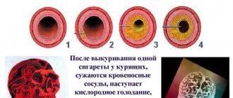
Poor blood flow. Nutrition and respiration of body cells is carried out by supplying blood to them. The retina of the eye is a delicate organ that suffers at the slightest circulatory disturbance. Actually, circulatory disorders are what ophthalmologists try to see by examining the fundus of the eye.
Conclusion. It is better to make regular visits to the ophthalmologist. After all, circulatory disorders of the retina lead to serious illnesses. If there is an existing predisposition to them, the doctor will prescribe medications to improve the condition of blood vessels. There are also certain diets, following which you can normalize blood circulation and maintain blood vessels in good condition. It would also be useful to take care of your blood vessels: it is better to avoid trips to the steam room or sauna, procedures in a pressure chamber, and other pressure changes.
Presbyopia
After 40-45 years, noticeable age-related changes begin not only in the entire body, but also in the visual apparatus. In most people at this age, the lens gradually loses its elasticity - this phenomenon is called presbyopia, or age-related farsightedness.
Near vision begins to get worse. Here is how the symptoms of this pathology manifest themselves:
- difficulty reading small letters close up, for example, on food packaging;
- eye fatigue;
- frequent headaches.
By the age of 60-70 years, the lens loses its ability to accommodate in most people.
Defocusing vision in elderly patients, unfortunately, cannot be treated.
To correct presbyopia, progressive glasses or multifocal contact lenses are used. In the case of a severe degree of this visual impairment, a lensectomy is performed - replacing the natural lens with an artificial intraocular lens. Good quality of vision after such an operation lasts for life. In addition, replacing the lens is a good prevention of cataracts , another common disease in old age, because the artificial implant cannot become cloudy.
Floaters before eyes
Atherosclerosis
Diabetes
62859 December 14
IMPORTANT!
The information in this section cannot be used for self-diagnosis and self-treatment.
In case of pain or other exacerbation of the disease, diagnostic tests should be prescribed only by the attending physician. To make a diagnosis and properly prescribe treatment, you should contact your doctor. Flashing of flies before the eyes: reasons for their appearance, what diseases they occur with, diagnosis and treatment methods.
Definition
The flickering of flies before the eyes is a subjective sensation, when describing it, people imply the presence in the field of vision of permanent or transient additional inclusions of various shapes and sizes.
The organ of vision is one of the most important sense organs providing orientation in space. The visual analyzer consists of the organ of vision (eye), which perceives and interprets the image into an electrical signal, the optic nerve, through which the signal enters the subcortical centers, and the cerebral cortex, where the signal is analyzed. The eye consists of the eyeball and accessory organs, and the eyeball is made up of three membranes: outer (cornea and sclera), middle (iris and choroid), and inner (retina). Behind the iris is the crystalline lens, a light-refracting lens. The space between the lens and the retina is filled with vitreous humor.
The vitreous body is a gel-like structure enclosed in a dense membrane. The gel contains water, hyaluronic acid, collagen fibers, proteins and other substances.
If the ratio of certain components of the vitreous body is disturbed, dense inclusions can form, perceived by a person as “spots”.
Types of flies before the eyes
“Flies” are dots, zigzags, worms, dust, dashes, strokes, scratches, cobwebs, commas, etc.
Floaters before the eyes, arising as a result of destructive processes in the vitreous, have clear descriptions and do not disappear from view:
- rings with an outer and inner rim or a dot inside, located in the field of view in large numbers, may indicate granular destruction of the vitreous body;
- threads that look like short or long strips with circles inside, of varying transparency, may indicate filamentous destruction of the vitreous body. The interweaving of threads indicates more severe destruction.
Destructive processes in the vitreous body Also, with the destruction of the vitreous body, spots and clots may appear, similar to “plastic film” or flakes, which greatly cloud the image.
When cholesterol crystals are included in the vitreous gel, shiny “snowflakes” appear in the field of view.
Any inclusions can move freely throughout the entire field of view, move within a limited range, or be stationary.
Possible causes of flies flashing before the eyes
The reasons for flies flashing before the eyes are quite varied. Floaters can be a sign of temporary functional disorders, for example, when lifting weights, overwork, or a sudden change in posture. However, inclusions in the visual field appear in some diseases of the eye structures - the vitreous body, choroid, retina, and also arise as a result of diseases of other organs and systems that affect the functioning of the visual analyzer.
What diseases cause floaters to appear before the eyes?
Destruction of the vitreous body
is the reason for the constant presence of additional inclusions in the field of view. It occurs due to a decrease in the ratio of fluid and other components of the vitreous gel during natural aging or after eye injury, eye surgery and other reasons. The characteristics of inclusions are described in the previous section.
The prognosis for vision is favorable, but the quality of life may suffer and the ability to work may decrease.
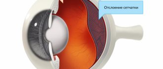
Retinal and/or vitreous detachment
, in addition to floaters, flashes and sparks, is characterized by a sharp deterioration in visual acuity and blurred vision. Without immediate medical attention, it can lead to blindness. The risk of detachment increases in people suffering from a high degree of myopia.
For vertebrobasilar insufficiency
blood flow through the vertebral arteries is disrupted, which affects the blood supply to certain areas of the brain, including those responsible for visual function. Clinically manifests itself as floaters, darkening before the eyes, short-term loss of vision.
The most common cause of acquired vertebrobasilar insufficiency is osteochondrosis of the cervical spine.
Choroiditis (inflammation of the choroid), retinitis (inflammation of the retina), chorioretinitis (inflammation of both structures)
can also cause the appearance of floaters. In addition, the patient experiences flashes of light (photopsia), visual acuity decreases, and vision in the dark is impaired. The disease of an infectious nature in advanced cases leads to blindness.
Retinopathy
(hypertensive, atherosclerotic, diabetic) occur when blood vessels are damaged by diseases of the same name. Retinopathy is characterized by transient visual impairment in the form of decreased visual acuity, floaters before the eyes, and the presence of large spots in the field of vision (scotomas). As the underlying disease is treated, the intensity of the symptoms decreases, and as it progresses, it can result in loss of vision.
Transient flashing of flies before the eyes, a feeling of a “noisy” picture can occur with low blood pressure.
Pressure surges in pregnant women can also lead to floaters. And if a slight decrease in pressure from the individual norm occurs due to physiological reasons, then the increase may indicate gestosis (late toxicosis) and requires careful additional examination.
Which doctors should I contact if I have spots flashing before my eyes?
Since the flickering of flies before the eyes can be caused not only by eye pathologies, but also by diseases of the internal organs, consultation is required not only with an ophthalmologist, but also. If necessary, an additional visit may be recommended, for pregnant women -.
Diagnostics and examinations for flies flashing before the eyes
To identify the causes of flies flashing before the eyes, it is necessary to examine and interview the patient, measure blood pressure levels, determine visual acuity and visual fields, assess the condition of the vitreous body and retina using biomicroscopy and ophthalmoscopy, and examine the fundus.
Additional laboratory and instrumental research methods are often recommended:
- clinical blood test;
Accommodative visual asthenopia
Asthenopia is the name given to rapid eye fatigue that occurs during intense visual work. In medical practice, it is classified not as an independent pathology, but as a disorder that precedes pathological deterioration of vision and in other cases leads to eye diseases.
Symptoms of asthenopia are expressed as follows:
- pain in the eyes, increased lacrimation, fatigue during visual work;
- headache;
- ghosting;
- sensation of film in the field of view.
The causes of accommodative asthenopia can be prolonged exposure to a computer monitor - currently this is the most common factor causing it. Eye strain often occurs when driving a car, reading for a long time or working with small parts, or watching TV for many hours. The development of this visual anomaly can also be caused by incorrect selection of contact optics for myopia or astigmatism. Blepharitis and conjunctivitis often develop from constant eye strain.
Prevention of accommodative asthenopia requires adherence to the regime, and a properly equipped and lit workplace is also important.
Every hour you should take breaks from work for 10-15 minutes.
Causes of blurred vision
Transient blurred vision in one eye often indicates an internal infection or mechanical irritation. For example, if something gets into the eye, it starts to itch. At the same time, scratching the eye too intensively can damage the mucous membrane. This often results in the release of mucus, which, when it comes into contact with the pupil, causes defects in the perception of visual information.
In this case, the surest way is to rinse the eye with clean running water and give it a good rest. No reading, working at close range and at the computer, no watching TV, etc. In a word, the eye needs to be given rest for the entire next day, protecting it from any stress.
Paralysis of accommodation
This pathology is less common. It can be triggered by several factors:
- Incorrect use of cycloplegic drops, in particular Atropine. In some cases, the pupil may dilate permanently.
- Eye injuries that affect the ciliary muscle.
- Infectious diseases, for example, botulism, influenza, diphtheria.
- Tumors or cysts in the brain, meningitis.
- Carelessly performed surgery that damages the eye muscles.
When paralysis of accommodation occurs, it is important to identify the exact cause as a result of which it occurred, and then begin treatment.
How does xerophthalmia develop?
One of the main links in the pathogenesis of dry eye syndrome is a violation of the secretory function of the glands that produce tears. Therefore, the film covering the cornea becomes unstable, dries out quickly, and is not renewed when blinking.
Changes in the anatomical structure of the film covering the surface of the cornea are facilitated not only by dysfunction of the tear-producing glands, but also by changes in the composition of tears and an increase in their evaporation. The development of xerophthalmia can be triggered by the following factors:
- ophthalmological diseases (blepharitis, conjunctivitis, entropion, ectropion, lagophthalmos, etc.);
- ophthalmological operations;
- taking antidepressants, oral contraceptives, antihypertensive drugs;
- hormonal imbalances;
- watching TV for a long time, reading, working at the computer;
- facial paralysis;
- injuries to the organ of vision.
Due to the rupture of the precorneal tear film, pathological deformations of the surface of the eyeball occur, as a result of which characteristic signs of dry eye syndrome appear (pain, a feeling of “sand” in the eyes, redness, itching, burning sensation), which cause discomfort. Due to moisture deficiency caused by dysfunction of the lacrimal glands, optical function is impaired. Therefore, many patients experience blurry vision with dry eye syndrome. If treatment is not started in a timely manner, the pathology progresses and can cause complications (tissue destruction, corneal clouding, decreased vision sharpness).
Common symptoms for all types of blurred vision
The reasons for blurred vision may be different, but it is characterized by general signs that can suggest the presence of this visual anomaly:
- blurriness of the image when looking from near to distant objects;
- frequent headaches;
- redness of the eyes, increased dryness in the evening;
- rapid fatigue during visual work;
- feeling of chronic fatigue.
If such conditions recur systematically, then it is necessary to visit an ophthalmologist who will diagnose the eyes. He will check visual acuity using tables, and also examine visual structures using computer methods and precision instruments. The doctor’s task in this case is to determine the reason why blurred vision occurs.
Additionally, an examination by other specialists may be prescribed if there is an assumption of other reasons that caused the deterioration of vision.
Causes
There are the most common reasons why the quality of vision deteriorates. Patients can view them before going to the doctor to have an idea about the diseases. But to make a diagnosis, a medical diagnosis is necessary.
Physiological
There are physiological reasons for temporary deterioration in the quality of vision. Their peculiarity is that they go away over time and do not require treatment:
- A temporary decrease in blood sugar that is not associated with diabetes. The reason in this case is a reduced intake of food into the body or starvation. The brain sends a signal that it needs glucose to function.
- A temporary increase in blood sugar not associated with diabetes. An increased amount of carbohydrates affects the brain, disrupting its function for a time.
- Changes in blood pressure associated with changes in weather. A person experiences loss of strength, dizziness, and blurred vision.
- Severe stress. Prolonged nervous tension causes the release of hormones into the blood and an increase in blood pressure. These factors affect the eye tissue and brain, causing blurred vision for a while.
- Senile changes in the internal structure of the eyes, which lead to the natural aging process and deterioration in the quality of vision.
If blurred vision occurs more than once in the presence of physiological factors, it is recommended to consult a doctor. It is possible to prescribe medications that will prevent this condition.
Disease related
There is a list of diseases in which visual function is most often impaired:
- damage to the vitreous body, which is caused by exposure to physical or chemical factors, as a result of which the eye loses the clarity of perception of the picture, which becomes foggy and blurry;
- clouding of the lens up to cataracts;
- formation of farsightedness and myopia;
- diseases associated with disorders of the retina, including retinal detachment;
- diseases that disrupt normal blood flow in the microcirculation vessels of the eyes (atherosclerosis, coronary artery disease, diabetic retinopathy);
- bacterial or viral conjunctivitis, causing excessive accumulation of secretions on the surface of the eyes;
- inflammatory condition of the cornea;
- entry of a foreign body under the eyelids;
- a benign or malignant tumor, which with its edges compresses the center of vision.
If any pathological condition is detected that impairs the functionality of the eyeball, treatment must begin. It can be conservative or surgical. The variety is selected by the doctor individually for each patient.
Treatment
The choice of treatment depends on the cause of blurred vision. In the case of presbyopia, this is the selection of correction means or surgery to replace the lens. For accommodative asthenopia, methods are prescribed to help the lens return to normal function. The doctor may prescribe drops that dilate the pupil and recommend special exercises for training the eye muscles. It will also be useful to take a course of vitamins containing elements important for the organs of vision. In case of paralysis of accommodation, the cause that caused it should be eliminated as far as possible.
For any manifestations of vision deterioration, you should definitely visit an ophthalmologist.
It is unacceptable to solve a problem by reading advice on the Internet - this way you can cause even greater harm to eye health.
Reason 1
The eye muscles don't work. The image of the objects we see depends on the retina of the eye - its light-sensitive shell, as well as on changes in the curvature of the lens - the natural eye lens, which certain muscles force to become either flatter or more convex, which depends on the distance to the object in question. If an object is constantly at the same distance (the text of a book or a computer screen), then the muscles that control the lens weaken. Like any muscle that doesn't work, it loses shape.
Conclusion. For good distance and near vision, it is necessary to constantly train the eye muscles by regularly performing the following exercise: concentrate your gaze alternately on distant and near objects.
Useful video
Clouding of the image in one eye may go away on its own and does not require specific therapy. The presence of a long-term decrease in the quality of vision is the basis for contacting a doctor.
Author's rating
Author of the article
Alexandrova O.M.
Articles written
2100
about the author
Was the article helpful?
Rate the material on a five-point scale!
( 14 ratings, average: 4.71 out of 5)
If you have any questions or want to share your opinion or experience, write a comment below.