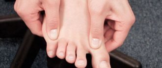
There are many diseases that can cause pain and sensory disturbances in the foot. One of them, although not the most common, is Morton's neuroma. Many people suffering from this pathology say that they live with constant pain and walk “as if on sharp stones.” This is due to the growth of the nerve sheath and the false hope that everything will go away on its own. Unfortunately, this does not happen, and orthopedists, neurologists and therapists are often forced to deal with advanced cases of Morton's neuroma. Although it is possible to solve a problem that has been poisoning the lives of many people for years and does not allow them to enjoy active recreation and even walks in just 15-60 minutes.
What is Morton's neuroma
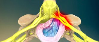
The foot is built in such a way that a thin metatarsal bone leads to each toe; their heads, together with the phalanges of the toes, form the metatarsophalangeal joints. In the immediate vicinity of the metatarsal bones, intermetatarsal nerves pass, branching into 2 branches in the area of the interdigital space. Each of these branches is responsible for transmitting nerve impulses to the lateral surfaces of neighboring fingers. In the area of the metatarsal heads, the interdigital ligament passes, touching the intermetatarsal nerves.
Morton's neuroma is a thickening of the intermetatarsal nerve sheath at the site of its division into 2 branches innervating two adjacent toes. Since it is located in close proximity to the heads of the metatarsal bones and the transverse ligament of the foot, these structures are capable of compressing the thickened nerve during walking and other types of physical activity, which leads to pain of varying degrees of intensity.
But the name of the disease does not accurately reflect its nature and essence. The concept of neuroma is widely used in oncology to describe tumors of the nerves, but in this case we are not talking about the formation of a tumor, and therefore the pathology has nothing to do with malignant processes. Since the formation of Morton's neuroma is accompanied by the formation of a thickening of the nerve, which can even be felt upon palpation, historically it came to be called a neuroma. The name has taken root in medicine, although it would be more correct to call this pathology Morton’s metatarsalgia.
Morton's neuroma is also called perineural fibrosis, intermetatarsal neuroma, plantar neuroma.
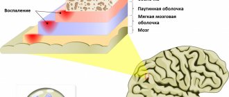
Morton's neuroma is one of the diseases accompanied by the development of carpal tunnel syndrome. In essence, it is a benign thickening of the nerve, formed as a result of thickening of its sheath and the formation of a connective tissue sheath around it. It was found that its dimensions can be 0.15-0.65 cm in width and 0.95-1.45 cm in length, i.e. the thickening has an elongated, spindle-shaped shape.
Most often, young women suffer from this pathology, and damage to the nerve trunk of only one of the feet is usually observed, although the development of a bilateral process is also possible. In most cases, the thickening is located between the 3rd and 4th toes, where the painful lump occurs. Somewhat less frequently, it forms in the 2nd interdigital space and very rarely in the 1st and 4th, i.e., at the big toe and little toe. Since the branches extending from the affected nerve innervate the lateral surfaces of the corresponding fingers, pain when irritated by the thickening tends to radiate to them.
Interestingly, Morton's neuroma almost never occurs in children. Their similar symptoms are usually caused by metatarsal fractures or metatarsophalangeal joint dislocations.
Thus, Morton's neuroma does not pose a threat to life, but can significantly reduce its quality, making it difficult to lead an active lifestyle and perform everyday tasks due to severe pain in the feet and fingers. Due to the resulting limitations in mobility, patients experience serious physical and psychological discomfort, which should not be tolerated. The possibilities of modern medicine make it possible to get rid of constant pain in the foot and restore the patient’s joy of life and movement.
Massage and Morton's neuroma
Nerve compression therapy
Morton's neuroma is one of the most common diseases associated with pinched nerves of the lower extremities. The term “neuroma” somewhat distorts the true meaning of this problem, since the cause of the development of this disease is not always a neuroma - a cyst or tumor of the nervous tissue. Morton's neuroma involves nerve damage in the forefoot. Strictly speaking, in most cases we are talking about nerve compression syndrome, despite the fact that this problem can be caused by fibrosis of the tissues surrounding the nerve. This disease is also called Morton's metatarsalgia (that is, pain in the metatarsal part of the foot) or interdigital neuritis. Morton's neuroma is a nerve compression problem, so massage can be a great help in managing symptoms.
ANATOMICAL REFERENCE
This nerve compression problem is sometimes confused with Morton's toe, a characteristic feature of the foot in which the second metatarsal of the foot is longer than the first. Due to this structure, the second toe is longer than the first (big). However, Morton's finger has nothing to do with the nerve compression that develops in Morton's neuroma. The plantar digital nerves provide innervation to the toes and forefoot. The digital nerves are branches of the medial and lateral plantar nerves, which in turn are branches of the tibial nerve. The tibial nerve is a continuation of the sciatic nerve (Fig. 1).
The study of the pathways of the plantar digital nerves made it possible to understand why, during the development of this disease, they are subject to compression between the heads of the metatarsal bones. The metatarsal heads are bound together by deep and superficial transverse metatarsal ligaments, which creates a small opening, a tunnel, between them. The plantar digital nerves pass through these narrow tunnels between the heads (Fig. 2). For the plantar digital nerves, the passage between the superficial and deep transverse metatarsal ligaments is quite narrow. At the same time, these nerves must pass between the heads, which narrows the tunnel even more.
Although these nerves can be compressed between any pair of heads, the plantar digital nerves are most commonly pinched between the third and fourth metatarsals. The distance between the heads of these metatarsal bones is especially small.
Another factor leading to pinching of the plantar digital nerves is an anatomical abnormality in the separation of nerve fibers. This anomaly implies the presence of a communicating branch between adjacent digital nerves. In the presence of this connecting branch, the likelihood of compression of the nerve increases due to a decrease in its mobility. Moreover, there are other key biomechanical factors leading to the development of Morton's neuroma.
BIOMECHANICS AND NERVE COMPRESSION
Compression of the plantar digital nerve is most common in women. According to a recent study, they are 10 times more likely to develop this disease than men. One of the reasons for this is wearing high-heeled shoes. If the heel lifts the back of the foot high relative to the front, it causes the forefoot to “press” into the forefoot of the shoe, compressing the metatarsal heads, which increases pressure on the structures located between these heads. Wearing shoes with a narrow front contributes significantly to the development of this problem - men should also pay attention to this.
Wearing such shoes is not always the key factor. Fractures and dislocations can also narrow the tunnel through which the nerve passes. In addition, the cause of compression may be pregnancy, diseases associated with impaired circulation of fluids in the body, fluid retention in the lower extremities or hygromas.
The biomechanics of foot movement also plays a significant role in the development of digital nerve compression. During the push-off phase of the gait cycle, hyperextension of the metatarsophalangeal joints occurs (Fig. 3). With toe hyperextension, the plantar digital nerves are greatly stretched. This foot position increases stress on the nerve, especially if it is already pinched by adjacent structures. As a result, clients often complain of increased pain during extension during the push-off phase.
KEY SYMPTOMS
The plantar digital nerves are sensitive and therefore, when they are compressed, symptoms such as loss of sensation, paresthesia and, most often, a tingling or burning sensation in the toes occur. The pain is often described as sharp and specific, rather than diffuse and dull in nature, which is typical for other diseases in this area.
The most common location for pinched digital nerves is between the third and fourth metatarsals, so most symptoms occur in the third and fourth toes. Compression of the digital nerves between other metatarsals is certainly possible, but is less common. Clients report an increase in the intensity of symptoms when wearing shoes with a narrow forefoot. Also, the severity of symptoms may increase when wearing tightly laced shoes. Due to the application of excessive stretching force to the nerve when the toes are extended, symptoms may also increase when walking up or down stairs.
Problems associated with nerve compression can sometimes be difficult to recognize with diagnostic testing. High-precision diagnostic methods, such as MRI, can show the spatial relationships of adjacent structures. For example, how close the nerve is to the bone. However, based on this alone, we cannot say with certainty that nerve compression is occurring. In this case, the most effective method of identifying the problem is clinical diagnosis. Multiple studies have shown that a thorough clinical examination is significantly more effective than MRI in detecting Morton's neuroma. This is good news for massage therapists as it places an emphasis on precise and detailed inspection and palpation of nearby tissue. Having the appropriate skills, you can save the client from the need to resort to MRI.
In addition to diagnosing specific clinical symptoms that may indicate a pinched digital nerve, there are also specific tests that can help identify pinched digital nerves. Because this nerve lies deep in the foot and is located directly between the metatarsal heads, it is impossible to induce symptoms by superficial palpation. The following specific tests are designed to carry a large load
It was the plantar digital nerves that were received, which, being pinched or damaged, when pressure is applied, will respond with the appearance of characteristic symptoms.
The first test is the interdigital compression test, which is most often used by doctors to detect digital nerve compression. One toe is placed on the dorsal surface of the foot in the space between the metatarsals (usually between the third and fourth). The big toe is located on the lower surface of the foot (Fig. 4). The therapist squeezes the foot between two fingers, applying moderate pressure. If this results in acute neurological symptoms in the toes, it is a sign of pinched digital nerves.
Doctors also often use the Morton test. The therapist places the palm of one hand on the medial side of the client's foot near the metatarsophalangeal joint. The other hand is on the lateral side of the foot at the metatarsophalangeal joint of the fifth toe (Fig. 5). The therapist then applies moderate-intensity pressure by squeezing the foot between the palms of the hands. Be careful not to bend the transverse arch of the foot. Squeeze the metatarsal heads evenly together. This position of the metatarsal bones resembles their position when wearing shoes with a narrow front part. The occurrence of neurological symptoms in the toes indicates the presence of a pinched digital nerve.
THERAPY STRATEGIES
The primary goal in the treatment of any type of nerve compression is to relieve or relieve pressure on the nerve. Tissue regeneration begins only after the impact of biomechanical forces (compression or tension) subsides.
First of all, it is worth strongly recommending that the client change his shoes, avoiding wearing shoes with a narrow front part, in order to give the foot the opportunity to adjust to its physiological position. Some shoe manufacturers are well aware of this problem, for example, Altra sneakers are shown in Fig. 6. The figure clearly shows that the foot takes a physiological position in them. Increasing the front part of the shoe not only reduces the degree of compression of the metatarsal heads, but also makes it possible to comfortably wear orthopedic insoles.
Don't underestimate this simple step. A pinched nerve due to wearing the wrong shoes can lead to nerve damage, which can cause sensitization of the entire nervous system. Morton's neuroma, while a relatively simple problem, can lead to the development of more serious nerve problems.
Traditional medicine strategies involve reducing irritating symptoms to avoid nerve sensitization. Corticosteroids and anesthetic injections are most commonly used. However, if you do not pay attention to the biomechanical factors leading to the development of this disease, pharmacological methods bring only short-term relief. Shock wave therapy, capsaicin applications, physical therapy or surgery are also used if conservative treatment is ineffective. The operation involves excision of the plantar digital nerve. Excision reduces the severity of pain, but also leads to loss of sensitivity in the fingers.
Massage can be effective, but never apply deep pressure to the area between the metatarsal heads. Applying pressure to this area can lead to increased compression. Massage therapists should be careful when stretching the toes by extending them.
The best results can be achieved by using techniques aimed at increasing the distance between the metatarsal heads and mobilizing them.
The first step is to apply the technique of mobilizing the metatarsal bones, which will lead to
significant reduction in the severity of symptoms. Stretch the metatarsal heads (not just the toes), spread them apart and hold in this position to stretch the foot muscles and other soft tissues (Fig. 7). Move your metatarsals back and forth from this position to increase their mobility. When combining this technique with other techniques aimed at increasing the distance between the heads of the metatarsal bones (I indicated one above), the effectiveness of therapy increases significantly.
Such techniques that increase joint mobility are certainly effective in treating this disease. However, it is difficult to achieve a lasting effect with soft tissue therapy alone; a multifaceted approach is required.
I repeat - first of all, you need to get rid of all biomechanical factors (wrong shoes), and then start working with soft tissues.
And one more thing - Morton's neuroma is often confused with sciatica or plantar fasciitis. Treatment will be effective only with correct diagnosis.
Whitney Lowe
Reasons for development
American surgeon Thomas George Morton in 1876 described a syndrome that he observed in 12 patients with unusual foot pain in the area of the 3rd interdigital space. He suggested that this is a consequence of mechanical compression of the nerve by the heads of the metatarsal bones. But today it is believed that in fact the disease is of a polyetiological nature.
The exact mechanism of formation of Morton's neuroma is not yet known, although various studies have been carried out in this direction, thanks to which a substantial amount of knowledge has been accumulated. As a result, there were suggestions that thickening of the nerve most likely becomes a consequence of constant overload of the forefoot, frequent repeated microtraumas, long-term, constantly repeated compression, i.e. squeezing, of the foot and, accordingly, the nerve. There is also evidence that nerve thickening can form as a result of ischemic phenomena, i.e., nutritional disorders, which can be observed when the walls of the blood vessels supplying a given area of the foot thicken or their lumen is completely blocked.
Therefore, today it is believed that nerve compression and constant microtrauma lead to a gradual thickening of the transverse intermetatarsal ligament of the foot with its disintegration and the development of edema. The formed pathological intermetatarsal ligament causes constant compression and displacement of the medial plantar nerve and the vessels that supply it, which provokes the occurrence of ischemic phenomena and compensatory thickening of the plantar nerve sheath.
Additional factors contributing to the development of the disease may be damage to bones and soft tissues, as well as the development of the inflammatory process, which is accompanied by the formation of an infiltrate. All this can lead to the nerve becoming firmly fused with the surrounding tissues, its sheath thickening to protect the delicate structure from damage, resulting in the formation of a space-occupying lesion. Since the distance from the intermetatarsal nerve to the metatarsal bones and interdigital ligament is extremely small, these anatomical structures compress the overgrown nerve sheath, i.e., its compression occurs, which causes the occurrence of tunnel syndrome with all its manifestations.
It is believed that genetic predisposition also plays a role in the occurrence of Morton's neuroma. Although the action of external and internal factors is still given leading importance.
Thus, the main reasons for the development of the disease are:
- transverse flatfoot and/or development of valgus deformity of 1 toe (Hallux Valgus);
- wearing shoes that are too tight, in particular narrow shoes with high heels, which explains the fact that the pathology is 4 times more common in women;
- injuries and formation of hematomas at the site of the nerves of the foot;
- gait disturbances with the foot turning inward, which leads to excessive tension on the nerve;
- vascular pathologies of the lower extremities, accompanied by fusion or closure of their lumen and acute circulatory disorders, i.e. obliteration (obliterating atherosclerosis, endarteritis of the lower extremities);
- obesity, leading to increased stress on the legs and feet in particular;
- increased loads on the forefoot;
- inflammatory diseases of the joints and ligaments of the feet (bursitis, tendovaginitis);
- some infectious and autoimmune diseases;
- lipomas of various types in the metatarsal area;
- changes in nerve anatomy.
Very often, Morton's neuroma occurs in women who constantly wear tight high-heeled shoes, as well as in people who actively engage in sports or are forced to stand for long periods of time. Excess weight and heavy physical labor or overly active sports are the main causes of pathology in men.
Pregnancy can act as a trigger for the rapid development of the disease. This is due to the relatively rapid increase in a woman’s body weight, which leads to an increase in the load on the foot. Therefore, it is extremely important from the first days of pregnancy to wear comfortable, non-squeezing shoes and avoid too much stress.
Thus, the risk group for developing Morton's neuroma includes:
- women who constantly wear narrow high-heeled shoes;
- people who are forced to stand or walk for a long time due to the nature of their profession (salespeople, teachers, flight attendants, waiters);
- people engaged in heavy physical labor (loaders, professional athletes, especially figure skaters, speed skaters, track and field athletes);
- patients with any diseases and deformities of the feet, especially flat feet, bursitis, arthrosis, tendinitis, tumors, hallux valgus.
Clinical Picture
Patients with Morton's neuroma experience pain in the forefoot, particularly in the ball of the foot and between the toes.
However, not every forefoot pain indicates Morton's neuroma. In fact, most chronic forefoot pain is not the result of Morton's neuroma; it is often the result of inflammation (synovitis) of the toe joints. Symptoms of Morton's neuroma involve irritation of one or more small nerves before they pass through the fingers (Figure 1). Symptoms of Morton's neuroma include pain, numbness (usually between the toes), and sometimes a burning sensation.
Rice. 1: Nerve location (between the base of the 2nd and 3rd fingers)
In many cases, a neuroma can develop as a result of overuse of the forefoot. Such overload, for example, can occur if you regularly wear high-heeled shoes with narrow toes. Sometimes the anatomical features of the patient’s foot structure contribute to overloading the forefoot.
Often neuroma develops spontaneously, for no apparent reason. However, as soon as the pressure on the nerve begins, namely the pressure that occurs when walking or the pressure of neighboring bone structures (metatarsal heads), constant pain occurs (Fig. 2). Patients begin to experience pain that intensifies when walking, especially when walking in narrow shoes with thin soles or high-heeled shoes. Such patients feel more comfortable without shoes.
Rice. 2: Location of the nerve under the skin (between the base of the 3rd and 4th fingers)
Symptoms of Morton's neuroma
While the neuroma is small in size, i.e., does not reach 5 mm, it usually does not manifest itself in any way and patients do not even suspect the presence of any problem in the foot. In such cases, pain or a feeling of tightness may appear when putting on uncomfortable shoes and quickly go away after they are removed. Sometimes there is discomfort between the toes after wearing such shoes or a slight tingling sensation.
However, provided that the action of the provoking factors continues, the thickening begins to increase in size, which first leads to the appearance of shooting and aching pain in the area of the 3rd interdigital space. They usually occur after exercise and walking, and may also be accompanied by the development of various sensory disorders, including:
- crawling sensation;
- burning;
- tingling;
- pain of varying intensity arising from mechanical or thermal irritation.
Moreover, Morton's neuroma is characterized by an almost complete absence of any discomfort in the foot at night.
If at this stage of the development of the disease a person does not consult a doctor, and the cause of pain, paresthesia, allodynia and other sensitivity disorders is not established and, accordingly, treatment with lifestyle correction is not carried out, the symptoms of the disease continue to intensify. The frequency and severity of pain, which can become throbbing, gradually increases; the interval between the start of physical activity and the appearance of the first discomfort is observed to decrease. Often, pain syndrome forces a person to take off their shoes and massage the affected area of the foot. This is due to the continued growth of the nerve thickening.
The disease can have a wave-like course, that is, with periods of intensification of symptoms and their temporary subsidence or even elimination. Sometimes manifestations of the disease may be absent even for several years.
In the later stages of pathology progression, when Morton's neuroma reaches large sizes, patients suffer from:
- sharp, burning pain that can occur even at rest and does not depend on the type of shoes the patient wears;
- sensations of the presence of a foreign object in shoes (as a rule, patients talk about the feeling of a pebble getting into their shoes);
- increasing intensity of sensitivity disorders up to its complete loss;
- changes in gait with forced lameness, since it is difficult to step on the affected leg, and the need to frequently stop while walking.
What is characteristic of this disease is the complete absence of external changes in the condition of the foot and the affected interdigital space. In this case, when you press on the area where the thickening of the nerve is formed, the pain increases sharply.
Severe pain syndrome that occurs during physical activity makes it impossible to engage in sports that involve stress on the legs, i.e. running, jumping, dancing, skiing, roller skating, skating, etc. Even normal walking becomes a challenge for people with this pathology .
Painful neuroma - symptoms
Neuralgic pain differs from regular pain in that it can occur immediately after an injury or long after it. In most cases, the pain is long-lasting and does not go away without treatment. As a result of nerve damage, the following symptoms may develop:
- Pain like a burning sensation, electrical burn, stab wound, or cut
- Feeling of tingling and numbness in the area
- Allodynia is the occurrence of pain due to exposure to irritants that usually do not cause it. For example, even minor irritation from wind or clothing can cause a sensation of pain in the affected area.
- Hyperalgesia - a stimulus that usually causes a sensation of tolerable pain, such as a needle puncture during blood sampling, results in terrible pain that cannot be tolerated
- Hyperpathy is a particularly prolonged and very strong pain reaction to a painful stimulus that does not go away with cessation of stimulation. External factors, such as changes in weather, touching clothes or shoes, etc., can aggravate central pain.
Diagnostics
If signs of Morton's metatarsalgia develop, you should consult an orthopedist or neurologist. Diagnosis of the disease is not difficult, and in most cases the doctor can suspect the presence of this particular pathology only on the basis of data obtained during a survey and examination of the patient.
Finding out the characteristics of the shoes that a person is used to wearing, as well as the length of time he or she wears them every day, is of great diagnostic importance. The specialist will definitely inquire about the presence of chronic and autoimmune diseases, vascular disorders, past injuries, etc. During the examination, the doctor draws attention to the presence of excess weight, flat feet and the presence of pain when palpating the area between the 3rd and 4th fingers or others, depending on the where the discomfort is felt.
A special test can also be performed, which consists of pressing the 3rd intermetatarsal space for 30-60 seconds. The presence of Morton's neuroma is indicated by the occurrence of numbness and burning at this point, which can spread to the lateral surfaces of the 3rd and 4th fingers. Additionally, a positive Tinnel's sign can indicate the presence of Morton's neuroma, i.e., the occurrence of pain when tapping the metatarsal bones of the 3rd and 4th toes, as well as the appearance of severe pain when squeezing the forefoot and toes.
However, in order to confirm the benign nature of the disease and exclude a number of other pathologies that can be accompanied by similar symptoms, patients are prescribed instrumental research methods. Indeed, much more often than the development of metatarsalgia, pain in the foot is caused by various deformations, in particular flat feet, which can be one of the causes of Morton's neuroma, as well as chronic tendinitis of the Achilles tendon. Differentiation is also required with hereditary spastic paraplegia, synovitis, arthritis of the metatarsophalangeal joints, stress fractures, osteonecrosis of the metatarsal bones, and tumor formation.
Some pathologies of the spine may also be accompanied by pain and sensory disturbances in the foot, in particular intervertebral disc herniations in the lumbar region.
To diagnose Morton's neuroma, the following may be performed:
- MRI or magnetic resonance imaging is an effective method for diagnosing a huge number of different soft tissue diseases. But in the case of Morton's neuroma, it is not always able to provide comprehensive data to confirm or refute the diagnosis, and often gives questionable results.
- CT or computed tomography - allows you to obtain accurate information about the condition of bone structures, especially at the site of thickening of the nerve, the presence of flat feet, and the consequences of injuries.
- Ultrasound is one of the main methods for diagnosing the disease, as it allows you to accurately determine the area of thickening of the intermetatarsal nerve, as well as detect degenerative-destructive changes in the joints.
The presence of Morton's neuroma can also be confirmed as the cause of the appearance of pain and sensitivity disorders of varying degrees of severity through a therapeutic and diagnostic blockade. This is the name given to the injection of a local anesthetic solution carried out in close proximity to the passage of the intermetatarsal nerve. A rapid reduction to complete extinction of pain is a convincing diagnostic criterion, indicating the development of Morton's metatarsalgia.
Diagnosis of Morton's neuroma
The diagnosis of Morton's neuroma is usually made based on patient complaints and examination.
The examination consists of provoking symptoms of Morton's neuroma. That is, the doctor tries to compress the nerve between the metatarsal bones by squeezing the foot with a hand. If the nerve is thickened and inflamed, then within 30-60 seconds the patient begins to experience numbness and a burning sensation in the 3rd interdigital space. This way the diagnosis is confirmed.
X-rays or MRI are performed to exclude other pathologies (consequences of trauma, osteoblastoclastoma and other tumors and soft tissue diseases).
Treatment of Morton's neuroma
For each patient, treatment tactics are developed individually, which largely depends on the stage of the disease, as well as the causes of its occurrence. In the initial stages of development, patients are usually recommended conservative therapy, which consists of:
- unloading the feet and abandoning tight shoes in favor of comfortable orthopedic ones with a small heel 2-4 cm high, ensuring proper distribution of the load on the foot;
- using metatarsal pads and lifts, which are special orthopedic inserts that are placed under the balls of the feet and fixed on the toes with elastic bands or silicone rings in order to separate the metatarsal bones and create an anatomically correct bend of the foot;
- wearing shoes with retrocapital support.
All these methods have one goal - reducing pressure on the affected nerve trunk. This will avoid further progression of the disease and reduce pain by reducing the load and compression of the intermetatarsal nerve.
In order to increase the effectiveness of the measures taken, patients are recommended to constantly wear orthopedic insoles specially made for them according to individual parameters. Thanks to this solution you will be able to:
- reduce the load on the forefoot and normalize the condition of the transverse arch, i.e., reduce flat feet;
- reduce the pressure of bones and ligaments on the modified nerve trunk;
- avoid the development or eliminate the inflammatory process in soft tissues, including those involving the nerve, which will make it possible to significantly reduce the intensity of pain;
- restore the anatomy of the foot, which will improve the gait and bring it closer to normal.
In the absence of severe pain, patients can be prescribed multicomponent compresses, which usually include NSAIDs, local anesthetics and dimexide, taking NSAID drugs orally in the form of tablets or capsules, or applying them directly to the interdigital space in the form of an ointment or gel. This helps to improve the condition, but does not have a pronounced therapeutic effect. In combination with compresses, muscle relaxants are usually prescribed, as well as manual therapy sessions, which can not only relieve muscle spasms and improve tissue nutrition by activating blood circulation, but also eliminate nerve compression by normalizing the position of the metatarsal bones and ligaments.
Additionally, you can massage your feet at home, as well as take relaxing baths with herbs.
Courses of physiotherapeutic procedures are also indicated. So, with Morton's neuroma, the following give a good effect:
- Magnetic therapy is a method that involves influencing the affected area with a pulsed magnetic field, which helps reduce inflammation, swelling and improve the course of metabolic processes in it;
- shock wave therapy (SWT) is a method of physical influence, through the use of which it is possible to improve blood circulation in the area of the thickening of the intermetatarsal nerve and thereby reduce the severity of ischemia, as well as accelerate the elimination of inflammation and achieve pain reduction;
- electrophoresis with the introduction of anti-inflammatory and analgesic drugs - ensures rapid and deeper penetration of drugs into tissues, and therefore obtains a more pronounced therapeutic effect;
- acupuncture – involves irritation of biologically active points, which helps to activate recovery processes.
Light therapeutic exercises are often additionally prescribed, the implementation of which does not take much time, but allows you to activate blood circulation in the legs, increase joint mobility, and also strengthen and stretch the muscles of the feet. But you need to do exercise therapy every day without haste at a pace that is convenient for you.
The main exercises include the following:
- Sitting on a chair with your feet firmly planted on the floor, you need to pull your socks towards you as much as possible, and then pull them forward.
- Standing facing the wall and leaning against it with outstretched arms, you need to take one leg back about 30-50 cm. The remaining leg in front is rhythmically bent at the knee, making sure that both heels are pressed firmly to the floor.
- Sitting on the floor with straight legs stretched out in front of you, use a towel to pull your feet towards you with your hands.
- Sitting on a chair, they make small movements with their toes, tearing them off and placing them on the floor, imitating playing the piano.
- Sitting on a chair, place one leg with the ankle joint on the knee of the other and perform rotational movements alternately in both directions, trying to make as wide a rotation as possible.
If these measures are not enough or a more severe course of the disease is initially observed, patients are prescribed corticosteroid injections, which are performed in the intermetatarsal space on the dorsum of the foot. In 50% of cases, this measure is enough to significantly improve well-being. Such blockades reduce swelling and inflammation, which leads to a decrease in pain.
In cases where concomitant orthopedic pathologies, such as deforming arthrosis, are detected in patients, treatment appropriate to the situation is prescribed.
However, conservative therapy is not always effective. Therefore, if patients, despite the measures taken, are still bothered by pain and other symptoms of Morton’s neuroma, they are recommended to undergo surgical treatment of the pathology.
Morton's neuroma: symptoms, diagnosis and treatment
Morton's neuroma is a specific foot disease. It is a benign thickening and occurs as a result of the proliferation of fibrous tissue. This occurs in the area of the plantar nerve of the foot. As a result, the bones and ligaments located next to the nerve begin to compress it. Generally, this tumor appears between the 3rd and 4th toes, in an area called the third intermetatarsal space. Very rarely, a neuroma can be found in another place - between the 2nd and 3rd fingers. Another characteristic of this disease is that it almost always occurs on only one foot. In most cases, nerve damage in neuroma is unilateral. Bilateral is very rare. Morton's neuroma may have other names. These are foot neuroma, interdigital neuroma, Morton's finger syndrome, Morton's syndrome, perineural fibrosis and intermetatarsal neuroma. This pathology occurs most often in women older than middle age.
Causes leading to the appearance of the disease
- Wearing tight and narrow shoes, as well as high-heeled shoes;
— Excess body weight; — Transverse flatfoot; — Mechanical impact, when the nerve is compressed by the bones of the metatarsus; — Increased loads experienced by the forefoot. Occurs when a person has to stand for a long time; — Various infections, hematomas and other foot injuries. Symptoms of the disease:
Morton's neuroma in the initial stage may have no visual signs.
The main symptom of the presence of pathology is pain that occurs when the forefoot is compressed transversely. The patient may complain of numbness in the toes, aching pain, and a feeling that there is a foreign object between the toes. All these symptoms are not pronounced, and the disease itself is sluggish. This may continue for several years. Exacerbations of the disease occur when wearing tight and narrow shoes. The pain appears while walking and subsides if you take off your shoes and massage the foot. When the disease enters its final stage, the pain becomes severe and shooting. Now it appears regardless of shoes and load size. Every year the pain becomes stronger. Diagnosis of Morton's neuroma
Diagnosis of the disease takes place in several stages. First, anamnesis is collected and the patient is interviewed. The doctor should listen to the patient's complaints and also find out what type of shoes he wears. In order to establish the possible cause of the disease, the doctor collects information about what leg diseases the patient has had in the past. These can be muscle diseases, arthritis and various types of injuries. The doctor then palpates the foot by applying pressure with their fingers to find out where the pain or numbness is. In cases where diagnosis is difficult, an x-ray or MRI may be taken. To determine the location of the neuroma, a method such as the administration of anesthetics is used.
Shock-absorbing inserts for the metatarsus Secret Orto Shock-absorbing inserts for the metatarsus Secret Price: 450 rub.
Orthopedic insoles Orto-Ultra Orto Orthopedic insoles Orto-Ultra Price: 2920 rub.
Available sizes: 36, 37, 38, 39, 40, 41, 42, 43, 44, 45, 46
Orthopedic insoles Optimum Green Orto Orthopedic insoles Optimum Green Price: 3960 rub.
Available sizes: 36, 37, 38, 39, 40, 41, 42, 43, 44, 45, 46
Treatment methods
Treatment of Morton's neuroma is carried out in two ways - conservative and surgical. Conservative treatment is usually used at the onset of the disease. All measures and procedures of this method serve to eliminate unnecessary pressure on the nerve area. In this case, shoes must be replaced. It should be loose and comfortable, with low heels. The best option is special orthopedic shoes. A pad should be used where the foot is compressed. Patients also need to systematically undergo physiotherapeutic procedures. To relieve pain and inflammation, the patient is usually prescribed non-steroidal anti-inflammatory drugs. This may be ibuprofen, indomethacin or others. If this treatment is ineffective, you can try other drugs - steroids. Together with an anesthetic, they are injected directly into the area of the neuroma. Drugs such as hydrocortisone, Kenalog or Diprospan may be used. Typically, conservative treatment is successful in 80% of cases. If conservative treatment does not give the desired result, surgical intervention is used. This procedure is done under local anesthesia. During it, the metatarsal canal must be opened, and then the neuroma itself must be dissected. Part of the nerve may also be removed. Often this surgery can cause numbness in part of the foot, which is temporary. After the operation, during the rehabilitation period, which lasts approximately 2 weeks, you should wear orthopedic shoes. The forefoot should be provided with the highest possible level of rest. You can walk the very next day after surgery, but the walking should not be long.
Home-prepared remedies allow you to cope with the initial forms of Morton's Neuroma in just 1-2 weeks. For making warming ointment 100 g. badger, pork or goose fat mixed with 1 tbsp. table salt. The resulting ointment is applied to the painful area and covered with a bandage. It would be a good idea to put a warm sock on top and leave the compress on until the morning. For foot treatment to be effective, the procedure must be carried out daily until the symptoms completely disappear, and during the daytime, wear shoes with orthopedic insoles.
Possible consequences that the disease can lead to:
If a progressive neuroma is not treated, the consequences can be very disappointing. This is the continued growth of the tumor, increasing discomfort and pain in the foot. Without treatment, physical activity such as running or dancing becomes impossible. You can also forget about sports. If you feel the slightest pain or discomfort in the forefoot, contact a specialist immediately. If Morton's neuroma is detected, treatment should begin immediately. We remind you that wearing correctly selected orthopedic shoes is the most important component of such treatment.
ATTENTION! All information posted on this site is advisory in nature. In each individual case, consultation with a specialist is necessary. January 11, 2019
Surgery for Morton's neuroma
The current level of development of orthopedics allows for effective and rapid treatment of Morton's neuroma at any stage of development. Today there are several techniques used for this purpose.
Often the thickening of the nerve trunk is removed, which is performed under local anesthesia. The surgeon individually chooses the type of access to the nerve: from the back or plantar side of the foot. He then makes a soft tissue incision to visualize the nerve and resects the thickened part. After this, the tissues are sutured and covered with a sterile bandage.
This operation lasts no more than an hour and involves the removal of a modified section of the nerve. This leads to a rapid improvement in the condition and complete disappearance of the pain syndrome with no risk of relapse of the disease, i.e. its re-development. But the method has a drawback - loss of sensitivity in the area of the foot for which the nerve was responsible for innervation.
The lack of sensitivity in such a small area does not in any way affect the supporting and motor function of the foot. It does not affect the patient’s condition and appears only when intentionally touching the area that has lost sensitivity. After such an operation, the recovery stage takes 2-4 weeks, during which it is recommended to reduce the load on the operated foot. You can get up and walk within a couple of hours after the operation, but you should limit the load on the forefoot, which is achieved through the use of special shoes with hard soles. The stitches are removed 2 weeks after the procedure.
However, loss of sensation even in a small area of the foot seems to many surgeons to be too high a price to pay for treating Morton's neuroma, especially as a primary measure. In such situations, such surgical intervention is considered as a last resort, which should be resorted to only in the absence of the expected effect from other measures. Therefore, they often initially try to cope with the problem and eliminate pain using less radical methods, for example, by cutting the transverse ligament between the metatarsal bones. The operation does not require special preparation and lasts only a few minutes. Dissection of the ligament will eliminate compression of the nerve trunk without compromising its integrity, and therefore preserve sensitivity. If the operation does not give the desired result, you should think about removing the neuroma by excision.
Also, to treat the disease, a surgical treatment technique such as osteotomy of the 4th metatarsal bone can be used. But today it is practically not used. The essence of this type of operation is to create space for the thickened nerve, which will allow it to be decompressed. This is achieved by performing an artificial fracture of the 4th metatarsal bone and displacing its head. Osteotomy and further manipulations are carried out under X-ray control through a minor soft tissue incision or even a puncture with a diameter of about 2 mm. In this case, the foot must be fixed with a plaster cast, and the recovery period increases to 1 month.
Recently, Morton's neuroma is increasingly removed by laser or radiofrequency ablation. The essence of both methods is approximately the same and consists in introducing, under local anesthesia, a thin cannula directly to the site of localization of the nerve thickening, through which a laser or radio wave probe is then immersed. Due to the receipt of thermal energy, the modified section of the nerve trunk is destroyed, which leads to the elimination of the pain syndrome, but also provokes a loss of sensitivity on the adjacent lateral surfaces of the 3rd and 4th fingers. Thus, laser and radio wave treatment of Morton's neuroma allows one to obtain the same effect as open surgery, but is not associated with the formation of scars on the skin, since the puncture size is only a few millimeters, is less traumatic and is easier to tolerate by patients. The duration of the operation is about 30 minutes.
Modern operations performed for Morton's neuroma allow patients to feel better in more than 90% of cases. Moreover, in 45% of cases there is a complete elimination of pain and neurological symptoms, in 32% there is a significant reduction in them. Only in 15% of cases the result was satisfactory, i.e., there was a decrease in pain and preservation of neurological disorders, and in only 8% of patients who underwent surgery, the situation did not change for the better. Negative results are usually due to the formation of a true amputation neuroma of the intermetatarsal nerve.
Metatarsalgia, forefoot pain
The pain that patients experience with metatarsalgia is usually localized in the forefoot at the base of the 2nd and 3rd toes (under the heads of the 2nd and 3rd metatarsals). Patients often describe the feeling as if they were “walking on small pebbles” or as if a sock had rolled under their fingers.
The pain is usually aching and worsens when standing or walking, especially on a hard surface. Patients may also notice a burning sensation that extends to the fingertips.
As the condition progresses, claw-like deformities of the fingers may develop. With such deformities, the plantar fat pad, located under the heads of the metatarsal bones and absorbing the load on the forefoot, moves forward, and the heads of the metatarsal bones along with the metatarsophalangeal joints (MTPs) remain “unprotected”.
Lack of protection and stress absorption leads to the formation of calluses called corns under the metatarsal heads and increased symptoms of metatarsalgia.
Unfortunately, in some patients, the causes of metatarsalgia are masked by the developing secondary claw-shaped deformity of the fingers, which aggravates the existing symptoms.
If we make an imprint of the foot in a standing position (plantogram), we will see signs of overload (more intense staining) in the area of the corresponding heads of the metatarsal bones.
Continued overloads lead to chronic damage to anatomical structures that are subject to such overloads. These anatomical structures are the capsule of the metatarsophalangeal joint, the plantar plate and the metatarsal bone (its head and/or neck).
Constant irritation of the metatarsophalangeal joint as a result of overload leads to its inflammation. The joint becomes swollen. This inflammation of the PFJ is called synovitis.
Patients are unable to fully bend their fingers. Often patients indicate the formation of “bones” along the edges of the foot.
In the early stages of the disease, a symptom of inflammation may be curvature of the small toes.
It is important to understand that many patients and doctors may mistakenly associate some form of metatarsalgia with Morton's neuroma.
Morton's neuroma can also cause metatarsalgia, but secondary neuritis (inflammation of the nerve) due to chronic repeated overload of the metatarsophalangeal joints is much more common.
In such a situation, removal of the neuroma will not lead to complete disappearance of symptoms, or the effect of the operation will be temporary.
Radiation diagnostics
On radiographs of patients with metatarsalgia, one can often see elongation of the 2nd or 3rd metatarsal bones relative to the first or fourth. Occasionally, true subluxation or even dislocation may occur in the metatarsophalangeal joints.
Signs of hallux valgus or midfoot instability may also be seen.
Prevention
Morton's neuroma is a disease whose development can be avoided by following simple rules. First of all, prevention of the formation of thickening of the intermetatarsal nerve consists of:
- wearing narrow high-heeled shoes only on special occasions and for a short period of time;
- preference for comfortable shoes as the main ones;
- prevention of flat feet, which consists of performing a special set of exercises, including rolling a ball with the foot and lifting small objects from the floor with the toes;
- performing a foot massage;
- weight control and maintaining it at optimal levels;
- timely treatment of orthopedic disorders;
- refusal to engage in traumatic sports;
- rational loads on the lower limbs;
- timely diagnosis and treatment of foot injuries;
- prevention of the development of atherosclerosis and other vascular disorders, which is mainly achieved through a healthy diet and rational physical activity.
Thus, Morton's neuroma is a harmless, but extremely unpleasant disease, the development of which is much easier to avoid than to then deal with its consequences. Treatment of pathology in the early stages can be carried out using conservative methods, but without correcting habits and footwear it will not be successful. The most reliable and effective method of combating already formed Morton's neuroma is surgery. Modern techniques make it possible to radically solve the problem of pain in less than an hour and return a person to the ability to move without pain. At the same time, operations of this kind are associated with minimal risks of complications, do not require long and complex rehabilitation, and also allow you to return to most everyday activities the very next day after the procedure.
When is Morton's neuroma surgery performed?
In order to correctly assess the patient’s condition and prescribe the correct treatment, Gelenk Clinic specialists will need to provide current MRI results. Based on the MRI images, the doctor determines the degree of need for surgical treatment of Morton's neuroma. If there are certain indications, the patient is prescribed the best surgical technique for him.
In the early stages, treatment of the disease is carried out using conservative methods, namely special insoles and gymnastic exercises for the feet. If the quality of life is significantly limited, and if the patient experiences severe pain despite conservative treatment, the doctor will refer him for surgery. If the thickening is 0.8 cm in size, a nerve-sparing operation (neurolysis) is performed. If the swelling of Morton's neuroma is greater than normal, the nerve is removed through neurectomy.